

Andrographolide Ameliorates Abdominal Aortic Aneurysm Progression by Inhibiting Inflammatory Cell Infiltration through Downregulation of Cytokine and Integrin Expression
- PMID: 26483397
- PMCID: PMC4702070
- DOI: 10.1124/jpet.115.227934
Andrographolide Ameliorates Abdominal Aortic Aneurysm Progression by Inhibiting Inflammatory Cell Infiltration through Downregulation of Cytokine and Integrin Expression
Abstract
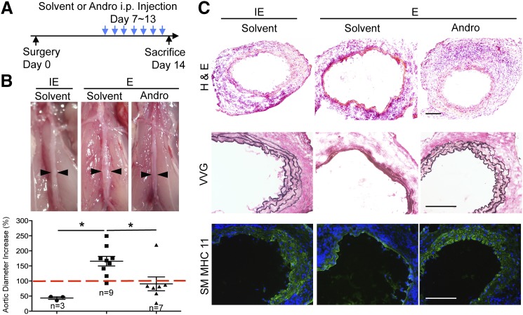
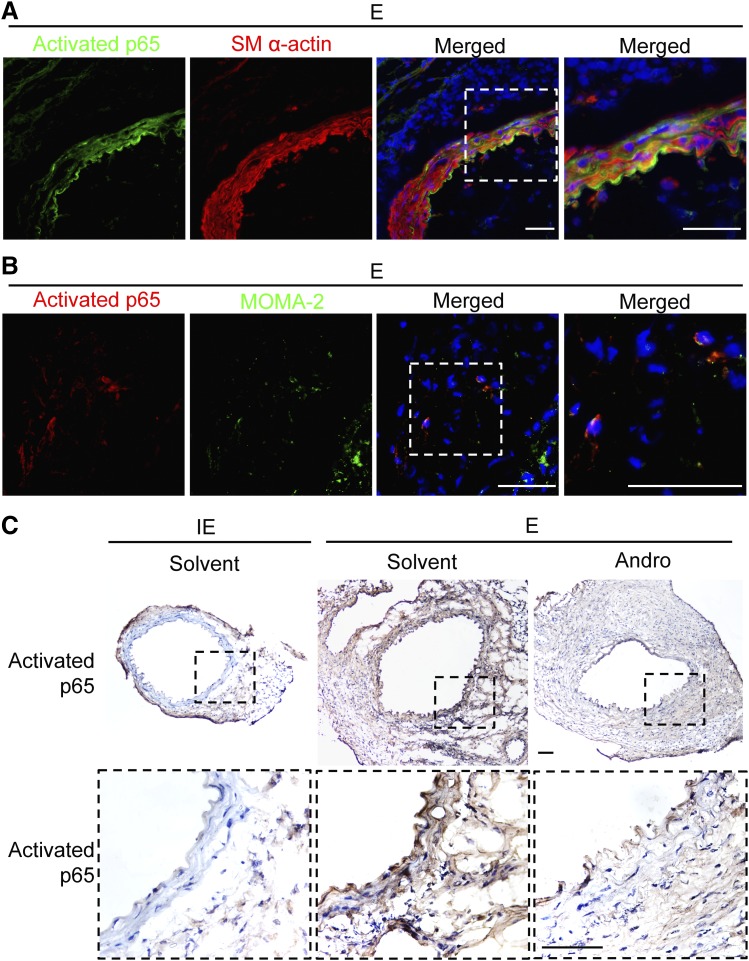
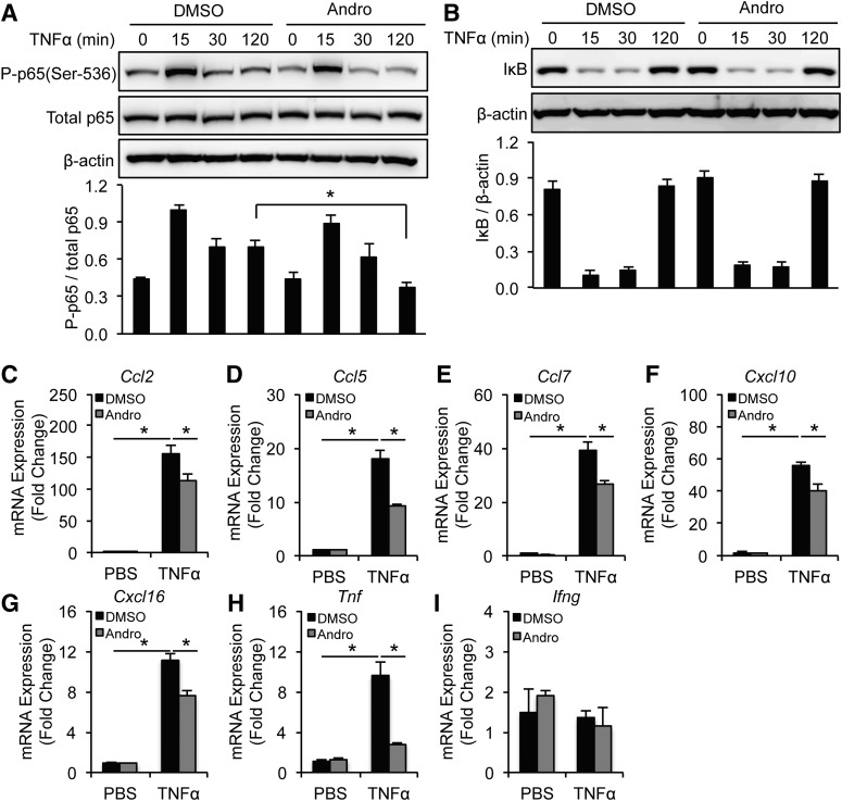
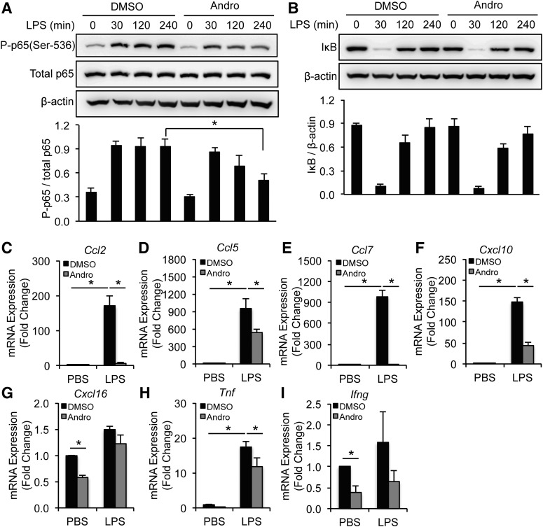
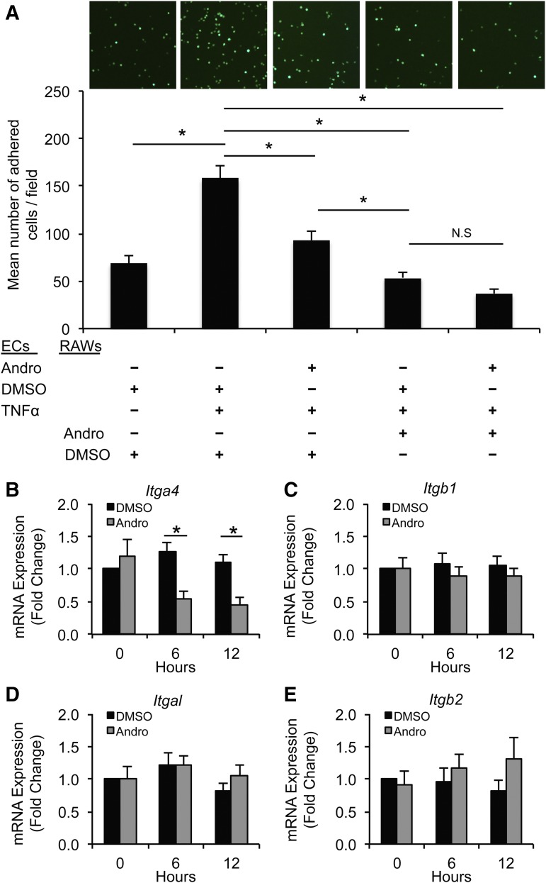
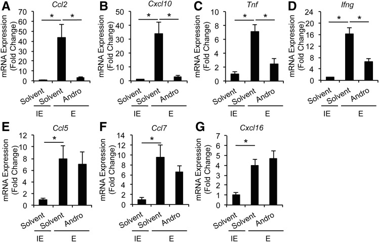
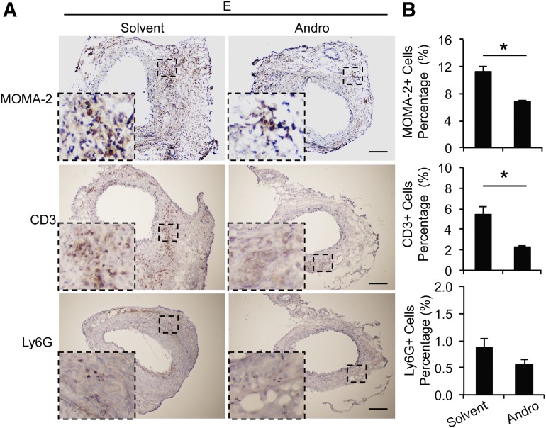
Abdominal aortic aneurysm (AAA), characterized by exuberant inflammation and tissue deterioration, is a common aortic disease associated with a high mortality rate. There is currently no established pharmacological therapy to treat this progressive disease. Andrographolide (Andro), a major bioactive component of the herbaceous plant Andrographis paniculata, has been found to exhibit potent anti-inflammatory properties by inhibiting nuclear factor κ-light-chain-enhancer of activated B cells (NF-κB) activity in several disease models. In this study, we investigated the ability of Andro to suppress inflammation associated with aneurysms, and whether it may be used to block the progression of AAA. Whereas diseased aortae continued to expand in the solvent-treated group, daily administration of Andro to mice with small aneurysms significantly attenuated aneurysm growth, as measured by the diminished expansion of aortic diameter (165.68 ± 15.85% vs. 90.62 ± 22.91%, P < 0.05). Immunohistochemistry analyses revealed that Andro decreased infiltration of monocytes/macrophages and T cells. Mechanistically, Andro inhibited arterial NF-κB activation and reduced the production of proinflammatory cytokines [CCL2, CXCL10, tumor necrosis factor α, and interferon-γ] in the treated aortae. Furthermore, Andro suppressed α4 integrin expression and attenuated the ability of monocytes/macrophages to adhere to activated endothelial cells. These results indicate that Andro suppresses progression of AAA, likely through inhibition of inflammatory cell infiltration via downregulation of NF-κB-mediated cytokine production and α4 integrin expression. Thus, Andro may offer a pharmacological therapy to slow disease progression in patients with small aneurysms.
Copyright © 2015 by The American Society for Pharmacology and Experimental Therapeutics.
Figures
Similar articles
-
Andrographolide inhibits inflammatory responses in LPS-stimulated macrophages and murine acute colitis through activating AMPK.Biochem Pharmacol. 2019 Dec;170:113646. doi: 10.1016/j.bcp.2019.113646. Epub 2019 Sep 20. Biochem Pharmacol. 2019. PMID: 31545974
-
Zoledronate attenuates angiotensin II-induced abdominal aortic aneurysm through inactivation of Rho/ROCK-dependent JNK and NF-κB pathway.Cardiovasc Res. 2013 Dec 1;100(3):501-10. doi: 10.1093/cvr/cvt230. Cardiovasc Res. 2013. PMID: 24225494
-
Agathis dammara Extract and its Monomer Araucarone Attenuate Abdominal Aortic Aneurysm in Mice.Cardiovasc Drugs Ther. 2025 Apr;39(2):239-257. doi: 10.1007/s10557-023-07518-0. Epub 2023 Nov 18. Cardiovasc Drugs Ther. 2025. PMID: 37979015
-
Andrographolide Ameliorates Inflammation and Fibrogenesis and Attenuates Inflammasome Activation in Experimental Non-Alcoholic Steatohepatitis.Sci Rep. 2017 Jun 14;7(1):3491. doi: 10.1038/s41598-017-03675-z. Sci Rep. 2017. PMID: 28615649 Free PMC article.
-
Andrographolide: A promising therapeutic agent against organ fibrosis.Eur J Med Chem. 2024 Dec 15;280:116992. doi: 10.1016/j.ejmech.2024.116992. Epub 2024 Oct 20. Eur J Med Chem. 2024. PMID: 39454221 Review.
Cited by
-
The Role of a Selective P2Y6 Receptor Antagonist, MRS2578, on the Formation of Angiotensin II-Induced Abdominal Aortic Aneurysms.Biomed Res Int. 2020 Apr 16;2020:1983940. doi: 10.1155/2020/1983940. eCollection 2020. Biomed Res Int. 2020. PMID: 32382533 Free PMC article.
-
N1-Methyladenosine (m1A) Regulation Associated With the Pathogenesis of Abdominal Aortic Aneurysm Through YTHDF3 Modulating Macrophage Polarization.Front Cardiovasc Med. 2022 May 10;9:883155. doi: 10.3389/fcvm.2022.883155. eCollection 2022. Front Cardiovasc Med. 2022. PMID: 35620523 Free PMC article.
-
Andrographolide Benefits Rheumatoid Arthritis via Inhibiting MAPK Pathways.Inflammation. 2017 Oct;40(5):1599-1605. doi: 10.1007/s10753-017-0600-y. Inflammation. 2017. PMID: 28584977
-
Andrographolide, a natural anti-inflammatory agent: An Update.Front Pharmacol. 2022 Sep 27;13:920435. doi: 10.3389/fphar.2022.920435. eCollection 2022. Front Pharmacol. 2022. PMID: 36238575 Free PMC article. Review.
-
TNF-α elicits phenotypic and functional alterations of vascular smooth muscle cells by miR-155-5p-dependent down-regulation of cGMP-dependent kinase 1.J Biol Chem. 2018 Sep 21;293(38):14812-14822. doi: 10.1074/jbc.RA118.004220. Epub 2018 Aug 13. J Biol Chem. 2018. PMID: 30104414 Free PMC article.
References
-
- Aoki T, Kataoka H, Ishibashi R, Nozaki K, Egashira K, Hashimoto N. (2009) Impact of monocyte chemoattractant protein-1 deficiency on cerebral aneurysm formation. Stroke 40:942–951. - PubMed
-
- Aoki T, Kataoka H, Shimamura M, Nakagami H, Wakayama K, Moriwaki T, Ishibashi R, Nozaki K, Morishita R, Hashimoto N. (2007) NF-kappaB is a key mediator of cerebral aneurysm formation. Circulation 116:2830–2840. - PubMed
-
- Bown MJ, Sweeting MJ, Brown LC, Powell JT, Thompson SG, RESCAN Collaborators (2013) Surveillance intervals for small abdominal aortic aneurysms: a meta-analysis. JAMA 309:806–813. - PubMed
-
- Burgos RA, Hancke JL, Bertoglio JC, Aguirre V, Arriagada S, Calvo M, Cáceres DD. (2009) Efficacy of an Andrographis paniculata composition for the relief of rheumatoid arthritis symptoms: a prospective randomized placebo-controlled trial. Clin Rheumatol 28:931–946. - PubMed
-
- Clowes AW, Clowes MM, Fingerle J, Reidy MA. (1989) Kinetics of cellular proliferation after arterial injury. V. Role of acute distension in the induction of smooth muscle proliferation. Lab Invest 60:360–364. - PubMed
Publication types
MeSH terms
Substances
Grants and funding
LinkOut - more resources
Full Text Sources
Other Literature Sources
