. 2015 Oct 19;84(1):127-37.
doi: 10.1128/IAI.00912-15. Print 2016 Jan.

Surface Polysaccharide Mutants Reveal that Absence of O Antigen Reduces Biofilm Formation of Actinobacillus pleuropneumoniae

Affiliations

Surface Polysaccharide Mutants Reveal that Absence of O Antigen Reduces Biofilm Formation of Actinobacillus pleuropneumoniae

S Hathroubi et al. Infect Immun. .

Abstract

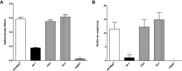
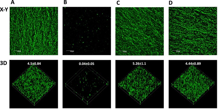
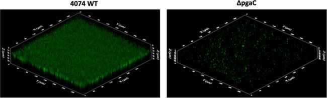
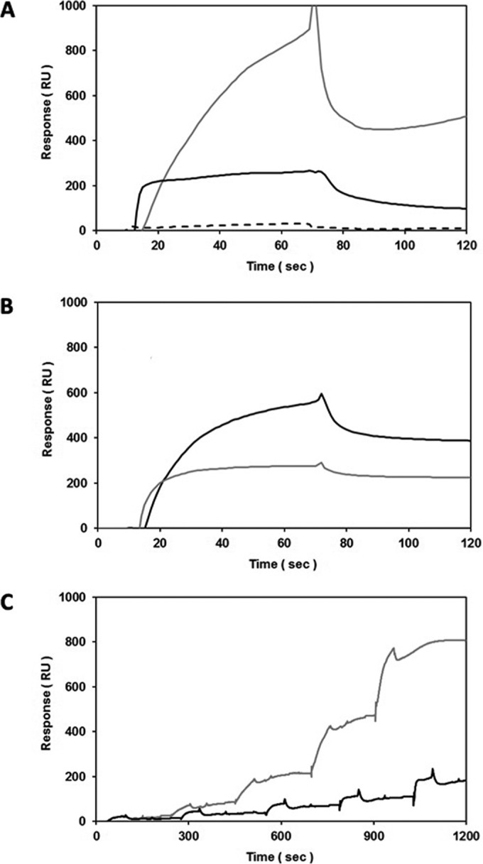
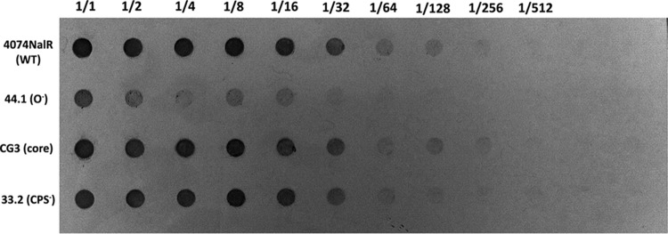
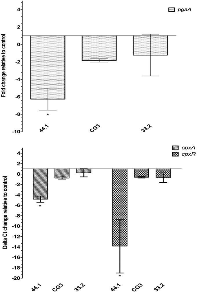
Actinobacillus pleuropneumoniae is a Gram-negative bacterium belonging to the Pasteurellaceae family and the causative agent of porcine pleuropneumonia, a highly contagious lung disease causing important economic losses. Surface polysaccharides, including lipopolysaccharides (LPS) and capsular polysaccharides (CPS), are implicated in the adhesion and virulence of A. pleuropneumoniae, but their role in biofilm formation is still unclear. In this study, we investigated the requirement for these surface polysaccharides in biofilm formation by A. pleuropneumoniae serotype 1. Well-characterized mutants were used: an O-antigen LPS mutant, a truncated core LPS mutant with an intact O antigen, a capsule mutant, and a poly-N-acetylglucosamine (PGA) mutant. We compared the amount of biofilm produced by the parental strain and the isogenic mutants using static and dynamic systems. Compared to the findings for the biofilm of the parental or other strains, the biofilm of the O antigen and the PGA mutants was dramatically reduced, and it had less cell-associated PGA. Real-time PCR analyses revealed a significant reduction in the level of pgaA, cpxR, and cpxA mRNA in the biofilm cells of the O-antigen mutant compared to that in the biofilm cells of the parental strain. Specific binding between PGA and LPS was consistently detected by surface plasmon resonance, but the lack of O antigen did not abolish these interactions. In conclusion, the absence of the O antigen reduces the ability of A. pleuropneumoniae to form a biofilm, and this is associated with the reduced expression and production of PGA.


Figures

FIG 1
FIG 1
(A) Optical density of biofilm formation in 96-well microtiter plates by A. pleuropneumoniae parental strain 4074Nalr, O-antigen mutant 44.1, LPS core mutant CG3, capsule mutant 33.2, and the pgaC mutant. (B) Dry weight of 24-h-old biofilms (average weight [in milligrams] ± SD from 3 independent experiments) formed on a glass slide in the drip-flow reactor system by A. pleuropneumoniae parental strain 4074Nalr (11.6 ± 2.5 mg), O-antigen mutant 44.1 (for which biofilm formation was barely detectable), LPS core mutant CG3 (12.2 ± 4.6 mg), capsule mutant 33.2 (14.9 ± 4.5 mg), and the pgaC mutant (for which biofilm formation was not detected). Statistical analysis was performed using ANOVA. *, P < 0.05 compared to parental strain 4074Nalr.
FIG 2
FIG 2
CLSM images of biofilms formed in 96-well microtiter plates by A. pleuropneumoniae parental strain 4074Nalr (A), O-antigen mutant 44.1 (B), LPS core mutant CG3 (C), and capsule mutant 33.2 (D) stained with WGA-Oregon Green 488. (Top) x-y planes of the biofilms; (bottom) three-dimensional (3D) images. The numerical values represent the biomass (in cubic micrometers per square micrometer) of each biofilm.
FIG 3
FIG 3
CLSM images of biofilms formed in 96-well microtiter plates by A. pleuropneumoniae parental wild-type (WT) strain 4074 and the pgaC mutant stained with FilmTracer FM1-43, a fluorescent probe that stains bacterial membranes. The three-dimensional images were obtained by using FluoView software.
FIG 4
FIG 4
Dispersion of biofilms formed in 96-well microtiter plates by A. pleuropneumoniae parental strain 4074Nalr, LPS core mutant CG3, and capsule mutant 33.2 by DNase I, proteinase K, and dispersin B (DspB). The means ± SDs from 3 independent experiments are shown. Statistical analysis was performed using ANOVA. *, P < 0.05 compared to the control (Ctr).
FIG 5
FIG 5
Specific, dose-dependent binding between PGA and the parental strain LPS or LPS from O-antigen mutant 44.1, as assessed by SPR using HPA sensors. (A) Screening of BSA (dashed line; 0.1 mg/ml = 1 μM; 66 kDa), parental strain LPS (black line; 1 mg/ml; 1 μM if the molecular mass is 1,000 kDa), and the LPS of mutant 44.1 (gray line; 1 mg/ml; 1 μM if the molecular mass is 1,000 kDa) binding to immobilized PGA (1,400 RU; in-line reference subtraction = 1,400 RU glucocerebrosides) at 25 μl/min in HBS-N running buffer; (B) screening of solution-phase PGA (1 mg/ml) binding to immobilized parental strain LPS and LPS of mutant 44.1 (1,000 RU each; black and gray lines, respectively; in-line reference subtraction = 1,000 RU immobilized PGA) at 25 μl/min in PBS-E running buffer; (C) single-cycle titrations of parental strain LPS and mutant 44.1 LPS (0.0313, 0.0625, 0.125, 0.25, and 0.5 mg/ml; black and gray lines, respectively) binding to immobilized PGA (1,000 RU; in-line reference subtraction = 1,000 RU BSA) at 25 μl/min in PBS-E running buffer.
FIG 6
FIG 6
Detection of PGA in the biofilm matrix isolated from parental strain 4074Nalr and mutant strains cultured in a drip-flow reactor for 24 h. Starting wells (1/1) contained 75 μg, and then the contents were serially diluted (1/2 to 1/512).
FIG 7
FIG 7
Expression of pgaA (top) and cpxA and cpxR (bottom) of O-antigen mutant 44.1, LPS core mutant CG3, and capsule mutant 33.2 relative to that of parental strain 4074Nalr. The means ± SDs from 3 independent experiments are shown. Statistical analyses were performed using 2−ΔΔCT values, and all results with an asterisk were statistically significant (P < 0.05).

Similar articles

Cited by

References

    1. Gottschalk M. 2012. Actinobacillosis, p 653–669. In Karriker L, Ramirez A, Schwartz K, Stevenson G, Zimmerman J (ed), Diseases of swine, 10th ed John Wiley & Sons, Inc, Hoboken, NJ.
    1. Jacques M. 2004. Surface polysaccharides and iron-uptake systems of Actinobacillus pleuropneumoniae. Can J Vet Res 68:81–85. - PMC - PubMed
    1. Bossé JT, Janson H, Sheehan BJ, Beddek AJ, Rycroft AN, Kroll JS, Langford PR. 2002. Actinobacillus pleuropneumoniae: pathobiology and pathogenesis of infection. Microb Infect 4:225–235. doi:10.1016/S1286-4579(01)01534-9. - DOI - PubMed
    1. Labrie J, Pelletier-Jacques G, Deslandes V, Ramjeet M, Auger E, Nash JH, Jacques M. 2010. Effects of growth conditions on biofilm formation by Actinobacillus pleuropneumoniae. Vet Res 41:3. doi:10.1051/vetres/2009051. - DOI - PMC - PubMed
    1. Altman E, Brisson JR, Perry MB. 1986. Structure of the O-chain of the lipopolysaccharide of Haemophilus pleuropneumoniae serotype 1. Biochem Cell Biol 64:1317–1325. doi:10.1139/o86-173. - DOI - PubMed

Publication types

MeSH terms