. 2015 Nov 3;112(44):E5936-43.
doi: 10.1073/pnas.1516319112. Epub 2015 Oct 19.

Erk signaling is indispensable for genomic stability and self-renewal of mouse embryonic stem cells

Affiliations

Erk signaling is indispensable for genomic stability and self-renewal of mouse embryonic stem cells

Haixia Chen et al. Proc Natl Acad Sci U S A. .

Abstract

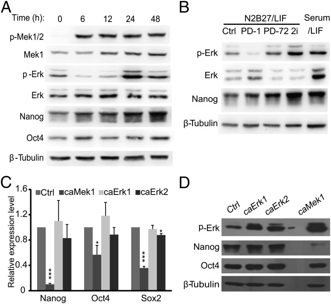
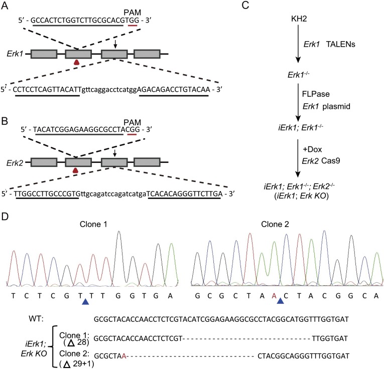
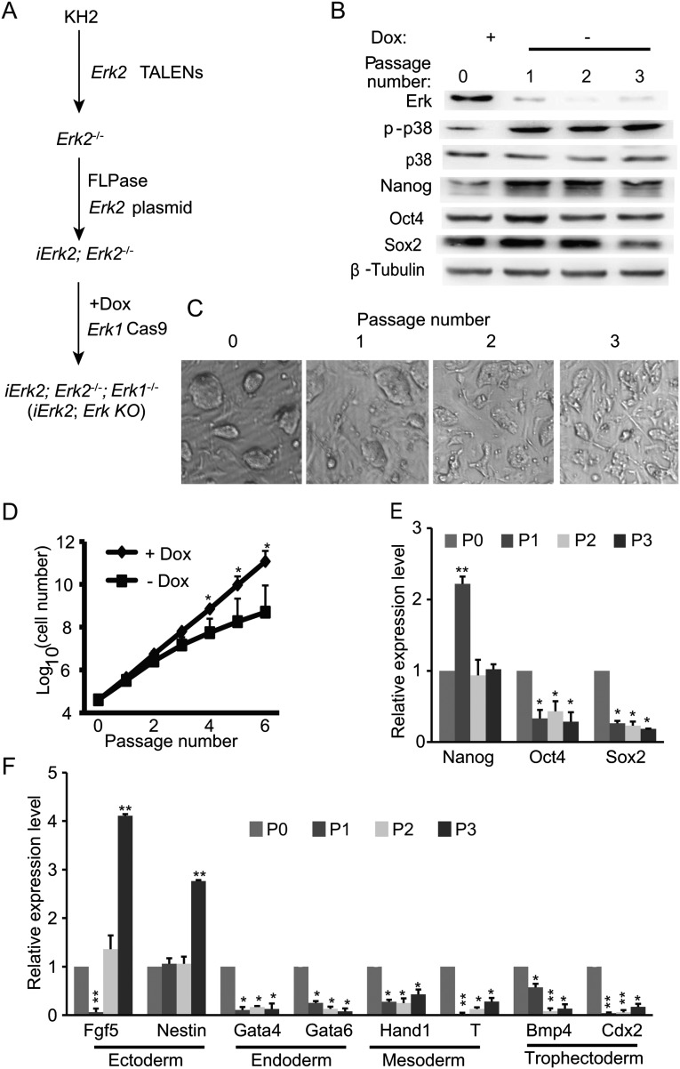
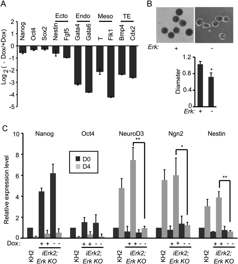
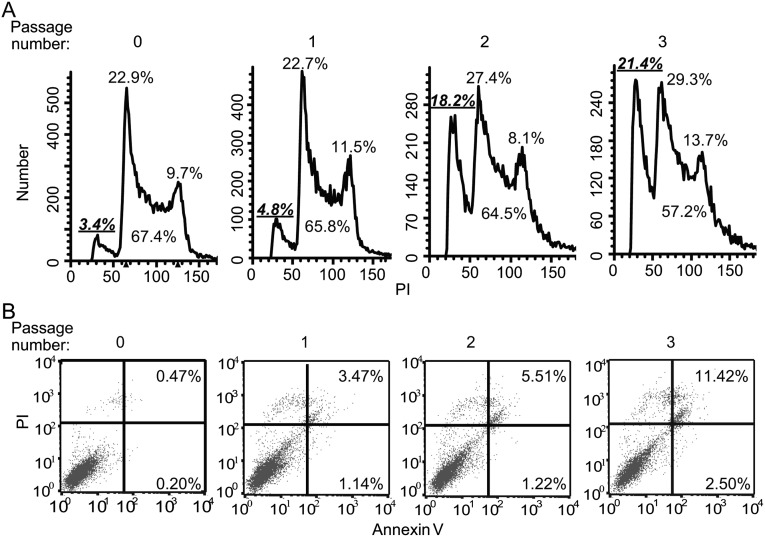
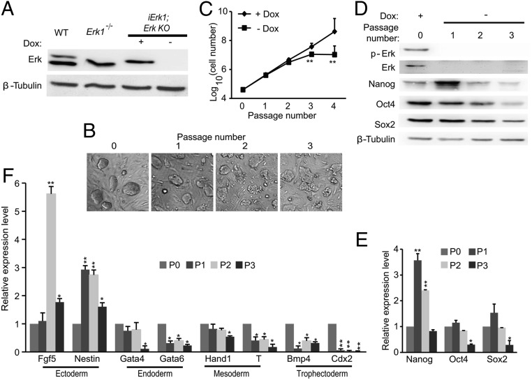
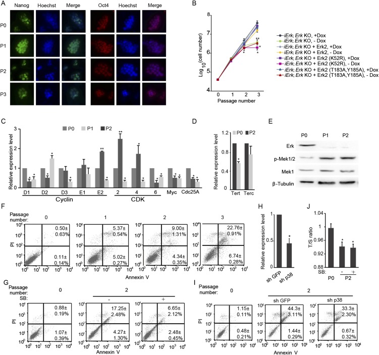
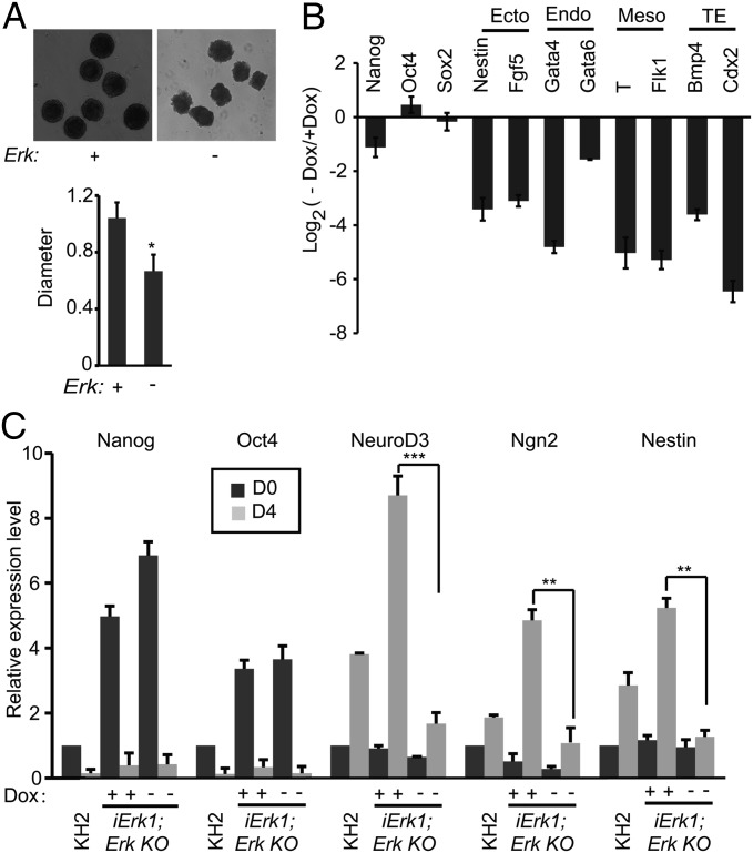
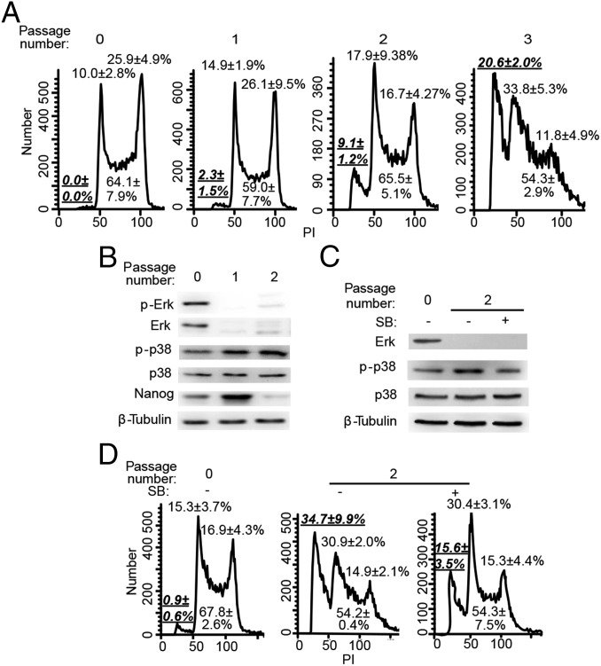
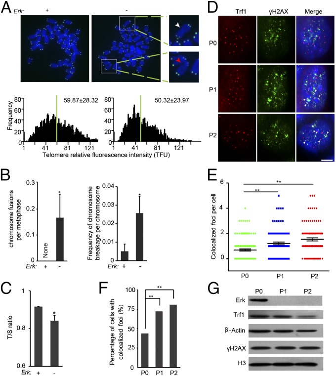
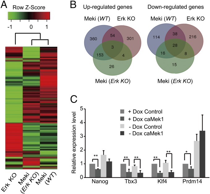
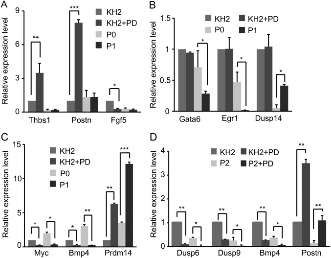
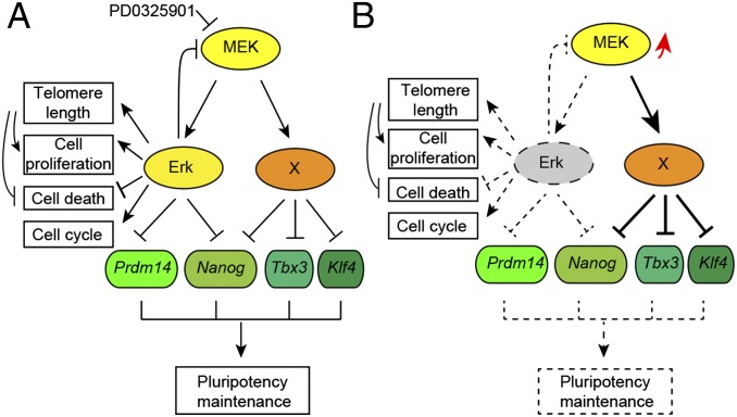
Inhibition of Mek/Erk signaling by pharmacological Mek inhibitors promotes self-renewal and pluripotency of mouse embryonic stem cells (ESCs). Intriguingly, Erk signaling is essential for human ESC self-renewal. Here we demonstrate that Erk signaling is critical for mouse ESC self-renewal and genomic stability. Erk-depleted ESCs cannot be maintained. Lack of Erk leads to rapid telomere shortening and genomic instability, in association with misregulated expression of pluripotency genes, reduced cell proliferation, G1 cell-cycle arrest, and increased apoptosis. Erk signaling is also required for the activation of differentiation genes but not for the repression of pluripotency genes during ESC differentiation. Furthermore, we find an Erk-independent function of Mek, which may explain the diverse effects of Mek inhibition and Erk knockout on ESC self-renewal. Together, in contrast to the prevailing view, Erk signaling is required for telomere maintenance, genomic stability, and self-renewal of mouse ESCs.

Keywords: Erk; Mek; embryonic stem cells; genomic stability; self-renewal.


Conflict of interest statement

The authors declare no conflict of interest.

Figures

Fig. 1.
Fig. 1.
Nanog expression is not negatively correlated with Erk phosphorylation. (A) Short-term PD treatment, but not prolonged PD treatment, reduces the level of p-Erk. V6.5 ESCs were cultured in a serum/LIF condition, and PD0325901 (1 μM) was added to the medium at the indicated time points before harvesting. Cells were harvested and subjected to Western blot analysis. (B) Expression of p-Erk of ESCs in N2B27 medium. V6.5 ESCs were switched from the serum/LIF condition to N2B27/LIF medium for 72 h (Ctrl). PD0325901 was added to the medium at 1 h (PD-1) or 72 h (PD-72) before harvesting cells. V6.5 ESCs cultured in serum/LIF or adapted to the 2i condition (2i) for more than five passages were also included. (C and D) caMeK1, but not caErk1 or caErk2, suppresses Nanog. Plasmids overexpressing caMek1, caErk1, and caErk2 were transfected into V6.5 ESCs. Cells were harvested 48 h after transfection. The expression of pluripotency genes Nanog, Oct4, and Sox2 was determined by quantitative RT-PCR (C) and Western blot (D). *P < 0.05; ***P < 0.001. Error bars are standard deviations (SDs).
Fig. S1.
Fig. S1.
Knockout of Erk1 and Erk2 by CRISPR/Cas and TALENs. (A) Schematic illustration of CRISPR/Cas and TALEN design for Erk1. (B) Schematic illustration of CRISPR/Cas and TALEN design for Erk2. CRISPR/Cas and TALEN targeting sites are labeled with red triangles and black arrows, respectively. Recognition sequences for CRISPR/Cas and TALENs are underlined. (C) Experimental outline for the construction of iErk1; Erk KO ESCs. (D) Sequencing results of two clones of iErk1; Erk KO ESCs demonstrate the disruption of the Erk2 alleles. Blue triangles mark the deletion sites. PAM, protospacer adjacent motif.
Fig. S2.
Fig. S2.
Erk signaling is essential for the self-renewal of ESCs, demonstrated by an independent cell line, iErk2; Erk KO. (A) Experimental outline for the construction of iErk2; Erk KO ESCs. (B) Western blots demonstrate the diminishment of Erk expression and the expression dynamics of pluripotency factors in iErk2; Erk KO ESCs after Dox withdrawal. (C) Colony morphology change upon Erk KO. iErk2; Erk KO ESCs were continuously cultured in the absence of Dox for three passages. Phase-contrast images of ESC colonies at each passage are shown. (D) Reduced proliferation of ESCs after Erk KO. iErk2; Erk KO ESCs were cultured in serum/LIF medium with or without Dox. The cell numbers were counted every passage, and equal amounts of ESCs were plated onto tissue-culture dishes. (E and F) Expression dynamics of pluripotency (E) and differentiation markers (F) after Erk KO. iErk2; Erk KO ESCs were cultured as described in C. *P < 0.05; **P < 0.01. Error bars are SDs.
Fig. S3.
Fig. S3.
Erk signaling is required for ESC differentiation, demonstrated by an independent cell line, iErk2; Erk KO ESCs. (A and B) iErk2; Erk KO ESCs were cultured with or without Dox for 48 h, and then the cells were used for EB differentiation, again with or without Dox. (A) RNA was isolated from day 4 EBs and analyzed by quantitative RT-PCR. (B) Images of day 4 EBs with or without Dox. The relative diameters of day 4 EBs with or without Dox (n > 10) are plotted. (C) iErk2; Erk KO ESCs were cultured with or without Dox for 48 h (day 0). These cells, as well as KH2 ESCs, were cultured in N2B27 with or without Dox to induce neural differentiation for 4 d. The expression of pluripotency genes and neural genes in the resulting cells was analyzed by quantitative RT-PCR. *P < 0.05; **P < 0.01. Error bars are SDs.
Fig. S4.
Fig. S4.
Loss of Erk signaling leads to G1 cell-cycle arrest and apoptosis of ESCs, demonstrated by an independent cell line, iErk2; Erk KO ESCs. Cell-cycle analysis (A) and cell apoptosis analysis (B) of iErk2; Erk KO ESCs at different passages after Dox withdrawal. Experiments were repeated three times. Results from a representative experiment are shown.
Fig. 2.
Fig. 2.
Erk signaling is essential for the self-renewal of ESCs. (A) Western blots demonstrate the diminishment of Erk expression in iErk1; Erk KO ESCs at 48 h after Dox withdrawal. (B) Colony morphology change upon Erk KO. iErk1; Erk KO ESCs were continuously cultured in the absence of Dox for three passages. Phase-contrast images of ESC colonies at each passage are shown. (C) Reduced proliferation of ESCs after Erk KO. iErk1; Erk KO ESCs were cultured in serum/LIF medium with or without Dox. Cell numbers were counted every passage, and equal amounts of ESCs were plated onto tissue-culture dishes. (DF) Expression dynamics of pluripotency and differentiation markers after Erk KO. Cells were cultured as described in C. The protein (D) and mRNA (E) levels of pluripotency markers Nanog, Oct4, and Sox2 were measured by Western blot and quantitative RT-PCR. (F) Expression of differentiation genes after Erk KO. *P < 0.05; **P < 0.01. Error bars are SDs.
Fig. S5.
Fig. S5.
Erk depletion compromises ESC self-renewal. iErk1; Erk KO ESCs were continuously cultured in the absence of Dox for three passages. (A) Immunofluorescence images of Nanog and Oct4. (B) Rescue slow proliferation rate in Erk KO ESCs by WT Erk2 but not by kinase-dead mutant (K52R) or phosphorylation-site mutant (T183A, Y185A). (C) Expression of cell-cycle regulators for G1-to-S transition after Dox withdrawal analyzed by quantitative RT-PCR. (D) The expression of Terc and Tert genes before and two passages after Dox withdrawal analyzed by quantitative RT-PCR. (E) The expression of Erk, Mek1, and p-Mek1 was analyzed by Western blot. (F) Cell apoptosis analysis of iErk1; Erk KO ESCs at different passages after Dox withdrawal. (G) Inhibition of p38 suppresses ESC apoptosis induced by Erk KO. iErk1; Erk KO ESCs were cultured in the absence of Dox for two passages, with or without the p38 inhibitor SB203580. Cell apoptosis analysis was performed with these cells. (H) Quantitative RT-PCR to measure the knockdown efficiency for p38. Two shRNA plasmids targeting p38 were pooled and transfected into iErk1; Erk KO ESCs. Forty-eight hours after transfection, cells were harvested and subjected to quantitative RT-PCR. (I) iErk1; Erk KO ESCs were cultured without Dox for 48 h, and then the control plasmid and p38 shRNA plasmids were transfected into these cells. Forty-eight hours after transfection, cell-cycle analysis was carried out for these cells. iErk1; Erk KO ESCs cultured with Dox were also included as a control. (J) iErk1; Erk KO ESCs were cultured in the absence of Dox for two passages, with or without the p38 inhibitor SB203580. Relative telomere length shown as T/S ratio in these cells was measured by quantitative PCR. *P < 0.05; **P < 0.01. Error bars are SDs.
Fig. 3.
Fig. 3.
Erk signaling is required for ESC differentiation. (A and B) iErk1; Erk KO ESCs were cultured with or without Dox for 48 h. The cells were then used for EB differentiation, again with or without Dox. (A) Images of day 4 EBs with or without Dox. The relative diameters of day 4 EBs with or without Dox (n > 10) are plotted. (B) RNA was isolated from day 4 EBs and analyzed by quantitative RT-PCR. TE, trophectoderm. (C) iErk1; Erk KO ESCs were cultured with or without Dox for 48 h (D0, day 0). These cells, as well as KH2 ESCs, were cultured in N2B27 with or without Dox to induce neural differentiation for 4 d. The expression of pluripotency genes and neural genes in the resulting cells was analyzed by quantitative RT-PCR. *P < 0.05; **P < 0.01; ***P < 0.001. Error bars are SDs.
Fig. 4.
Fig. 4.
Loss of Erk signaling leads to G1 cell-cycle arrest and apoptosis of ESCs. (A) Cell-cycle analysis of iErk1; Erk KO ESCs at different passages after Dox withdrawal. (B) p38 is activated upon Erk KO. The expression of p38 and p-p38 in iErk1; Erk KO ESCs at different passages after Dox withdrawal was determined by Western blot. (C and D) Inhibition of p38 suppresses ESC apoptosis induced by Erk KO. iErk1; Erk KO ESCs were cultured in the absence of Dox for two passages, with or without the p38 inhibitor SB203580. (C) Expression of p38 and p-p38 in Erk KO ESCs treated with or without SB203580 (SB). (D) Cell-cycle analysis was performed with these cells. (A and D) Percentages of sub-G1 cells in total cells (in bold and underlined) and percentages of G1, S, and G2/M cells in nonapoptotic cells are shown.
Fig. 5.
Fig. 5.
Rapid telomere shortening occurs in Erk KO ESCs. iErk1; Erk KO ESCs with Dox or two passages after Dox withdrawal were subjected to telomere Q-FISH analysis (A) and telomere/single copy gene 36B4 (T/S) ratio analysis (C). (A, Upper) Telomere Q-FISH images of chromosome spreads. Blue, DAPI-stained chromosomes; green dots, telomeres. Blown-up images are examples of chromosome fusion (marked with a white arrowhead) and chromosome breakage (indicated by a red arrowhead). (A, Lower) Histograms showing the distribution of relative telomere length displayed as TFU by Q-FISH. Green bars mark the average telomere length. Mean ± SD of telomere length is shown in each panel. Heavy black bars on the y axis show the frequency of telomere signal-free ends. Fifteen chromosome spreads were quantified for each group. (B) Quantification of chromosome fusion and chromosome breakage frequencies from the Q-FISH data in A. Mean ± SE are plotted (n ≥ 15). (C) Relative telomere length shown as T/S ratio measured by quantitative PCR. (DG) iErk1; Erk KO ESCs cultured with Dox, 48 and 96 h after Dox withdrawal, were subjected to immunofluorescence and Western blot analyses. (D) Immunofluorescence images of TRF1 (red) and γH2AX (green). Colocalized foci are indicated by arrowheads. (Scale bar, 5 μm.) Approximately 100 nuclei from images captured in D were quantified for colocalized TRF1 and γH2AX foci per cell (E) and percentage of cells with TRF1 and γH2AX colocalized foci (F). Mean ± SE are shown in E. (G) Protein levels of TRF1 and γH2AX as measured by Western blot. *P < 0.05; **P < 0.01.
Fig. 6.
Fig. 6.
Erk-dependent and -independent functions of Mek. (A and B) RNA-seq analysis of six samples: WT KH2 ESCs treated with or without PD for 48 h (KH2+PD and KH2, respectively), iErk1; Erk KO ESCs cultured in the presence of Dox (P0) and 48 and 96 h after Dox withdrawal (P1 and P2, respectively), and iErk1; Erk KO ESCs cultured without Dox for 96 h and treated with PD in the last 48 h (P2+PD). Meki (WT), Meki (Erk KO), and Erk KO are paired comparisons of KH2+PD and KH2, P2+PD and P2, and P1 and P0, respectively. Differentially expressed genes were identified in these three comparisons with the criteria of more than twofold change and false discovery rate (FDR) <0.001 (Dataset S1). (A) Clustering analysis of differentially regulated genes. (B) Venn diagrams of up- and down-regulated genes from three conditions. (C) caMek1 represses the expression of Klf4, Tbx3, and Nanog in the absence of Erk signaling. iErk1; Erk KO ESCs with Dox or 48 h after Dox withdrawal were transfected with empty vector or caMek1 overexpression vector. Forty-eight hours after transfection, cells were harvested for quantitative RT-PCR analysis. *P < 0.05; **P < 0.01. Error bars are SDs.
Fig. S6.
Fig. S6.
Validation of RNA-seq results for selected genes. (A) Genes affected in Meki (WT) but not in Erk KO. (B) Genes affected in Erk KO but not in Meki (WT). (C) Genes affected in both Meki (WT) and Erk KO. (D) Genes affected in both Meki (WT) and Meki (Erk KO). *P < 0.05; **P < 0.01; ***P < 0.001. Error bars are SDs.
Fig. 7.
Fig. 7.
Working model for Mek/Erk signaling in mouse ESCs. (A) Mek might suppress the expression of pluripotency genes through Erk-dependent and -independent pathways. In addition to its role in repressing Prdm14 and Nanog, Erk signaling is required for cell proliferation, cell-cycle progression, and telomere-length maintenance as well as suppression of apoptosis. (B) Upon Erk KO, Mek and its Erk-independent pathway are activated (marked by a red arrow) to perturb the pluripotency regulation network.

Similar articles

Cited by

References

    1. Jaenisch R, Young R. Stem cells, the molecular circuitry of pluripotency and nuclear reprogramming. Cell. 2008;132(4):567–582. - PMC - PubMed
    1. Young RA. Control of the embryonic stem cell state. Cell. 2011;144(6):940–954. - PMC - PubMed
    1. Boyer LA, et al. Core transcriptional regulatory circuitry in human embryonic stem cells. Cell. 2005;122(6):947–956. - PMC - PubMed
    1. Loh YH, et al. The Oct4 and Nanog transcription network regulates pluripotency in mouse embryonic stem cells. Nat Genet. 2006;38(4):431–440. - PubMed
    1. Raz R, Lee CK, Cannizzaro LA, d’Eustachio P, Levy DE. Essential role of STAT3 for embryonic stem cell pluripotency. Proc Natl Acad Sci USA. 1999;96(6):2846–2851. - PMC - PubMed

Publication types

Substances

Associated data