. 2015 Nov 3;112(44):13447-54.
doi: 10.1073/pnas.1514944112. Epub 2015 Oct 19.

Survival of human lymphoma cells requires B-cell receptor engagement by self-antigens

Affiliations

Survival of human lymphoma cells requires B-cell receptor engagement by self-antigens

Ryan M Young et al. Proc Natl Acad Sci U S A. .

Abstract

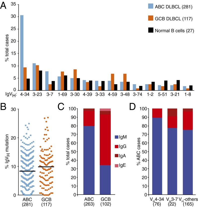
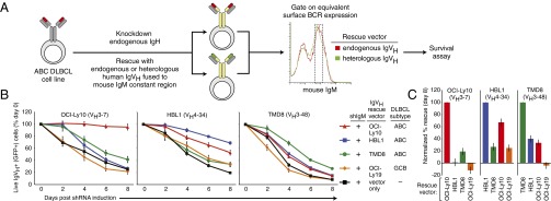
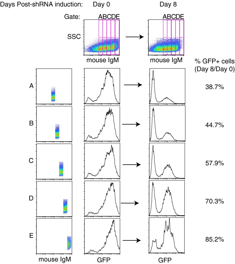
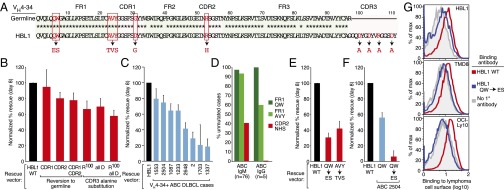
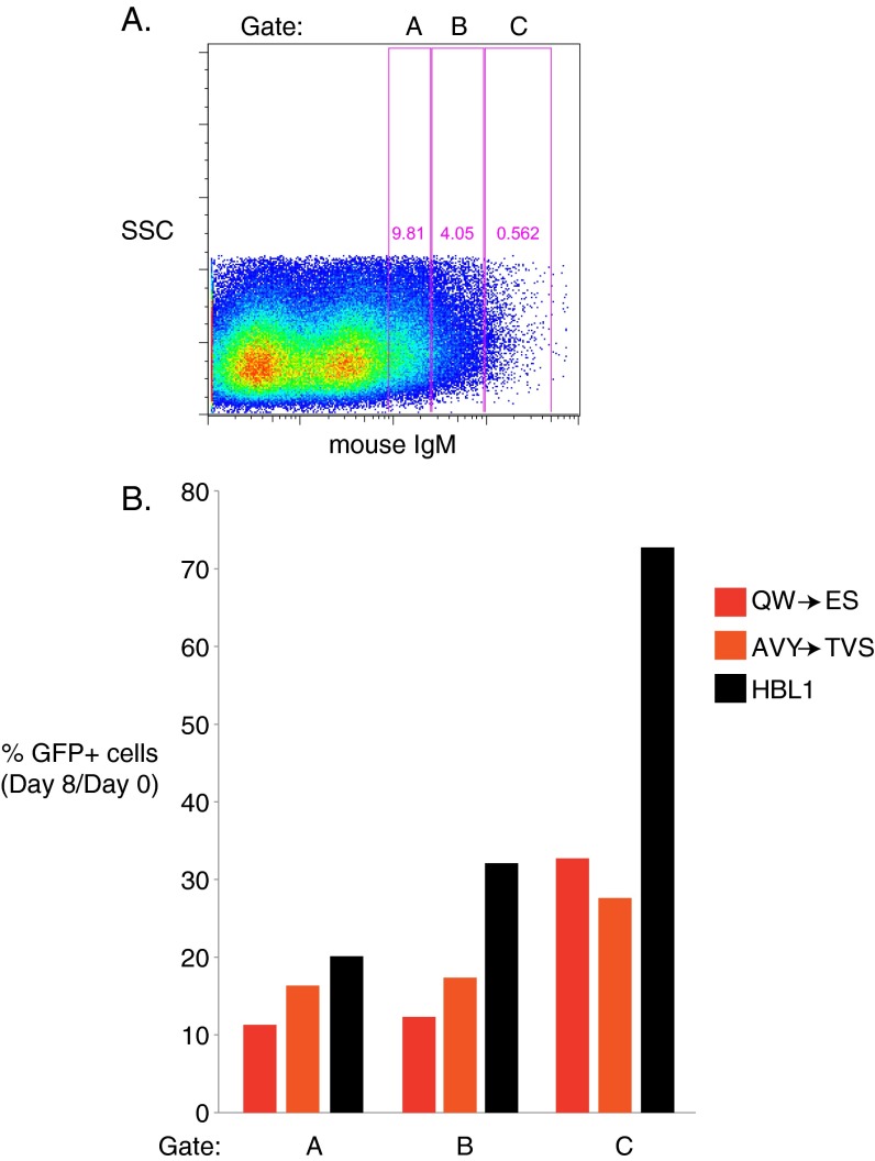
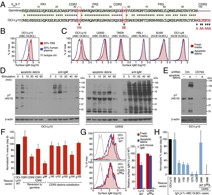
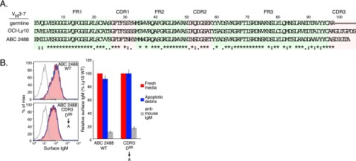
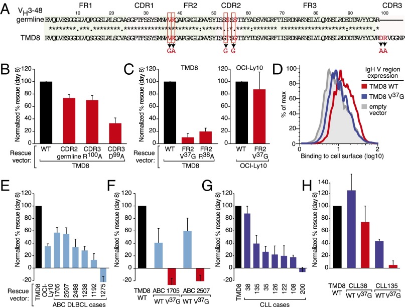
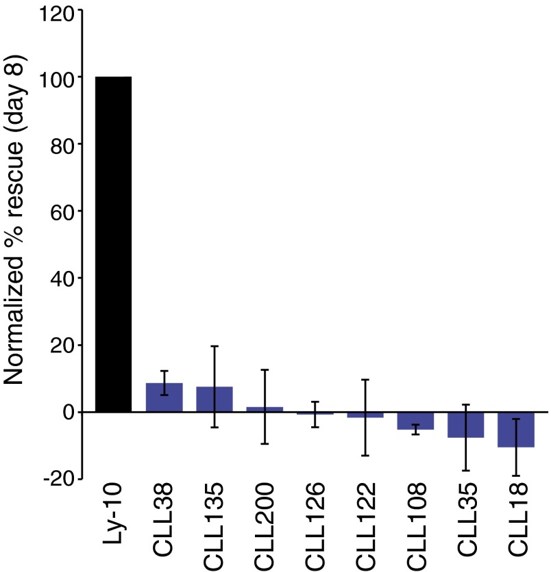
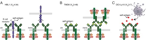
The activated B-cell-like (ABC) subtype of diffuse large B-cell lymphoma (DLBCL) relies on chronic active B-cell receptor (BCR) signaling. BCR pathway inhibitors induce remissions in a subset of ABC DLBCL patients. BCR microclusters on the surface of ABC cells resemble those generated following antigen engagement of normal B cells. We speculated that binding of lymphoma BCRs to self-antigens initiates and maintains chronic active BCR signaling in ABC DLBCL. To assess whether antigenic engagement of the BCR is required for the ongoing survival of ABC cells, we developed isogenic ABC cells that differed solely with respect to the IgH V region of their BCRs. In competitive assays with wild-type cells, substitution of a heterologous V region impaired the survival of three ABC lines. The viability of one VH4-34(+) ABC line and the ability of its BCR to bind to its own cell surface depended on V region residues that mediate the intrinsic autoreactivity of VH4-34 to self-glycoproteins. The BCR of another ABC line reacted with self-antigens in apoptotic debris, and the survival of a third ABC line was sustained by reactivity of its BCR to an idiotypic epitope in its own V region. Hence, a diverse set of self-antigens is responsible for maintaining the malignant survival of ABC DLBCL cells. IgH V regions used by the BCRs of ABC DLBCL biopsy samples varied in their ability to sustain survival of these ABC lines, suggesting a screening procedure to identify patients who might benefit from BCR pathway inhibition.

Keywords: B-cell receptor; cancer biology; lymphoma.


Conflict of interest statement

The authors declare no conflict of interest.

Figures

Fig. 1.
Fig. 1.
IgH analysis of DLBCL tumors. (A) The IgVH repertoire in ABC and GCB DLBCL samples compared with normal naïve B cells. (B) Percentage of IgVH bases mutated in ABC and GCB tumors. (C) IgH constant region isotypes in ABC and GCB DLBCL samples. (D) IgH constant region isotypes in ABC DLBCL cases expressing the indicated VH segments.
Fig. 2.
Fig. 2.
Ectopic expression of IgH chains to determine the role of IgH V regions in maintaining the viability of ABC DLBCL cell lines. (A) A knockdown-rescue strategy to create isogenic ABC cell lines that express different IgH V regions. See Results for details. (B) Ability of the indicated IgH V regions to sustain viability of ABC cell lines following knockdown of endogenous IgM expression. For each indicated ABC cell line, ectopic re-expression of the endogenous IgH V region was most efficient at sustaining survival (mean ± SEM, n ≥ 8). (C) Data from B plotted as the percent rescue of survival at 8 d after shIgM induction: 100% is scaled to the degree of rescue by re-expression of the endogenous IgH V region and 0% is scaled to the degree of rescue by an empty vector.
Fig. S1.
Fig. S1.
Levels of ectopic mouse IgM expression determine rescue of human IgM knockdown. (A–E) HBL1 cells were cotransduced with a vector expressing shIgM, marked by GFP, and the HBL1 IgH V region fused to mouse IgM. Cells were gated on increasing levels of surface mouse IgM. The amount of GFP+ cells was analyzed on day 0 and day 8, and the percentage of GFP+ cells on day 8, relative to day 0, is shown. Higher levels of surface mouse IgM correlated to increased rescue from toxicity induced by IgM knockdown.
Fig. 3.
Fig. 3.
Reactivity of VH4-34+ BCRs with self-antigens in ABC DLBCL. (A) The HBL1 IgH V region sequence compared with the germ-line VH4-34 sequence. Mutations introduced in this study are highlighted in red. (B) Substitutions of CDR residues in the HBL1 IgH V region affect HBL1 viability (mean ± SEM, n = 3). (C) Ability of VH4-34+ V regions derived from ABC DLBCL biopsies to sustain HBL1 survival (mean ± SEM, n ≥ 3). (D) Frequency of mutations in ABC DLBCL biopsies targeting the Q6W7 and A24V25Y26 FR1 motifs and the N52H53S54 CDR2 motif in the germ-line VH4-34 gene segment, for ABC cases using either an IgM or IgG constant region. (E) Mutation of the FR1 Q6W7 and A24V25Y26 motifs in the HBL1 IgH V region fail to sustain viability of HBL1 cells (mean ± SEM, n = 3). (F) Mutation of the FR1 Q6W7 motif in a VH4-34+ ABC DLBCL biopsy impairs rescue of HBL1 cells (mean ± SEM, n = 2). (G) FACS analysis of a soluble human IgG1 antibody bearing the HBL1 V region binding to the surface of the indicated ABC DLBCL cell lines. The IgH V region of the recombinant antibody was either identical to the HBL1 IgH V region (WT) or was mutated in the FR1 Q6W7 motif (representative data from three experiments).
Fig. S2.
Fig. S2.
The wild-type HBL IgVH sequence rescues better than FR1 mutant HBL1 IgVH sequences at all levels of ectopic mouse IgH expression. HBL1 cells were cotransduced with shIgM, marked by GFP, and mouse IgM with a human V region of either wild-type HBL1, HBL1 FR1 QW mutated to ES, or HBL1 FR1 AVY mutated to TVS. (A) Gates were placed on increasing levels of surface mouse IgM. (B) The percentage of GFP+ cells on day 8, relative to day 0, was plotted for each construct in each of the three gates.
Fig. 4.
Fig. 4.
Reactivity of ABC DLBCL BCRs to antigens present in apoptotic debris. (A) Sequence of the OCI-Ly10 IgH V region compared with the germ-line VH3-7 sequence. Mutations introduced in this study are highlighted in red. (B) FACS analysis of surface IgM in OCI-Ly10 cells cultured in FBS or human plasma. (C) FACS analysis of surface IgM expression in the indicated DLBCL cell lines cultured with or without apoptotic cell debris (see SI Materials and Methods for details). (D) Western blot analysis of tyrosine phosphorylated proteins (pY 4G10) in OCI-Ly10 (Left) and U2932 (Right) following stimulation with apoptotic debris or anti-IgM (0.5 μg/mL) for the indicated times. (E) Western blot analysis of pY in OCI-Ly10 cells exposed to apoptotic debris as indicated. Cells expressed either an shRNA targeting CD79A or a control shRNA, as indicated. (F) Substitutions of CDR residues affect rescue of OCI-Ly10 viability (mean ± SEM, n ≥ 3). (G) FACS analysis of OCI-Ly10 cells ectopically expressing mouse IgM fused to either the OCI-Ly10 IgH V region (WT) or this V region with a K98A substitution, in the indicated culture conditions (Left); relative mean fluorescence intensity is plotted (Right) (mean ± SEM, n ≥ 3). (H) Rescue by VH3-7+ V regions derived from ABC DLBCL biopsies in OCI-Ly10 cells (mean ± SEM, n = 3). Right two bars indicate the degree of rescue by ABC DLBCL case 2488 (WT) compared with this V region with a D99A substitution in its CDR3 region (mean ± SEM, n = 3).
Fig. S3.
Fig. S3.
BCRs containing the VH3-7 ABC 2488 IgH V region rescues do not respond to antigens present in apoptotic debris. (A) Alignment of the germ-line VH3-7 sequence with IgH V region sequences from OCI-Ly10 cells and the ABC 2488 biopsy sample. (B) FACS analysis of IgM expression on the cell surface in OCI-Ly10 cells engineered to express an IgH V region derived from the ABC 2488 biopsy sample or the same V region with a D99A substitution in its CDR3 region. Binding was evaluated in fresh media plus or minus apoptotic debris, or following BCR cross-linking with anti-mouse IgM antibodies. (Left) Representative data from three experiments. (Right) Relative mean fluorescent intensity (error bars indicated SEM).
Fig. 5.
Fig. 5.
Anti-idiotypic reactivity of ABC DLBCL BCRs. (A) Sequence of the TMD8 IgH V region compared with the germ-line VH3-48 sequence. Mutations introduced in this study are highlighted in red. (B) Substitutions of CDR residues in the TMD8 IgH V region affect TMD8 viability (mean ± SEM, n = 3). (C, Left) Substitutions of FR2 residues in the TMD8 IgH V region fail to sustain TMD8 viability. (Right) Substitution of a FR2 residue in the OCI-Ly10 IgH V region maintains OCI-Ly10 viability (mean ± SEM, n = 5). (D) Surface binding of a soluble human IgG1 antibody bearing the TMD8 V region to HBL1 cells transduced with either the IgH V region from TMD8 cells (WT), the TMD8 FR2 V37G, or empty vector, as indicated (FACS plots representative of three experiments). (E) Rescue by VH3-7+ V regions derived from ABC DLBCL biopsies and OCI-Ly10 in TMD8 (mean ± SEM, n ≥ 3). (F) Rescue by ABC DLBCL biopsies with the FR2 V37G substitutions in TMD8 (mean ± SEM, n ≥ 3). (G) Rescue by IgH V regions derived from CLL patient samples in TMD8 (mean ± SEM, n ≥ 3). (H) Effect of a FR2 V37G substitution in two CLL patient samples on rescue in TMD8 (mean ± SEM, n ≥ 3).
Fig. S4.
Fig. S4.
Ability of IgH V regions derived from CLL patient samples to sustain OCI-Ly10 survival (error bars indicate SEM of three experiments).
Fig. 6.
Fig. 6.
Models of self-antigen–dependent survival signaling in ABC DLBCL. (A) HBL1 (VH4-34) recognizes self-antigens expressed on the surface of malignant B cells, either in a cis or trans fashion. Binding to self-antigens is potentially mediated through IgH FR1 and CDR3 interactions with n-acetyl-lactosamine glycans attached to cell surface proteins. (B) TMD8 (VH3-48) binds to an idiotope in the IgH FR2 region of its BCR, either in a cis or trans fashion. (C) The OCI-Ly10 (VH3-7) BCR binds to self-antigens released from apoptotic cells.

Similar articles

Cited by

References

    1. Dameshek W, Schwartz RS. Leukemia and auto-immunization—Some possible relationships. Blood. 1959;14:1151–1158. - PubMed
    1. Küppers R. Mechanisms of B-cell lymphoma pathogenesis. Nat Rev Cancer. 2005;5(4):251–262. - PubMed
    1. Quinn ER, et al. The B-cell receptor of a hepatitis C virus (HCV)-associated non-Hodgkin lymphoma binds the viral E2 envelope protein, implicating HCV in lymphomagenesis. Blood. 2001;98(13):3745–3749. - PubMed
    1. Wotherspoon AC, et al. Regression of primary low-grade B-cell gastric lymphoma of mucosa-associated lymphoid tissue type after eradication of Helicobacter pylori. Lancet. 1993;342(8871):575–577. - PubMed
    1. Agathangelidis A, et al. Stereotyped B-cell receptors in one-third of chronic lymphocytic leukemia: A molecular classification with implications for targeted therapies. Blood. 2012;119(19):4467–4475. - PMC - PubMed

Publication types

MeSH terms