. 2015 Nov 3;112(44):13675-80.
doi: 10.1073/pnas.1508355112. Epub 2015 Oct 19.

Subdiffusive motion of bacteriophage in mucosal surfaces increases the frequency of bacterial encounters

Affiliations

Subdiffusive motion of bacteriophage in mucosal surfaces increases the frequency of bacterial encounters

Jeremy J Barr et al. Proc Natl Acad Sci U S A. .

Abstract

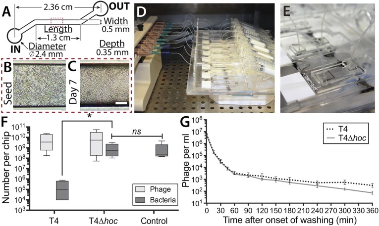
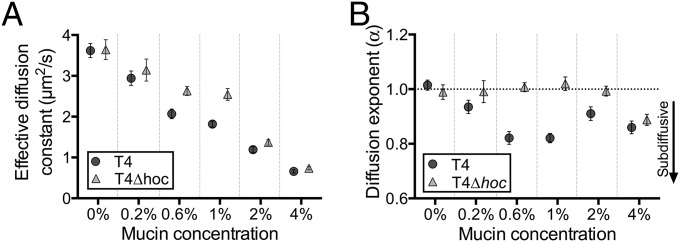
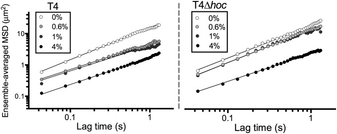
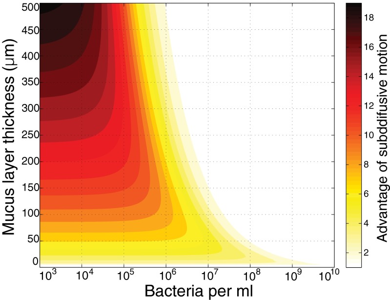
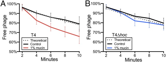
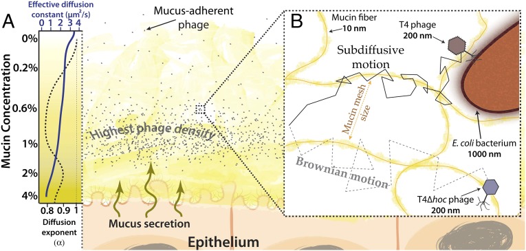
Bacteriophages (phages) defend mucosal surfaces against bacterial infections. However, their complex interactions with their bacterial hosts and with the mucus-covered epithelium remain mostly unexplored. Our previous work demonstrated that T4 phage with Hoc proteins exposed on their capsid adhered to mucin glycoproteins and protected mucus-producing tissue culture cells in vitro. On this basis, we proposed our bacteriophage adherence to mucus (BAM) model of immunity. Here, to test this model, we developed a microfluidic device (chip) that emulates a mucosal surface experiencing constant fluid flow and mucin secretion dynamics. Using mucus-producing human cells and Escherichia coli in the chip, we observed similar accumulation and persistence of mucus-adherent T4 phage and nonadherent T4∆hoc phage in the mucus. Nevertheless, T4 phage reduced bacterial colonization of the epithelium >4,000-fold compared with T4∆hoc phage. This suggests that phage adherence to mucus increases encounters with bacterial hosts by some other mechanism. Phages are traditionally thought to be completely dependent on normal diffusion, driven by random Brownian motion, for host contact. We demonstrated that T4 phage particles displayed subdiffusive motion in mucus, whereas T4∆hoc particles displayed normal diffusion. Experiments and modeling indicate that subdiffusive motion increases phage-host encounters when bacterial concentration is low. By concentrating phages in an optimal mucus zone, subdiffusion increases their host encounters and antimicrobial action. Our revised BAM model proposes that the fundamental mechanism of mucosal immunity is subdiffusion resulting from adherence to mucus. These findings suggest intriguing possibilities for engineering phages to manipulate and personalize the mucosal microbiome.

Keywords: BAM; mucus; search strategy; subdiffusion; virus.


Conflict of interest statement

The authors declare no conflict of interest.

Figures

Fig. 1.
Fig. 1.
Microfluidic devices (chips) simulating a lifelike in vitro mucosal surface. (A) Schematic of chip design. (B) Mucus-producing lung tissue culture cells seeded into the main channel of a chip. (C) Tissue culture cells in the main channel after perfusion for 7 d. (D) Multiplex syringe pump and scaffold perfusing nine chips simultaneously. (E) Close-up of a single chip bonded to a glass microscope slide with microfluidic tubing attached to the in and out ports. (F) Phage therapy experiment. Cell layers were pretreated with T4 or T4∆hoc phage for 12 h, washed for 1 h, and then inoculated with E. coli. Cells with mucus layer were harvested 18 h later, and both the phages and bacteria present were titered (PFU and CFU). (G) Phage detachment. Cell layers were pretreated with T4 or T4∆hoc phage for 12 h and then washed with phage-free media for 6 h, during which time the number of released phages was monitored by titering (PFU).
Fig. 2.
Fig. 2.
(A) Effective diffusion constants (μm2/s) calculated at 43.5-ms time intervals for T4 and T4∆hoc phage in 0% (buffer), 0.2%, 0.6%, 1%, 2%, and 4% mucin solutions (wt/vol). (B) Diffusion exponents (α) of T4 and T4∆hoc phage in 0% (buffer), 0.2%, 0.6%, 1%, 2%, and 4% mucin solutions (wt/vol). Brownian diffusion α ∼ 1, subdiffusion α < 1.
Fig. 3.
Fig. 3.
Log-log graph of the ensemble-averaged MSD (μm2) of T4 and T4∆hoc phage in 0% (buffer), 0.6%, 1%, and 4% mucin solutions (wt/vol). Solid lines indicate line of best fit from which the diffusion exponent (α) is determined. For full MSD plots, see SI Appendix, Fig. S1.
Fig. 4.
Fig. 4.
Theoretical advantage of a phage–host encounter for a phage particle, using subdiffusive motion compared with Brownian motion across a range of bacterial concentrations and mucus layer thickness. Color scale indicates the ratio of probabilities of bacterial encounter for a subdiffusive phage (α = 0.82) versus a normally diffusing phage (α = 1). The ratio ranges from 19 (black) to 1 (white).
Fig. 5.
Fig. 5.
Adsorption assay measuring the percentage of free phage remaining during a 10-min period in control (0%) and 1% mucin solutions. Error bars show SD (n = 6). Theoretical values were calculated from the T4 phage adsorption constant (k) = 2.4 × 10⁻⁹ mL/min, phage concentration (2 × 10⁵ mL–1), and bacterial concentration (1 × 10⁷ mL–1). (A) T4 phage. (B) T4∆hoc phage.
Fig. 6.
Fig. 6.
BAM model with subdiffusion. (A) The mucus layer is a dynamic gradient of mucin glycoproteins. Closer to the epithelium, the mucin concentration increases. The corresponding decrease in the mucin mesh size reduces phage diffusion constants. Subdiffusive motion of mucus-adherent phage particles at lower mucin concentrations enriches phage particles within an optimal zone of the mucus layer. (Left) Qualitative representations of the effective diffusion constant (K) (solid blue line) and diffusion exponent (α) (dashed black line) for T4 phage from Fig. 2. (B) Transient binding of mucus-adherent phage to mucin glycoproteins facilitates subdiffusive motion over a range of mucin concentrations present within a mucus layer.

Similar articles

Cited by

References

    1. Johansson MEV, et al. The inner of the two Muc2 mucin-dependent mucus layers in colon is devoid of bacteria. Proc Natl Acad Sci USA. 2008;105(39):15064–15069. - PMC - PubMed
    1. Linden SK, Sutton P, Karlsson NG, Korolik V, McGuckin MA. Mucins in the mucosal barrier to infection. Mucosal Immunol. 2008;1(3):183–197. - PMC - PubMed
    1. d’Hérelle F. The Bacteriophage: Its Role in Immunity. Masson et Cie; Paris: 1921.
    1. Brüssow H. Phage therapy: The Escherichia coli experience. Microbiology. 2005;151(Pt 7):2133–2140. - PubMed
    1. Sulakvelidze A, Alavidze Z, Morris JG., Jr Bacteriophage therapy. Antimicrob Agents Chemother. 2001;45(3):649–659. - PMC - PubMed

Publication types