. 2015 Nov 3;112(44):13555-60.
doi: 10.1073/pnas.1510100112. Epub 2015 Oct 19.

Targeted binding of nucleocapsid protein transforms the folding landscape of HIV-1 TAR RNA

Affiliations

Targeted binding of nucleocapsid protein transforms the folding landscape of HIV-1 TAR RNA

Micah J McCauley et al. Proc Natl Acad Sci U S A. .

Abstract

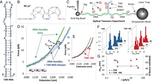
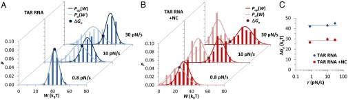
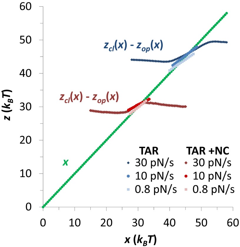
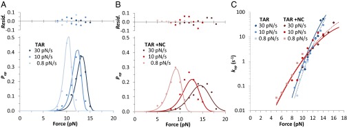
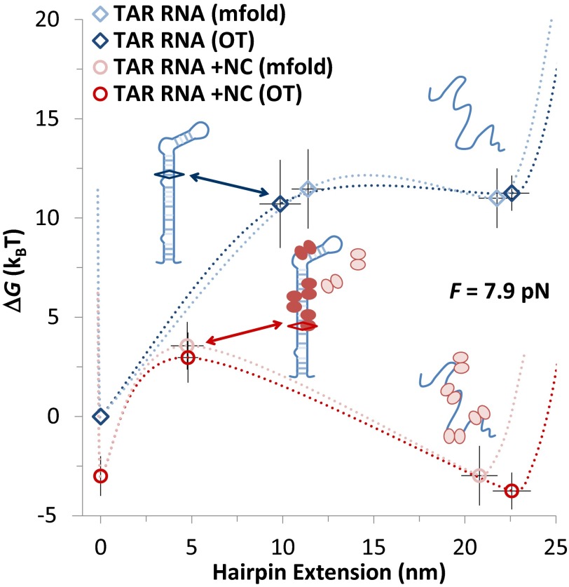
Retroviral nucleocapsid (NC) proteins are nucleic acid chaperones that play a key role in the viral life cycle. During reverse transcription, HIV-1 NC facilitates the rearrangement of nucleic acid secondary structure, allowing the transactivation response (TAR) RNA hairpin to be transiently destabilized and annealed to a cDNA hairpin. It is not clear how NC specifically destabilizes TAR RNA but does not strongly destabilize the resulting annealed RNA-DNA hybrid structure, which must be formed for reverse transcription to continue. By combining single-molecule optical tweezers measurements with a quantitative mfold-based model, we characterize the equilibrium TAR stability and unfolding barrier for TAR RNA. Experiments show that adding NC lowers the transition state barrier height while also dramatically shifting the barrier location. Incorporating TAR destabilization by NC into the mfold-based model reveals that a subset of preferential protein binding sites is responsible for the observed changes in the unfolding landscape, including the unusual shift in the transition state. We measure the destabilization induced at these NC binding sites and find that NC preferentially targets TAR RNA by binding to specific sequence contexts that are not present on the final annealed RNA-DNA hybrid structure. Thus, specific binding alters the entire RNA unfolding landscape, resulting in the dramatic destabilization of this specific structure that is required for reverse transcription.

Keywords: RNA binding; RNA stretching; force spectroscopy; single molecule.


Conflict of interest statement

The authors declare no conflict of interest.

Figures

Fig. 1.
Fig. 1.
Probing the interaction of the TAR RNA hairpin and NC. (A) The 59-nt sequence and predicted secondary structure of HIV-1 TAR RNA include 24 bp. (B) NC consists of two zinc fingers and a basic N terminus. Basic residues are shown in blue and acidic residues in red, and black denotes zinc-coordinating amino acids. Aromatic residues Phe and Trp are marked. (C) An optical tweezers experiment tethers a single RNA hairpin between two beads through long DNA handles. The micropipette is translated to increase the tension. (D and E) Control constructs excluding the hairpin show an elastic response typical of DNA for three cycles of extension/release (solid/dotted green lines). Experimental constructs incorporating TAR RNA hairpins reveal the same elastic response as the DNA handles until interrupted by sudden hairpin opening at ∼12 pN (solid/dotted blue lines for TAR and solid/dotted red lines for TAR with NC). Thick solid lines are fits to polymer elasticity models from SI Materials and Methods, section 1. Unfolding is characterized by a measured force (Fop) and length increase (Δxop), and the hairpin closing force (Fcl) is identified where the release and extension data overlap. The shaded region represents the net work done by the instrument to open the hairpin (Wop) and represents the energy required to extend the handles as the hairpin remains folded (ΔWd) minus the energy required to extend the handles and the unfolded hairpin construct over the same extension range (ΔWd+r). (F) Histograms of measured opening lengths for pulling rates shown in pN/s. (G) At each pulling rate the average length (solid symbols) shows a variability, which disappears when corrected for polymer elasticity (open symbols), giving the number of bases unfolded: n = 47.8 ± 1.3 for the TAR hairpin and NNC = 48.4 ± 0.5 in the presence of NC.
Fig. 2.
Fig. 2.
Equilibrium energies of hairpin opening. (A) Normalized probability densities of measured work during unfolding [Pop(W), blue] and closing [Pcl(W), cyan] for the TAR RNA hairpin over pulling rates of 30 pN/s, 10 pN/s, and 0.8 pN/s (n = 250 opening events). Solid lines are fits to Gaussian distributions to guide the eye. Distributions cross at the equilibrium work (W = ΔGo), marked by solid circles. (B) Distributions of work done during unfolding (red) and folding (pink) in the presence of NC (n = 162 cycles). (C) TAR (blue) and TAR with NC (red) unfolding free energies for distributions shown in A and B. Averaged over all rates, the measured free energy and SE are ΔGo = 43.3 ± 0.9 kBT in the absence and ΔGo,NC = 28.3 ± 0.9 kBT in the presence of NC. Details of the free-energy measurement are discussed in SI Materials and Methods, section 2.
Fig. S1.
Fig. S1.
Bennett’s analysis of TAR RNA hairpin unfolding. Plots of Eq. S5 at pulling rates of 30 pN/s, 10 pN/s, and 0.8 pN/s for TAR hairpins alone (blue) and in the presence of 50-nM NC (red). The intersection of zcl(x) − zop(x) with the line z = x/kBT gives the estimate of the energy of unfolding at that rate, and these are shown in Table S1. Averaged, these values give ΔGo = 44.2 ± 1.6 kBT for TAR hairpins and ΔGo,NC = 30.3 ± 0.7 kBT after NC is added.
Fig. 3.
Fig. 3.
Kinetics and thermodynamics of TAR unfolding and folding. (A) Normalized TAR RNA opening probabilities Pop(F) recast as points for pulling rates of 30 pN/s, 10 pN/s, and 0.8 pN/s (dark blue, blue, and cyan) globally fitted to Eq. S7 (solid lines) described in SI Materials and Methods, section 3, and including residuals. Standard histogram bin uncertainties are omitted for clarity, and fits shown are for shape factor ν = 0.5, as discussed in the text. Averaged fitted parameters (with SE) were found: Δxop = 9.9 ± 1.1 nm, ΔGop = 27.0 ± 2.2 kBT, and kopo = (8 ± 5) × 10−9 s−1 for TAR with n = 250 and χυ2 ∼ 1 for all fits. (B) In the presence of 50 nM NC, global fits over 30 pN/s, 10 pN/s, and 0.8 pN/s (dark red, red, and pink) yield Δxop,NC = 4.8 ± 0.6 nm, ΔGop,NC = 14.3 ± 1.3 kBT, and kop,NCo = (1.2 ± 0.8) × 10−4 s−1, where n = 162 and χυ2 ∼ 0.8 for all fits. Histogram bin widths were scaled for direct comparisons between A and B. (C) TAR RNA hairpin opening rate as a function of unzipping force, kop(F), omitting/including NC, calculated according to Eqs. S9 and S10 in SI Materials and Methods, section 4. Solid lines represent the fit to Pop(F), and dotted lines are direct fits of kop(F) to Eq. S6. The full list of fitted values is compared in Table S2.
Fig. S2.
Fig. S2.
Stretching force rate dependence of Pop(F) and Pcl(F). (A) Normalized probability of opening/closing (blue/cyan) vs. force for TAR RNA (n = 250) and (B) distributions of opening/closing (red/pink) for TAR in the presence of 50-nM NC (n = 162). Opening forces are fitted in the main text and shown in Fig. 3.
Fig. S3.
Fig. S3.
Fitting minima for opening force data, Pop(F). Values of χ2 for the fits of Eq. S7 to the data of Pop(F), for the shape factor v = ½. Variations about the minima for (A) TAR RNA and (B) TAR with NC, for each free parameter, with the other two held fixed. Minimized values are shown in Table S2, along with fitted uncertainties determined from the parameter values that increase χ2 by 8, which represents the 95% confidence interval for three fitting parameters.
Fig. S4.
Fig. S4.
Stretching force dependence of rates kop(F) and kcl(F). (A) kop(F) (solid symbols) and kcl(F) (open symbols) for TAR RNA pulled at rates of 30 pN/s, 10 pN/s, and 0.8 pN/s (dark blue, blue, and cyan), calculated from the data of Fig. S2, using Eqs. S9 and S10. (B) kop(F) (solid symbols) and kcl(F) (open symbols) for TAR RNA with 50-nM NC pulled at rates of 30 pN/s, 10 pN/s, and 0.8 pN/s (dark red, red, and pink). In both cases the opening rate is pulling rate independent while the closing rate depends on the pulling rate, reflecting multistate TAR RNA closing kinetics. Fits of Eq. S3 to the opening data are discussed in the text.
Fig. 4.
Fig. 4.
Transition state predictions for the TAR hairpin in the absence and presence of NC. (A) Theoretical energy profiles determined from mfold. Horizontal and vertical lines indicate Δxop and ΔGo for TAR RNA (blue), for the lowest energy state seen in these experiments, as discussed in the text. (B) Free-energy profiles at F1/2, where folded and unfolded state free energies are equal, with corrections for ssRNA elasticity and added potentials for open and closed states and probability distributions for the two states (green) (SI Materials and Methods, section 5). Horizontal and vertical lines indicate theoretical estimates of xop and ΔG1/2 for TAR RNA (blue): F1/2 = 10.1 ± 0.1 pN, xop = 11.3 ± 0.9 nm, and ΔGop = 30.7 ± 2.0 kBT. (C and D) Predicted destabilization of TAR in the presence of NC (for four bound NCs). The resulting landscapes (red) give F1/2,NC = 7.9 ± 0.1 pN, xop,NC = 4.7 ± 0.9 nm, and ΔGop,NC = 14.8 ± 1.2 kBT. (E) Potential NC binding sites located at defect-adjacent G-containing base pairs are circled in red for four test cases. Sites marked with solid circles were uniformly destabilized by a total δGo = 16 kBT. (F) Values of the transition state location and height calculated from mfold for each case shown in E. A 2D Z-test of the data with the various models gives a probability of 0.74 for the four-site model and 0.08 for the five-site model, whereas the three- and six-site models each have probability of less than 0.001.
Fig. 5.
Fig. 5.
Specific binding of NC transforms the energy landscape of the TAR RNA hairpin. Shown is a summary of experimental and computational results for TAR RNA hairpin force-unfolding, combining the overall opening length and unfolding free energy with the transition state location into a unified free-energy landscape. Theory and experiment are compared in the absence (cyan/blue) and the presence (pink/red) of 50 nM NC and presented at a common external force of F1/2,NC = 7.7 pN. Lines are interpolations to guide the eye. Transition state distances and energies are shown relative to a folded state where the bottom part of the stem is frayed as described in the text. The zero-force TAR and TAR + NC data are offset by a qualitative preference of NC for ssRNA (SI Materials and Methods, section 9). Diamonds locate the transition state for TAR RNA and for TAR in the presence of NC (ovals highlight binding sites). NC dramatically enhances the force-free rate of TAR unzipping by specifically destabilizing the interrupted upper part of the TAR stem, thereby shortening the region at the bottom of the TAR hairpin that has to open before reaching the transition state.
Fig. S5.
Fig. S5.
RNA/DNA construct preparation. (A) RNA/DNA construct (not to scale), where the TAR RNA (blue) includes 12-nt RNA flanking sequences (highlighted in red and yellow). These sequences are annealed/ligated to short DNA oligos (green), and then to long, labeled DNA handles, as described in SI Materials and Methods. (B) Sequences of all oligonucleotides used, with bases shaded to match A. The T7 promoter for RNA synthesis is also shown (purple).

Similar articles

Cited by

References

    1. Levin JG, Guo J, Rouzina I, Musier-Forsyth K. Nucleic acid chaperone activity of HIV-1 nucleocapsid protein: Critical role in reverse transcription and molecular mechanism. Prog Nucleic Acid Res Mol Biol. 2005;80:217–286. - PubMed
    1. Berkhout B, Jeang KT. Functional roles for the TATA promoter and enhancers in basal and Tat-induced expression of the human immunodeficiency virus type 1 long terminal repeat. J Virol. 1992;66(1):139–149. - PMC - PubMed
    1. Vo MN, Barany G, Rouzina I, Musier-Forsyth K. Mechanistic studies of mini-TAR RNA/DNA annealing in the absence and presence of HIV-1 nucleocapsid protein. J Mol Biol. 2006;363(1):244–261. - PubMed
    1. Cruceanu M, Gorelick RJ, Musier-Forsyth K, Rouzina I, Williams MC. Rapid kinetics of protein-nucleic acid interaction is a major component of HIV-1 nucleocapsid protein’s nucleic acid chaperone function. J Mol Biol. 2006;363(5):867–877. - PubMed
    1. Wu H, et al. Aromatic residue mutations reveal direct correlation between HIV-1 nucleocapsid protein’s nucleic acid chaperone activity and retroviral replication. Virus Res. 2013;171(2):263–277. - PMC - PubMed

Publication types

MeSH terms

LinkOut - more resources