. 2015 Dec 4;290(49):29567-77.
doi: 10.1074/jbc.M115.685065. Epub 2015 Oct 19.

Structural and Functional Studies of a Newly Grouped Haloquadratum walsbyi Bacteriorhodopsin Reveal the Acid-resistant Light-driven Proton Pumping Activity

Affiliations

Structural and Functional Studies of a Newly Grouped Haloquadratum walsbyi Bacteriorhodopsin Reveal the Acid-resistant Light-driven Proton Pumping Activity

Min-Feng Hsu et al. J Biol Chem. .

Abstract

Retinal bound light-driven proton pumps are widespread in eukaryotic and prokaryotic organisms. Among these pumps, bacteriorhodopsin (BR) proteins cooperate with ATP synthase to convert captured solar energy into a biologically consumable form, ATP. In an acidic environment or when pumped-out protons accumulate in the extracellular region, the maximum absorbance of BR proteins shifts markedly to the longer wavelengths. These conditions affect the light-driven proton pumping functional exertion as well. In this study, wild-type crystal structure of a BR with optical stability under wide pH range from a square halophilic archaeon, Haloquadratum walsbyi (HwBR), was solved in two crystal forms. One crystal form, refined to 1.85 Å resolution, contains a trimer in the asymmetric unit, whereas another contains an antiparallel dimer was refined at 2.58 Å. HwBR could not be classified into any existing subgroup of archaeal BR proteins based on the protein sequence phylogenetic tree, and it showed unique absorption spectral stability when exposed to low pH values. All structures showed a unique hydrogen-bonding network between Arg(82) and Thr(201), linking the BC and FG loops to shield the retinal-binding pocket in the interior from the extracellular environment. This result was supported by R82E mutation that attenuated the optical stability. The negatively charged cytoplasmic side and the Arg(82)-Thr(201) hydrogen bond may play an important role in the proton translocation trend in HwBR under acidic conditions. Our findings have unveiled a strategy adopted by BR proteins to solidify their defenses against unfavorable environments and maintain their optical properties associated with proton pumping.

Keywords: bacteriorhodopsin; crystal structure; lipid cubic phase; membrane protein; proton pump; rhodopsin; site-directed mutagenesis; spectral-tuning; structure.


Figures

FIGURE 1.
FIGURE 1.
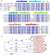
Sequence alignment of the light-driven proton pumps in halobacteria. A, 13 amino acid sequences of BR-like proteins were aligned. The key residues are annotated with different symbols. Circle, retinal-binding pocket; diamond, proton reuptake residue; square, proton releasing group. The key residue, Arg82 (HwBR), in this study is marked by the red box. The secondary-structural information of HwBR is shown above the alignment. B, phylogenomics analysis of the amino acid sequences of the light-driven proton pumps in halobacteria. The analysis classified HwBR from a quadrate-shaped bacteria into a new separate superfamily, qR.
FIGURE 2.
FIGURE 2.
Optical property and overall structure of wild-type and D93N HwBR proteins. A, the UV-visible spectra of purple membrane (trimeric HsBR) (gray dashed line), monomeric HsBR (black dashed line), and HwBR (purple solid line). The UV-visible spectra were measured in the buffer solution containing 50 mm MES (pH 5.8), 4 m NaCl, 0.02% DDM. AU, absorbance units. B, overall structure of monomeric HwBR. Nter, N terminus; Cter, C terminus. C, top view of wild-type trimeric structure. D, top view of wild-type antiparallel dimeric structure. E, three-dimensional structure alignment of BR-like proteins. Superimposition of aR-1 (1UAZ; green), aR-2 (1VGO; yellow), bR (1C3W; cyan), dR3 (4FBZ; purple), cR-3 (4L35; blue), and qR (4QI1; pink) structures is shown.
FIGURE 3.
FIGURE 3.
Buffer and detergent selection of HwBR using size exclusion column. The HwBR protein was loaded into the size exclusion column (Superdex 200 10/30 GL) and eluted by six combinations of two buffers and three detergents. The gray line is the absorption spectra at 280 nm, and the purple line is at 552 nm. AU, absorbance units.
FIGURE 4.
FIGURE 4.
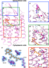
The structure and proton translocation path of HwBR. A, the proton outward cap region is drawn in a blue box, and residues Arg82 and Thr201 are shown as sticks. The hydrogen bonds are represented by black dashed lines. B, the residues involved in the proton-releasing group are represented by sticks in a green box. C, the retinal binding pocket and proton re-uptake residue Asp93 are shown in a red box. The proton-pumping flow is directed from the cytoplasmic site through the Schiff base to the proton-releasing complex, with protons exiting from the proton outward cap. The waters are shown as red sphere. A–C, have enlarged view on the right site labeled with key residues and waters. D, electron density maps of retinal and the surrounding region. The 2FoFc electron density map contoured at 1 σ is shown in blue. The all-trans retinal is shown in orange stick, and the key residues surrounded the binding pocket are shown in magenta stick.
FIGURE 5.
FIGURE 5.
Light-driven proton translocation activity assay using photocurrent measurements. A and B, an indium tin oxide-based photocurrent device was adopted to measure the light-driven photocurrent generation in both wild-type HwBR (A) and D104N/HwBR (D96N/HsBR-corresponding mutant) (B) at pH 5.8 with 0.1% DDM. A continuous 532-nm green laser was turned on at 0 s and turned off at 1.85 s while the photocurrent was continuously recorded. The light green shading indicates the light-on duration. The recovery of photocurrent traces started at time 1.85 s represented the proton reuptake step during the light-driven proton pumping. The recovery half-time (t½) values of the wild type and D104N/HwBR were around 0.05 and 0.75 s, respectively.
FIGURE 6.
FIGURE 6.
Comparison of proton outward caps. The proton outward caps of structures from five BR proteins are presented for qR (magenta), bR (cyan), aR-1 (green), aR-2 (yellow), dR3 (purple), and cR-3 (pink). The residues related to Arg82 and Thr201 of HwBR are shown as blue sticks.
FIGURE 7.
FIGURE 7.
pH-dependent transitions of wild type and R82E/HwBR in 0. 1% DDM and 100 mm NaCl. In A and B, the red curves (pH 2) and blue curves (pH 8) indicate the spectra of wild type and R82E/HwBR, respectively. The spectra of the putative fully protonated mutant D93N/HwBR (brown curve) and R82E/D93N/HwBR (magenta curve) are shown in panels A and B, respectively. AU, absorbance units. C, pH dependence of absorption maximum of HwBR (solid circle) and R82E/HwBR (open circle) upon increasing the pH from 1.3 to 8, respectively. Each spectrum was obtained at pH 1.3, 1.5, 2, 2.5, 3, 3.5, 4, 4.5, 5, 5.5, 6, 6.5, 7, 7.5, 8. The pH under 1.3 was inapplicable because of the protein denaturation.
FIGURE 8.
FIGURE 8.
The light-driven proton pump activity assay of wild-type HwBR and R82E/HwBR. A and B, light-driven proton transport by HwBR (A) and by R82E/HwBR (B) in E. coli cells. Red and green lines indicate the pump activities measured before and after the addition of CCCP, respectively. The arrows show the 1-watt 532-nm continuous green laser stimulation period. The results indicated that the R82E mutant did not interrupt the overall light-driven proton pump activity in HwBR.
FIGURE 9.
FIGURE 9.
pH-dependent thermal stability of wild type and R82E/HwBR. The absorbance of wild type and R82E/HwBR was determined at 0, 5, 15, 30, 45, 60, 90, 120, and 180 min at 75 °C in the buffers at pH 4 and 8, respectively. The time versus residual pigment was plotted for wild-type (solid line) and R82E (dashed line) at pH 4 (yellow) and pH 8 (blue) to determine the k value. AU, absorbance units.
FIGURE 10.
FIGURE 10.
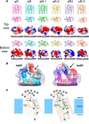
Electrostatic analysis of different BR structures and the proposed schematic of important proton translocation path residues and BC loop effect of HsBR and HwBR. A, top (extracellular) and bottom (cytoplasmic) views of known BR structures analyzed with respect to their electrostatic charge distribution, using PyMOL. The residues shown in sticks are important during light-driven proton pumping, including the proton-releasing group (Arg82, Glu194, Glu204-corresponding residues), Asp85, Asp212-corresponding residues, retinal residues, and Asp96-corresponding residues numbered in HsBR. B, electrostatic analysis of Glu74 and Ser193 for HsBR and Arg82 and Thr201 for HwBR. A negatively charged hook region in the BC loop was observed in HsBR but not in HwBR. A flat region in the HwBR BC loop showed a positive charge corresponding to the Arg82–Thr201 hydrogen-bonding region. C, a schematic of HsBR and HwBR with their proton translocation path-related residues under acidic conditions. The red solid and dashed arrows indicate better or lower accessibility for protons, respectively. Under low pH conditions, the high proton concentration in the extracellular region might access the outlet of proton releasing group (Arg82, Glu194, Glu204) in HsBR (left) via diffusion, whereas the Arg82–Thr201 hydrogen-binding network in HwBR (right) can shield the access from the outlet of proton releasing group (Arg90, Glu202, Glu212), thus maintaining the protonation status of the interior and the retinal-binding pocket.

References

    1. Spudich J. L., Yang C. S., Jung K. H., and Spudich E. N. (2000) Retinylidene proteins: structures and functions from archaea to humans. Annu. Rev. Cell Dev. Biol. 16, 365–392 - PubMed
    1. Schopf J. W. (2006) Fossil evidence of Archaean life. Philos. Trans. R. Soc. Lond. B Biol. Sci. 361, 869–885 - PMC - PubMed
    1. Béjà O., Spudich E. N., Spudich J. L., Leclerc M., and DeLong E. F. (2001) Proteorhodopsin phototrophy in the ocean. Nature 411, 786–789 - PubMed
    1. Man D., Wang W., Sabehi G., Aravind L., Post A. F., Massana R., Spudich E. N., Spudich J. L., and Béjà O. (2003) Diversification and spectral tuning in marine proteorhodopsins. EMBO J. 22, 1725–1731 - PMC - PubMed
    1. Balashov S. P., Imasheva E. S., Boichenko V. A., Antón J., Wang J. M., and Lanyi J. K. (2005) Xanthorhodopsin: a proton pump with a light-harvesting carotenoid antenna. Science 309, 2061–2064 - PMC - PubMed

Publication types

MeSH terms

LinkOut - more resources