. 2015 Dec 4;290(49):29167-77.
doi: 10.1074/jbc.M115.683268. Epub 2015 Oct 19.

Structural Insights into the MMACHC-MMADHC Protein Complex Involved in Vitamin B12 Trafficking

Affiliations

Structural Insights into the MMACHC-MMADHC Protein Complex Involved in Vitamin B12 Trafficking

D Sean Froese et al. J Biol Chem. .

Abstract

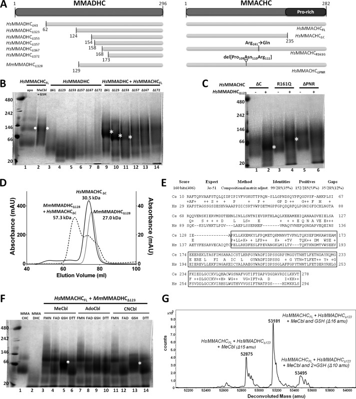
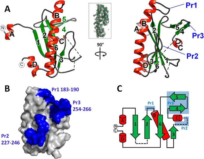
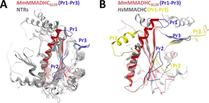
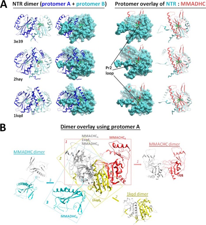
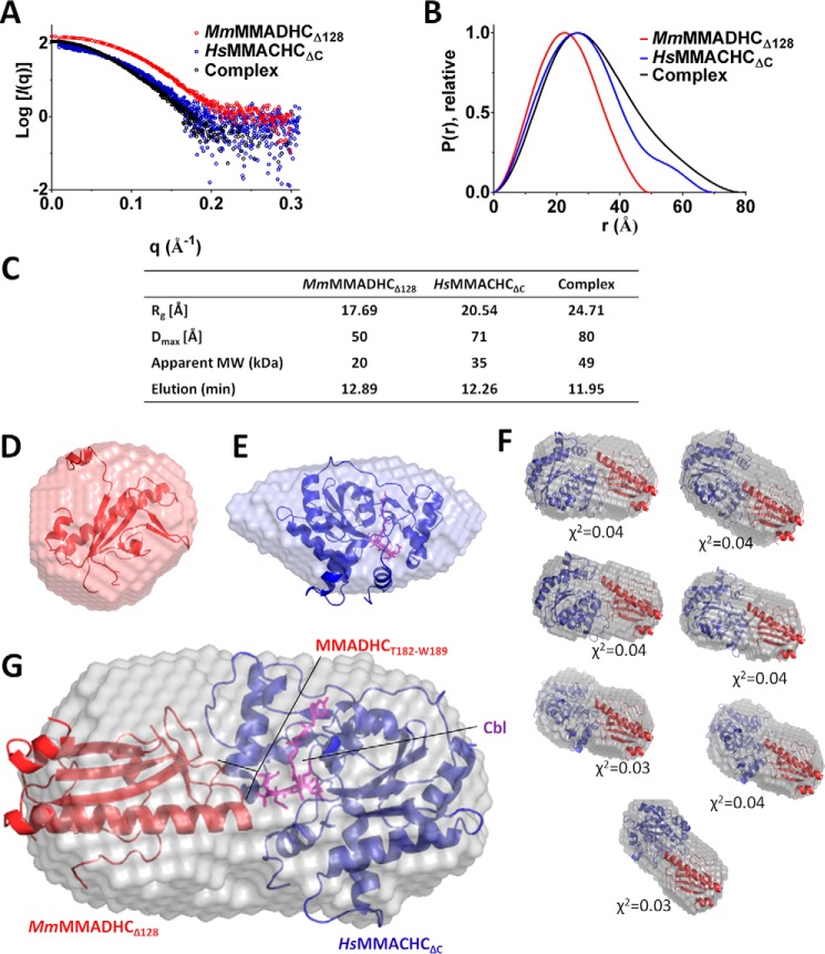
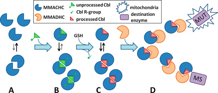
Conversion of vitamin B12 (cobalamin, Cbl) into the cofactor forms methyl-Cbl (MeCbl) and adenosyl-Cbl (AdoCbl) is required for the function of two crucial enzymes, mitochondrial methylmalonyl-CoA mutase and cytosolic methionine synthase, respectively. The intracellular proteins MMACHC and MMADHC play important roles in processing and targeting the Cbl cofactor to its destination enzymes, and recent evidence suggests that they may interact while performing these essential trafficking functions. To better understand the molecular basis of this interaction, we have mapped the crucial protein regions required, indicate that Cbl is likely processed by MMACHC prior to interaction with MMADHC, and identify patient mutations on both proteins that interfere with complex formation, via different mechanisms. We further report the crystal structure of the MMADHC C-terminal region at 2.2 Å resolution, revealing a modified nitroreductase fold with surprising homology to MMACHC despite their poor sequence conservation. Because MMADHC demonstrates no known enzymatic activity, we propose it as the first protein known to repurpose the nitroreductase fold solely for protein-protein interaction. Using small angle x-ray scattering, we reveal the MMACHC-MMADHC complex as a 1:1 heterodimer and provide a structural model of this interaction, where the interaction region overlaps with the MMACHC-Cbl binding site. Together, our findings provide novel structural evidence and mechanistic insight into an essential biological process, whereby an intracellular "trafficking chaperone" highly specific for a trace element cofactor functions via protein-protein interaction, which is disrupted by inherited disease mutations.

Keywords: crystal structure; metabolic disease; nitroreductase fold; protein-protein interaction; site-directed mutagenesis; small-angle x-ray scattering (SAXS); vitamin B12.


Figures

FIGURE 1.
FIGURE 1.
Interaction study of MMACHC and MMADHC. A, domain diagram showing recombinant human MMACHC and MMADHC constructs described in this figure. For reference, mouse construct MmMMADHCΔ128 used to generate the crystal structure is also shown. B, interaction of human MMACHC full-length and MMADHC truncated proteins by BN-PAGE. Lanes 1–8: single protein controls including MMACHCFL alone (lane 1) and with MeCbl and GSH (lane 2); and MMADHC truncations including MMADHCΔ61 (lane 3), MMADHCΔ123 (lane 4), MMADHCΔ153 (lane 5), MMADHCΔ157 (lane 6), MMADHCΔ167 (lane 7), and MMADHCΔ172 (lane 8). Lanes 9–14: combined protein incubation of MMACHCFL in presence of MeCbl and GSH with MMADHCΔ61 (lane 9), MMADHCΔ123 (lane 10), MMADHCΔ153 (lane 11), MMADHCΔ157 (lane 12), MMADHCΔ167 (lane 13), and MMADHCΔ172 (lane 14). C, interaction of MMACHC mutants and MMADHCΔ123 by BN-PAGE, in the presence of MeCbl and GSH. Lanes 1–6: MMACHCΔC alone (lane 1) and with MMADHCΔ123 (lane 2); MMACHCR161Q alone (lane 3) and with MMADHCΔ123 (lane 4); and MMACHCΔPNR alone (lane 5) and with MMADHCΔ123 (lane 6). For B and C, white dots indicate MMACHC homodimer, and white asterisks indicate MMACHC-MMADHC heterodimer. D, analysis of complex formation by size-exclusion chromatography including chromatographs of MmMMADHCΔ123 (gray line), HsMMACHCΔC + MeCbl + GSH (black line), and MmMMADHCΔ123 + HsMMACHCΔC + MeCbl + GSH (black dotted line). x axis: elution volume; left y axis: absorbance (milliabsorbance units (mAU)) for HsMMACHCΔC + MeCbl + GSH; right y axis: absorbance (mAU) for MmMMADHCΔ123 and MmMMADHCΔ123 + HsMMACHCΔC + MeCbl + GSH. Molecular weights of each peak are calculated from a calibration curve using molecular weight protein standards (Sigma-Aldrich). E, sequence alignment of MMADHC from H. sapiens (HS, NP_056517.1, human) and predicted MMADHC protein (Y76A2B.5) from C. elegans (Ce) using the BLAST server (http://blast.ncbi.nlm.nih.gov/Blast.cgi). The boxed region represents the smallest interacting construct of HsMMADHC for HsMMACHC. F, BN-PAGE of HsMMACHCFL and HsMMADHCΔ123 under various conditions. White asterisks indicate MMACHC-MMADHC heterodimer. G, native mass spectrometry of HsMMACHCFL and HsMMADHCΔ123 incubated with MeCbl and GSH. Expected sizes: HsMMACHCFL, 31818.6 Da; HsMMADHCΔ123, 19727.7 Da; MeCbl, 1343.6 Da; and GSH, 307.3 Da.
FIGURE 2.
FIGURE 2.
Structure of the MMACHC interaction module of MMADHC. A, graphic representation of the MmMMADHCΔ128 structure in orthogonal views. Secondary structures are colored green for β-sheets and red for α-helices. The first (aa 132) and last (aa 296) residues observed in the structure are labeled with N and C, respectively. Dotted lines indicate disordered regions. Inset: view of the σ-weighted (2FoFc) electron density map of MmMMADHCΔ128 aa region 190–217, contoured at σ = 1. B, surface representation of MmMMADHCΔ128 (same orientation as A, right panel) highlighting the three protrusions (Pr1–Pr3) in blue. C, topology diagram of the MmMMADHCΔ128 secondary structure with the same coloring and labeling as in A and B. Disordered regions are shown as dashed lines.
FIGURE 3.
FIGURE 3.
Structural comparison of MMADHC and proteins of the nitroreductase family. A, structural superposition of MmMMADHCΔ128 (red) and two other NTRs (Protein Data Bank (PDB): 2HAY, 2ISL; gray). B, superposition of MmMMADHCΔ128 (red) and MMACHC (PDB: 3SOM; gray), highlighting the absence of the HsMMACHC four-helix cap domain (aa 185–234) in MmMMADHCΔ128 as well as the different orientations of protrusions Pr1 and Pr2 (HsMMACHC: Pr1 aa 69–77; Pr2 aa 104–116; MmMMADHCΔ128: Pr1 aa 183–190, Pr2 aa 227–246) between the proteins. Protrusions (Pr1–Pr3) are colored blue for MmMMADHCΔ128 and yellow for HsMMACHC.
FIGURE 4.
FIGURE 4.
Comparison of MMADHC to other NTR dimers. A, overlay of the MmMMADHCΔ128 structure (red) with nitroreductases from Desulfovibrio desulfuricans (PDB: 3E39), Streptococcus pyogenes (PDB: 2HAY), and Enterobacter cloacae (PDB: 1KQD). The canonical FMN/FAD binding site of NTRs (left, indicated by the green stick ligand), located at the conventional dimer interface, is not present in MMADHC (right) due to disruption by its protrusion loop Pr2. B, structural alignment of a single subunit (protomer A) of MmMMADHCΔ128 (MMADHC), HsMMACHCFL (MMACHC, PDB: 3SOM), and an NTR (PDB: 1KQD) in gray, with the second subunit (protomer B) of their crystallographic dimers color-coded (MMADHC in blue; NTR in yellow; MMACHC in red), demonstrating the lack of a conserved dimeric orientation and interface among the three structures.
FIGURE 5.
FIGURE 5.
Structural and biochemical analysis of MMADHC missense mutations. A, domain diagram of MMADHC, with missense mutations color-coded according to their cellular defects (10–12), namely MMA (green), combined HC+MMA (stretch 1, red), or HC (stretches 2 and 3, blue) phenotypes. Purple line indicates aa region observed in the crystal structure. B, graphic (left) and surface (right) representations of MmMMADHCΔ128 mapped with stretches 1–3 and individual mutations found within these regions. Dotted lines indicate disordered regions. C, analysis of thermal unfolding curves of various protein constructs reveals a melting temperature (Tm) of 62 °C (right) for HsMMADHCΔ123, which is relatively unchanged for all mutants with the exception of HsMMADHCΔ123-D226A where the Tm is slightly decreased as compared with wild type. Error bars indicate means ± S.E. D, interaction of HsMMACHCFL with wild-type or mutant HsMMADHCΔ123 studied by BN-PAGE. Depicted are: HsMMACHCFL with MeCbl and GSH alone (lane 1), and in combination with HsMMADHCΔ123 (lane 2), HsMMADHCΔ123-T182N (lane 3), HsMMADHCΔ123-M186A (lane 4), HsMMADHCΔ123-W189A (lane 5), HsMMADHCΔ123-D226A (lane 6), and HsMMADHCΔ123-L259P (lane 7). White asterisks indicate the MMACHC-MMADHC heterodimer.
FIGURE 6.
FIGURE 6.
SAXS analysis of the MMACHC-MMADHC interaction. A, raw scattering curves for MmMMADHCΔ128 (red), HsMMACHCΔC (blue), and their complex (black). B, P(r) plots, calculated with Scatter (27) and normalized to peak height, show differences between the single proteins and the complex. Guinier plots indicate aggregation-free data. C, table of radius of gyration (Rg), maximal intraparticle dimension (Dmax) as calculated from the Guinier plot, apparent molecular weight (MW), and elution time from the in-line HPLC column. D and E, ab initio bead models calculated with 13 runs of DAMMIF (39) for MmMMADHCΔ128 fitted with its x-ray structure (this study) (D) and HsMMACHCΔC fitted with its x-ray structure (PDB: 3SOM) (E). F, top seven models of the HsMMACHCΔC-MmMMADHCΔ128 complex produced by ZDOCK with fitted χ2 values shown. The SAXS envelope is overlaid with modeled orientations of HsMMACHCΔC (blue) and MmMMADHCΔ128 (red) structures. MmMMADHCΔ128 is presented in the same orientation on all models for better comparison. G, HsMMACHCΔC-MmMMADHCΔ128 complex fitted with a representative example of the ZDOCK (35) complex model. In E and G, Cbl is shown as purple sticks.
FIGURE 7.
FIGURE 7.
Proposed role of the MMACHC-MMADHC complex in Cbl targeting. A, MMACHC (blue sector) in the unbound state exists primarily as a monomer. B, upon binding unprocessed Cbl with an intact upper axial ligand (triangle), it can shift toward the homodimeric state. C, following processing of Cbl (green to red triangle) via GSH-mediated removal of its upper axial ligand (green stick and square), MMACHC is available for interaction with MMADHC (orange sector). D, MMADHC binds MMACHC in a 1:1 heterodimer and escorts Cbl-laden MMACHC either toward the mitochondria (star) for use by methylmalonyl-CoA MUT or to MS in the cytosol.

References

    1. Watkins D., and Rosenblatt D. S. (2014) Inherited disorders of folate and cobalamin transport and metabolism. in The Online Metabolic and Molecular Bases of Inherited Disease (Valle D., Beaudet A. L., Vogelstein B., Kinzler K. W., Antonarakis S. E., Ballabio A., Gibson M., and Mitchell G., eds), McGraw-Hill, New York
    1. Froese D. S., and Gravel R. A. (2010) Genetic disorders of vitamin B metabolism: eight complementation groups–eight genes. Expert Rev. Mol. Med. 12, e37. - PMC - PubMed
    1. Gherasim C., Lofgren M., and Banerjee R. (2013) Navigating the B12 road: assimilation, delivery, and disorders of cobalamin. J. Biol. Chem. 288, 13186–13193 - PMC - PubMed
    1. Baumgartner M. R. (2013) Vitamin-responsive disorders: cobalamin, folate, biotin, vitamins B1 and E. Handb. Clin. Neurol. 113, 1799–1810 - PubMed
    1. Harding C. O., Arnold G., Barness L. A., Wolff J. A., and Rosenblatt D. S. (1997) Functional methionine synthase deficiency due to cblG disorder: a report of two patients and a review. Am. J. Med. Genet. 71, 384–390 - PubMed

Publication types

MeSH terms