. 2015 Dec 11;290(50):29931-40.
doi: 10.1074/jbc.M115.675603. Epub 2015 Oct 19.

Salt Bridge Swapping in the EXXERFXYY Motif of Proton-coupled Oligopeptide Transporters

Affiliations

Salt Bridge Swapping in the EXXERFXYY Motif of Proton-coupled Oligopeptide Transporters

Nanda G Aduri et al. J Biol Chem. .

Abstract

Proton-coupled oligopeptide transporters (POTs) couple the inward transport of di- or tripeptides with an inwardly directed transport of protons. Evidence from several studies of different POTs has pointed toward involvement of a highly conserved sequence motif, E1XXE2RFXYY (from here on referred to as E1XXE2R), located on Helix I, in interactions with the proton. In this study, we investigated the intracellular substrate accumulation by motif variants with all possible combinations of glutamate residues changed to glutamine and arginine changed to a tyrosine, the latter being a natural variant found in the Escherichia coli POT YjdL. We found that YjdL motif variants with E1XXE2R, E1XXE2Y, E1XXQ2Y, or Q1XXE2Y were able to accumulate peptide, whereas those with E1XXQ2R, Q1XXE2R, or Q1XXQ2Y were unable to accumulate peptide, and Q1XXQ2R abolished uptake. These results suggest a mechanism that involves swapping of an intramotif salt bridge, i.e. R-E2 to R-E1, which is consistent with previous structural studies. Molecular dynamics simulations of the motif variants E1XXE2R and E1XXQ2R support this mechanism. The simulations showed that upon changing conformation arginine pushes Helix V, through interactions with the highly conserved FYING motif, further away from the central cavity in what could be a stabilization of an inward facing conformation. As E2 has been suggested to be the primary site for protonation, these novel findings show how protonation may drive conformational changes through interactions of two highly conserved motifs.

Keywords: membrane transport; molecular dynamics; peptide transport; protein structure; site-directed mutagenesis.


Figures

FIGURE 1.
FIGURE 1.
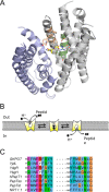
Overall architecture and conformations of POTs. A, crystal structure of the inward open PepTSo2·Ala-Ala-Ala complex (Protein Data Bank code 4TPJ) (42). The N- and C-terminal domains are colored white and blue, respectively. Side chains shown as sticks correspond to the E1XXE2RFXYY motif (green) and the FYING motif (wheat). The substrate is represented as yellow sticks. B, schematic representation of possible conformational changes in POTs during proton/peptide translocation; the proton is represented as a black sphere with spikes, and the peptide is represented by a black rhombus. C, multiple sequence alignment of E1XXE2RFXYY and FYXXINXG motifs using mafft from GkPOT (Q5KYD1), YjdL (P39276), YdgR (P77304), YbgH (P75742), hPepT1 (P46059), PepTSo (Q8EHE6), PepTSt (Q5M4H8), and NRT1.1 (Q05085).
FIGURE 2.
FIGURE 2.
A, fluorescence quenching of β-Ala-Lys(AMCA). 0.5 mm β-Ala-Lys(AMCA) was incubated with cells harboring YjdL-WT (A600 of 10 for 30 min). The cells were then centrifuged and washed using ice-cold uptake buffer. The fluorescence was measured before and after sonication on a Safire 2 fluorometer at an excitation wavelength and emission wavelength of 340 and 460 nm, respectively. The data shown were normalized to the fluorescence values shown by β-Ala-Lys(AMCA) in intact cells. Error bars indicate S.E. (n = 3). B, Western blots of the motif mutants (n = 3). Only a representative blot is shown.
FIGURE 3.
FIGURE 3.
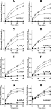
Accumulation profiles of motif variants Q1XXE2Y (A), E1XXQ2Y (B), E1XXE2Y (C), Q1XXQ2Y (D), E1XXE2R (E), E1XXQ2R (F), Q1XXE2R (G), and Q1XXQ2R (H). The ratio of substrate accumulated inside the cell with respect to the substrate concentration outside the cell was plotted as a function of time. The assay was performed at pHo 5.5 (open squares), 6.5 (open circles), and 7.5 (open triangles). The closed symbols represent empty vector. The error bars indicate the S.E. (n = 3).
FIGURE 4.
FIGURE 4.
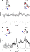
Molecular dynamics simulations probing the effect of E2 to Q change. The shortest side chain N–O distances (Å) from Arg36 to Glu32 (gray) and Glu35 (black), respectively, as a function of time in GkPOT-WT (E1XXE2R) (A) and E35Q-GkPOT (E1XXQ2R) (B) are shown. Insets represent the position of arginine at 0 and 35 ns.
FIGURE 5.
FIGURE 5.
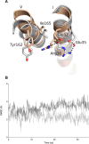
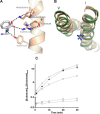
Movement of Helix V as a consequence of salt bridge swapping. A, the movement in Helix V of E35Q-GkPOT accompanying the shift of Arg36 on Helix I. The helices of E35Q-GkPOT at 35 ns (wheat) were aligned to GkPOT-WT at 35 ns (white). B, the root mean square deviation (RMSD) (Å) of the Cα atom of Tyr162 on Helix V plotted as a function of time in GkPOT-WT (gray) and E35Q-GkPOT (black).
FIGURE 6.
FIGURE 6.
Schematic representation of salt bridge swapping in the E1XXE2R motif shown in GkPOT corresponding to the presence of substrate Ala-Ala and proton (red sphere). A, absence of both substrate and proton. B, presence of Ala-Ala alone. C, presence of both substrate and proton.
FIGURE 7.
FIGURE 7.
A, hydrogen bonding network in the PepTSt·AF complex (Protein Data Bank code 4D2C) (17) between the E1XXE2R residues in Helix V and Lys126 in PepTSt (wheat) and the dipeptide AF (white). The amino acid residue numbering corresponds to PepTSt followed by GkPOT and YjdL. B, superposition of Helices I and V from the 35-ns E35Q-GkPOT (E1XXQ2R) structure in green and NRT1.1 protomer B (Protein Data Bank code 4OH3) (43) in wheat, respectively. Arginine is shown as sticks. C, accumulation profiles of V159G-YdgR (open squares), G146V-YjdL (open circles), YjdL-WT (open rhombuses), and YdgR-WT (closed rhombuses). The ratio of substrate accumulated inside the cell with respect to the substrate concentration outside the cell was plotted as a function of time. The assay was performed at pHo 6.5. The open downward facing triangles represent empty vector. Error bars indicate S.E. (n = 3).
FIGURE 8.
FIGURE 8.
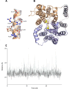
Further destabilization of the binding site as a consequence of E2 protonation. A, Helix I of E35Q-GkPOT after 35 ns (wheat) superimposed with Helix I of the GkPOT-WT after 35 ns (gray) indicating the change in conformation of Tyr40. B, crystal structure of GkPOT displaying the salt bridge between N- (wheat) and C (blue)-domains, i.e. between residues Arg43 and Glu310 (yellow). C, the shortest side chain N–O distance (Å) between Arg43 and Glu310 as a function of time in GkPOT-WT (black) and E35Q-GkPOT (gray).

References

    1. Fei Y. J., Kanai Y., Nussberger S., Ganapathy V., Leibach F. H., Romero M. F., Singh S. K., Boron W. F., and Hediger M. A. (1994) Expression cloning of a mammalian proton-coupled oligopeptide transporter. Nature 368, 563–566 - PubMed
    1. Liang R., Fei Y. J., Prasad P. D., Ramamoorthy S., Han H., Yang-Feng T. L., Hediger M. A., Ganapathy V., and Leibach F. H. (1995) Human intestinal H+/peptide cotransporter. Cloning, functional expression, and chromosomal localization. J. Biol. Chem. 270, 6456–6463 - PubMed
    1. Weitz D., Harder D., Casagrande F., Fotiadis D., Obrdlik P., Kelety B., and Daniel H. (2007) Functional and structural characterization of a prokaryotic peptide transporter with features similar to mammalian PEPT1. J. Biol. Chem. 282, 2832–2839 - PubMed
    1. Fang G., Konings W. N., and Poolman B. (2000) Kinetics and substrate specificity of membrane-reconstituted peptide transporter DtpT of Lactococcus lactis. J. Bacteriol. 182, 2530–2535 - PMC - PubMed
    1. Theis S., Döring F., and Daniel H. (2001) Expression of the myc/His-tagged human peptide transporter hPEPT1 in yeast for protein purification and functional analysis. Protein Expr. Purif. 22, 436–442 - PubMed

Publication types

LinkOut - more resources