. 2015 Oct 26;211(2):435-53.
doi: 10.1083/jcb.201502032. Epub 2015 Oct 19.

Rootletin organizes the ciliary rootlet to achieve neuron sensory function in Drosophila

Affiliations

Rootletin organizes the ciliary rootlet to achieve neuron sensory function in Drosophila

Jieyan V Chen et al. J Cell Biol. .

Abstract

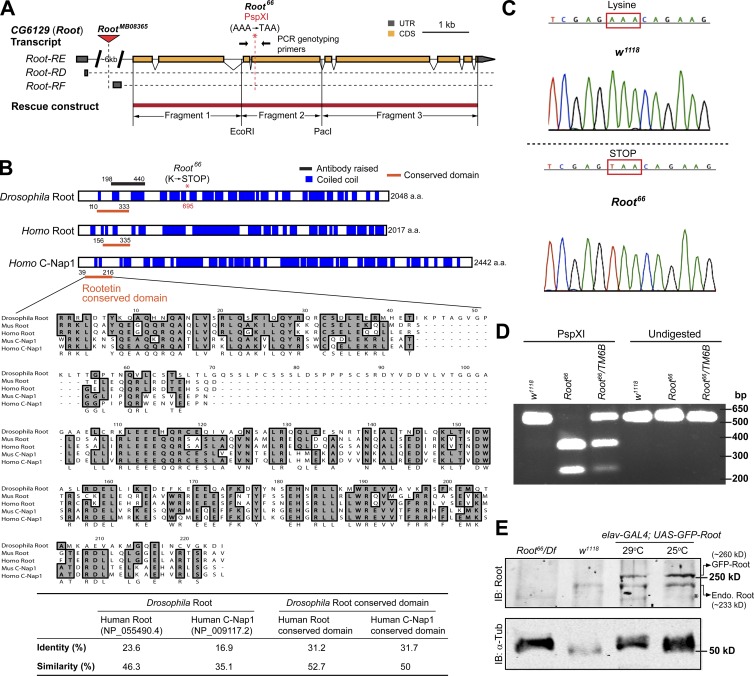
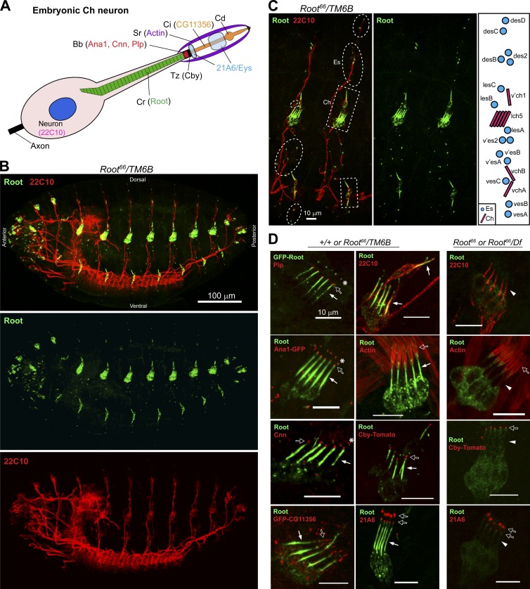
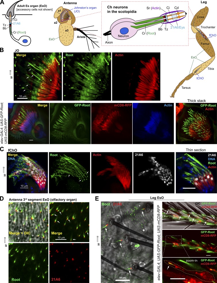
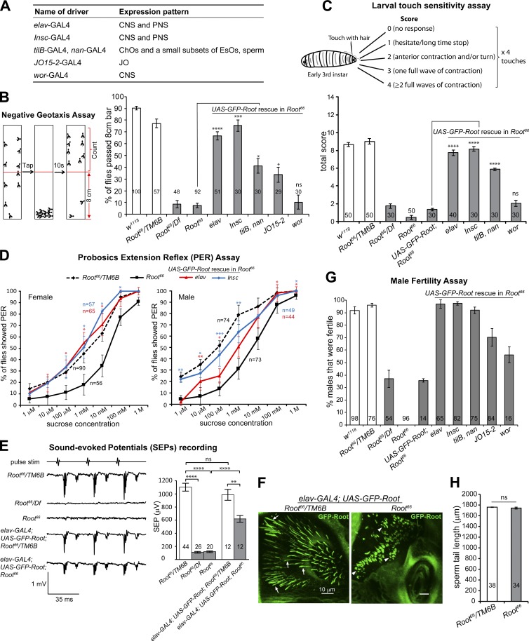
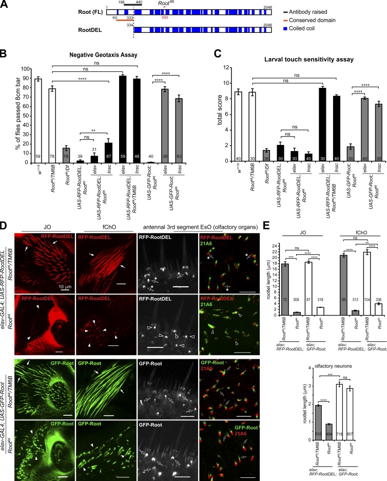
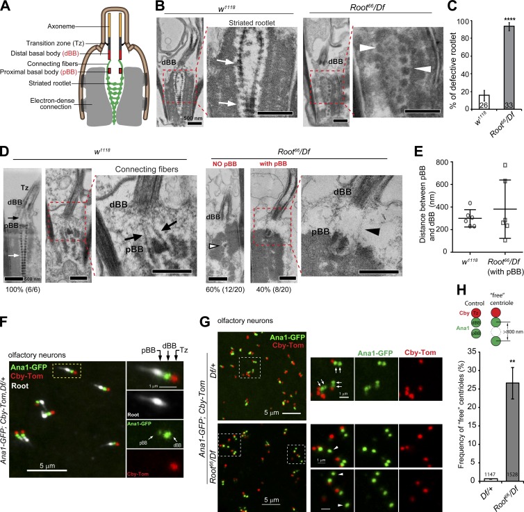
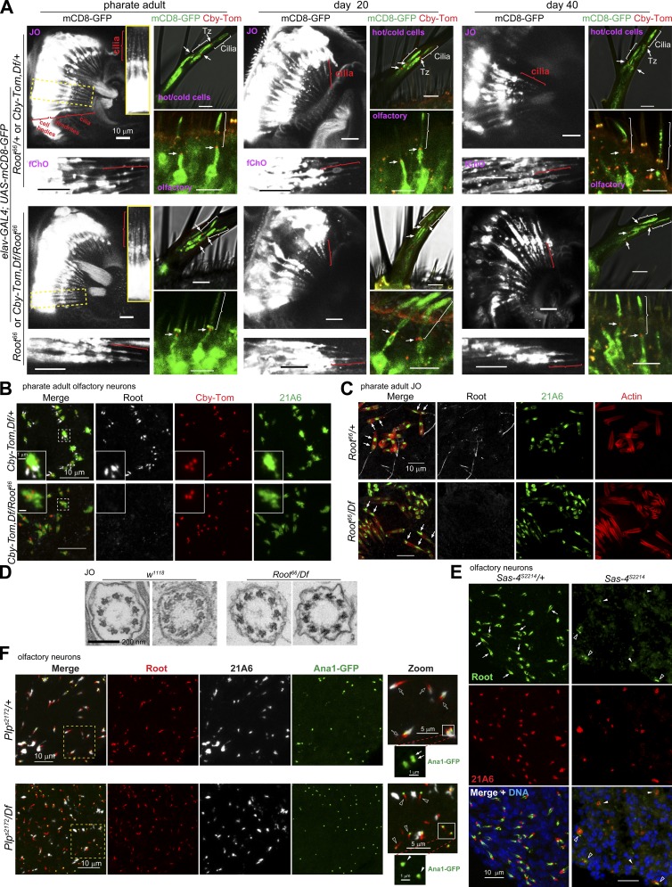
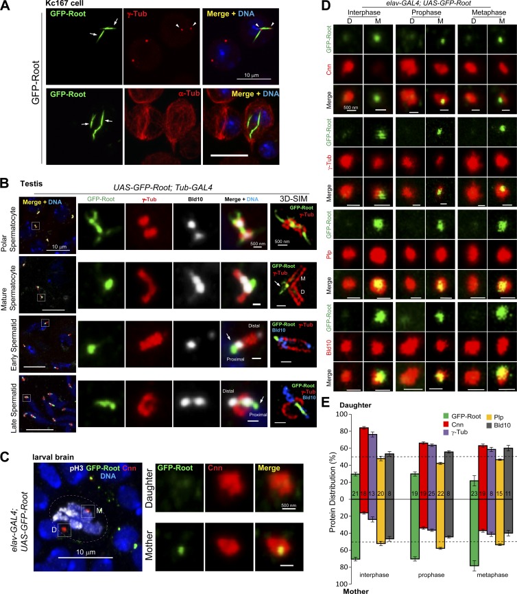
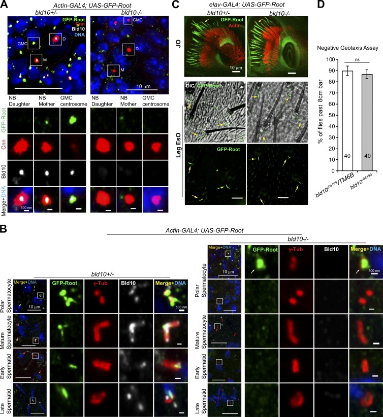
Cilia are essential for cell signaling and sensory perception. In many cell types, a cytoskeletal structure called the ciliary rootlet links the cilium to the cell body. Previous studies indicated that rootlets support the long-term stability of some cilia. Here we report that Drosophila melanogaster Rootletin (Root), the sole orthologue of the mammalian paralogs Rootletin and C-Nap1, assembles into rootlets of diverse lengths among sensory neuron subtypes. Root mutant neurons lack rootlets and have dramatically impaired sensory function, resulting in behavior defects associated with mechanosensation and chemosensation. Root is required for cohesion of basal bodies, but the cilium structure appears normal in Root mutant neurons. We show, however, that normal rootlet assembly requires centrioles. The N terminus of Root contains a conserved domain and is essential for Root function in vivo. Ectopically expressed Root resides at the base of mother centrioles in spermatocytes and localizes asymmetrically to mother centrosomes in neuroblasts, both requiring Bld10, a basal body protein with varied functions.


Figures

Figure 1.
Figure 1.
Drosophila Root is the orthologue of mammalian Root and C-Nap1. (A) Diagram showing the Drosophila Root transcripts RE, RD, and RF, which differ only in their 5′UTRs (only the 5′UTR is shown for RD and RF); the transposon insertion in RootMB08365; the nonsense mutation in Root66 that also introduces a PspXI restriction site; the primers used for PCR genotyping; and the Root rescue construct, which was cloned by ligating three genomic fragments together. (B) Drosophila Root, human Root, and C-Nap1 are large proteins with extensive coiled coils, and they share a highly conserved Root domain near the N terminus. The conserved domain in mouse Root and C-Nap1 is also shown in the sequence homology. The Root66 mutation and the region used to raise the Root antibody are also indicated. The table shows the percentage identity and similarity between Drosophila Root with human Root and C-Nap1, using ClustalW alignment. (C) DNA sequencing confirms the nonsense mutation in Root66. (D) PspXI restriction digest analysis of a PCR product spanning the mutation site in Root66. PCR primers are as shown in A. The size of the uncut PCR products is 602 bp. DNA products amplified from wild-type w1118 cannot be digested by PspXI, whereas all products from the Root66 homozygote and about half from the Root66/TM6B heterozygote are cut by PspXI into smaller fragments of expected sizes (∼350 bp and 250 bp). TM6B is a balancer chromosome and is wild type for Root. (E) Western blot of isolated antenna shows that Root is absent from the Root66 mutant and the GFP-Root transgene expresses slightly higher levels compared with endogenous (Endo) Root. *, nonspecific bands. Lysates from 40–50 antenna pairs were loaded in each lane. Df is Df(3R)Exel6197, a deficiency line with chromosome deletion covering the entire Root gene. See Fig. S1 C for the whole blot. See also Fig. S1.
Figure 2.
Figure 2.
Root localizes to the ciliary rootlet in embryonic Ch and Es neurons. (A) Schematic view of the embryonic Ch neuron with structural protein markers. Bb, basal body; Cd, ciliary dilation; Ci, cilium; Cr, ciliary rootlet; Sr, scolopale rod; Tz, transition zone. (B) Endogenous Root is expressed in the embryonic PNS. Antibody 22C10 recognizes Futsch, a PNS neuron marker. (C) Root is expressed in both Ch and Es neurons. Boxed and circled areas indicate Ch neurons and Es neurons, respectively, which are illustrated as pink bars and blue circles in the cartoon on the right. The schematic is adapted from Orgogozo and Grueber (2005) and the names of type I sensory neurons are indicated. (D) Root localizes to the rootlet in Ch neurons. Endogenous Root or GFP tagged Root is counterstained with the PNS marker 22C10 or the following ciliary markers (open arrows): Plp, Ana1, and Cnn mark the Bb; Cby marks the Tz; CG11356 marks the axoneme; and 21A6/Eys marks the cilium proximal end and the extracellular region right below the ciliary dilation. Root also localized to a focus, together with Cnn and Ana1, distal to the basal body, in an unknown structure that might be the ciliary dilation (asterisks). In control +/+ or Root66/TM6B, Root resides at a structure consistent with the ciliary rootlet (solid arrows) that extends from the base of the basal body, passes through the dendrite, and reaches the cell body. In homozygous Root66 or hemizygous Root66/Df, Root is absent from the rootlet (solid arrowheads). However, the morphologies of mutant neurons and scolopale rods appear normal, as marked by 22C10 and actin, respectively; the localizations of Cby and 21A6 are also unaffected (open arrows). Mutant embryos are from Root heterozygous parents and distinguished by Root antibody staining. Bars: (B) 100 µm; (C and D) 10 µm.
Figure 3.
Figure 3.
Root localizes to the ciliary rootlet in adult Ch and Es neurons. (A) Illustration of ChOs and EsOs in the antenna and leg. The JO in the antennal a2 segment is a specialized ChO composed of arrays of scolopidia, each containing two or three neurons. In the leg, scolopidia, each containing two neurons, are present in bundles in the fChO and tChO. EsOs, consisting of Es neurons and supporting cells, usually associate with external bristles. Bb, basal body; Cd, ciliary dilation; Ci, cilium; Cr, ciliary rootlet; Sr, scolopale rod; Tz, transition zone. (B) In Ch neurons of the JO, endogenous Root (upper panel) or GFP-Root (bottom panel) localizes to the ciliary rootlet, typically 15–25 µm long. The rootlet stretches from the base of the cilium to the neuron cell body (arrows). Upper panel shows immunostaining of antenna cryosections. Actin marks scolopale rods; mCD8-RFP localizes to plasma membranes and outlines the neurons. (C) Endogenous Root localizes to the approximately 20-µm rootlet in Ch neurons of the fChO. Each scolopidium has two Ch neurons and hence two rootlets (arrows). Actin marks scolopale rods, 21A6 marks the cilium base and the region right below the ciliary dilation. (D) Endogenous Root resides at the ∼2- to 10-µm rootlet in Es neurons (olfactory neurons) in the antennal a3 segment. Each set of olfactory organs has one to four neurons, as indicated by different numbers of rootlets. 21A6 marks the cilium base. (E) In leg Es neurons, endogenous Root (left) or GFP-Root driven by elav-GAL4 (right) localizes at the ∼2- to 8-µm rootlet. 21A6 marks the cilium base, mCD8-RFP outlines the neurons. Bars, 10 µm.
Figure 4.
Figure 4.
Root is essential for neuron-specific behaviors. (A) GAL4 drivers used for Root rescue and their expression patterns. (B) Root is essential for negative geotaxis. The percentages of Root66 mutant flies that passed the negative geotaxis assay are significantly lower compared with controls (white bars). The mutants are rescued, to different degrees, by expressing GFP-Root with different GAL4 drivers. Expression in the entire nervous system (elav, insc) or mainly in the ChOs (tilB+nan, JO15-2) conferred complete or significant rescue, whereas expression limited to the central nervous system (CNS; wor) did not. (C) Root66 larvae lack sensitivity to touch, compared with the controls (white bars). Touch sensitivity is best restored by expressing GFP-Root in both ChOs and EsOs (elav, insc), expression limited to mainly ChOs (tilB+nan) also rescues the defect significantly, but expression of GFP-Root in CNS (wor) did not rescue the phenotype. (D) Root66 flies show reduced taste responses to sucrose compared with Root66/TM6B. The proboscis extension reflex (PER) taste response is significantly restored by driving GFP-Root expression in the entire PNS (elav, Insc), although the rescue was more thorough in females than in males. Significance was measured between Root66 and rescue groups at each sucrose concentration. (E) Root66 flies show significantly reduced hearing response indicated by SEPs, compared with Root66/TM6B (white bars). GFP-Root expression driven by elav-GAL4 rescues the hearing impairment of Root66 significantly but not completely. (F) In the JO, ectopically expressed GFP-Root localizes to the normal-sized, ∼15- to 25-µm rootlets in Root66/TM6B (arrows); however, the GFP-Root rescue in the Root66 mutant organizes much shorter rootlets at ∼2–8 µm (arrowheads). Bars, 10 µm. (G) Root66 males have impaired fertility. More than 90% of the control males are fertile (white bars); in contrast, none of Root66 and less than 40% of Root66/Df males produce progeny. UAS-GFP-Root; Root66 showed some fertility, probably because of leaky expression of the transgene. The fertility of Root66 males is fully rescued by expressing GFP-Root in the nervous system with elav-, Insc-, or tilB+nan-GAL4. (H) Root66 males produce mature sperm with normal tail length. For all charts, numbers of flies/larvae/sperm assayed are indicated in/near the bars, and error bars represent SEM. ns, P > 0.05; *, P ≤ 0.05; **, P ≤ 0.01; ***, P ≤ 0.001; ****, P ≤ 0.0001. See also Video 1 and Fig. S2.
Figure 5.
Figure 5.
The Rootletin conserved domain is essential for Root function and rootlet assembly, but not for localization to basal bodies. (A) Schematic view showing RootDEL, which has a deletion of the N-terminal 333 amino acids, including the entire conserved Root domain. (B) elav-GAL4 driving RFP-RootDEL expression does not rescue Root66 locomotor defects. Flies with RFP-RootDEL expression driven by Insc-GAL4 performed better than the Root66, but the level of rescue is low compared with that of GFP-Root (full length). RootDEL does not introduce dominant effects, as flies with RFP-RootDEL expression in the Root66/TM6B background do not show locomotor defects. (C) RFP-RootDEL expression driven by Insc- or elav-GAL4 failed to rescue the larval touch insensitivity phenotype in Root66. RFP-RootDEL expression in Root66/TM6B background does not cause defects, indicating no dominant effects are associated with RootDEL. (D) RFP-RootDEL localizes to basal bodies but does not assemble normal rootlets. GFP-Root is included as a positive control. When expressed in control Root66/TM6B Ch neurons, RFP-RootDEL localizes to rootlets and does not affect their assembly (solid arrows in top panels), similar to GFP-Root (solid arrows in lower panels). For unknown reason, in Root66/TM6B Es neurons, RootDEL localizes to rootlets shorter than those labeled by GFP-Root (open arrows with asterisks), see also E. While GFP-Root supports assembly of short rootlets in Root66 Ch neurons (solid arrowheads) and normal ones in Es neurons (open arrows), RFP-RootDEL localizes to basal bodies but does not form proper rootlets in both Ch and Es neurons (solid and empty arrowheads). Note that the signal of RFP-RootDEL may not be a proper rootlet but rather a focus of RFP-RootDEL accumulated at the basal body. The black and white panels are live images of the signal for GFP-Root or RFP-RootDEL taken through the pupal cuticle. Bars, 10 µm. (E) Quantification of rootlet lengths in control and Root66 neurons expressing GFP-Root or RFP-RootDEL. Rootlets are measured from at least four antennae for each genotype. Rightmost panels in D show images of the antenna squash. For all graphs, numbers of males/larvae/rootlets assayed are indicated in/near the bars, and error bars represent SEM. ns, P > 0.05; **, P ≤ 0.01; ***, P ≤ 0.001; ****, P ≤ 0.0001. See also Fig. S2.
Figure 6.
Figure 6.
Root is essential for rootlet formation and basal body cohesion. (A) Schematic view of the rootlet and its connection to the basal bodies in the JO. (B) Root66 JO neurons lack striated rootlets. Representative transmission EM longitudinal section images show that in control w1118, the rootlet is organized as a characteristic striated fiber (arrows), but in Root66/Df, this organization is lost; instead, some disconnected electron-dense clumps are observed at the rootlet region (arrowheads). (C) Quantification of defective rootlets in Root66 JO. Rootlet structures were observed in longitudinal and cross-sections. Numbers of rootlets analyzed are indicated; ****, P ≤ 0.0001. (D) The connecting fibers are normally found between the pBB and the dBB as thread-like electron-dense structures in the control (black arrows); they are lost in Root66 (black arrowhead). The striated rootlet is present in wild type (white arrow) but disrupted in the mutant (white arrowhead). Examined by serial sections, 60% of mutant JO neurons appear to lack a pBB. (E) Quantification of the edge-to-edge distance between the dBB and pBB in JO Ch neurons. Single data points, mean, and standard deviation are indicated. There is no significant difference between mean values of w1118 and Root66, using Mann-Whitney test. But F-distribution analysis indicated that distances in Root66 are significantly more variable than in w1118. (F) Immunostaining of olfactory neurons for basal bodies (Ana1-GFP), the transition zone (Cby-Tomato), and Root. Root localizes into the space between dBB and pBB. (G) In the control olfactory neurons (upper panels), basal bodies (Ana1-GFP) are in tandem pairs (arrows) with Cby located near one of them. In Root66 (lower panels), one of the basal bodies is frequently more distant, or “free” (arrowheads). (H) Quantification of loss of basal body cohesion in Root66. A basal body (centriole), marked with Ana1-GFP, is scored as “free” if it is not directly associated with the Tz (Cby-Tomato) and located more than 800 nm (center-to-center distance, which is about twice the diameter of an Ana1-GFP dot) from the nearest dBB that is associated with Cby. The frequency of “free centrioles” is defined by the ratio of the number of “free” Ana1 dots to the number of Cby dots. Numbers near/in the bars indicate numbers of Cby dots assayed from at least four antennae for each genotype. **, P ≤ 0.01. F and G show images of the antenna squash. Bars: (B and D) 500 nm; (F and G) 5 µm; (F and G, zoom) 1 µm.
Figure 7.
Figure 7.
Cilium structure is normal and maintained with age in Root mutant neurons, and centrioles but not cilia are required for rootlet assembly. (A) Various ciliated neurons at indicated age expressing mCD8-GFP to label ciliary membranes and Cby-Tomato to mark the transition zone (Tz). Brackets indicate the cilia, and arrows the Tz. Cilium morphology appears normal in Root66 through aging. hot/cold cells: temperature-sensing neurons in the arista, olfactory: olfactory neurons in the antennal third segment. For the hot/cold neuron images, transmitted light images are overlaid to show the morphology of the arista. (B) Olfactory neurons stained for 21A6 to label the cilium base and Cby-Tom to label the Tz. Cby and 21A6 localizations appear normal in Root66. (C) Ch neurons in the JO stained for actin to label the scolopale rods and 21A6 to label cilia. 21A6 localizes both to the cilium base and a distal region in the cilium (arrows), and this localization appears normal in Root66. (D) Cross-section of JO cilia by transmission electron microscopy shows that Root66 axoneme ultrastructure appears normal. (E) Sas-4 mutant lacking centrioles fails to organize rootlets in olfactory neurons. Compared with the control, where rootlets project from cilium base marked by 21A6 (arrows), rootlet structures are absent in most Sas-4 olfactory neurons (solid arrowheads), although sometimes abnormal tiny fibers are associated with 21A6 (open arrowheads). Endogenous Root is stained in green. (F) In control olfactory neurons, all rootlets are associated with the cilium base marker 21A6 (empty arrows), and the basal bodies (dBB and pBB) marked by Ana1-GFP are in tandem (solid arrows). In Plp mutant that lacks cilia and has the basal bodies displaced from the dendrite tip, most of the rootlets (empty arrowheads) are associated with a single basal body (solid arrowheads), which is not attached with 21A6. B, C, E, and F show images of the antenna squash. Bars: (A–C and E) 10 µm; (B, inset) 1 µm; (D) 200 nm; (F, main) 10 µm; (F, zoom) 5 µm; (F, zoom inset) 1 µm. See also Fig. S3.
Figure 8.
Figure 8.
Ectopic Root localizes to mother centrioles in testes and distributes asymmetrically to NB centrosomes. (A) Ectopic expression of Root in Kc167 cells forms rootlet-like fibers (arrows) that associate with the centrioles marked by γ-Tub (arrowheads) but not the MTs marked by α-Tub. (B) During spermatogenesis, GFP-Root associates with centrioles and has localization patterns that vary with cell type. In polar spermatocytes, GFP-Root forms fibrous structures both inside and between the centriole pairs. In mature spermatocytes, which have long, engaged centriole pairs, GFP-Root localizes at the base of the mother centrioles (arrow). 3D-structured illumination microscopy images distinguish the mother centriole from the daughter (the daughter grows from the side of the mother) and show that GFP-Root localizes at the entrance to the mother centriole lumen. In spermatids, GFP-Root localizes to the proximal end of the centriole (arrows). γ-Tub and Bld10 mark the centrioles. (C) GFP-Root localizes asymmetrically to NB centrosomes: higher at the mother than the daughter. The mother and daughter centrosomes are distinguished by higher level of the pericentriolar material protein Cnn at the daughter. Phospho-Histone H3 (pH3) marks the mitotic cells. (D) Representative images showing distributions of GFP-Root, Cnn, γ-Tub, Plp, and Bld10 in NB centrosomes. Cnn or γ-Tub that distributes significantly more to daughter centrosomes is used to distinguish the mother and daughter centrosomes. (E) Quantification of asymmetric distribution of proteins between the mother and the daughter centrosomes in NBs. Total signal intensity of the mother plus the daughter centrosome is 100%. The distribution of protein in the daughter or the mother centrosome was calculated as 100% × D/(D + M) or 100% × M/(D + M), where D was the signal intensity in the daughter centrosome and M was the intensity in the mother. Numbers of NBs measured are indicated in the bars. Bars: (A–C) 10 µm; (B and C, zoom) 500 nm; (D) 500 nm. See also Fig. S4.
Figure 9.
Figure 9.
Bld10 is required for GFP-Root localization to brain and testis centrosomes/centrioles but is dispensable for rootlet assembly in ciliated neurons. (A) bld10 null mutant abolishes GFP-Root localization to centrosomes in NBs and ganglion mother cells (GMCs). The mother and the daughter centrosomes in the NB are distinguished by the pericentriolar material protein Cnn, which distributes more in the daughter than the mother. (B) bld10 mutant abolishes GFP-Root localization to centrioles in mature spermatocytes and spermatids, though some polar spermatocytes still have GFP-Root localizing at the centrioles (arrows). γ-Tub marks the centrioles. (C) In the bld10 JO or leg EsOs, GFP-Root localization to rootlets appears unaffected, with normal length and morphology (arrows). Actin marks scolopale rods. (D) bld10 (null) mutant flies show normal climbing activities in the negative geotaxis assay. Numbers of males assayed are indicated inside the bars. ns, P > 0.05. Bars: (A and B) 10 µm; (zoom) 500 nm; (C) 10 µm. See also Fig. S5.

References

    1. Ali Y.O., Escala W., Ruan K., and Zhai R.G.. 2011. Assaying locomotor, learning, and memory deficits in Drosophila models of neurodegeneration. J. Vis. Exp. 49:2504. - PMC - PubMed
    1. Bahe S., Stierhof Y.D., Wilkinson C.J., Leiss F., and Nigg E.A.. 2005. Rootletin forms centriole-associated filaments and functions in centrosome cohesion. J. Cell Biol. 171:27–33. 10.1083/jcb.200504107 - DOI - PMC - PubMed
    1. Basto R., Lau J., Vinogradova T., Gardiol A., Woods C.G., Khodjakov A., and Raff J.W.. 2006. Flies without centrioles. Cell. 125:1375–1386. 10.1016/j.cell.2006.05.025 - DOI - PubMed
    1. Blachon S., Cai X., Roberts K.A., Yang K., Polyanovsky A., Church A., and Avidor-Reiss T.. 2009. A proximal centriole-like structure is present in Drosophila spermatids and can serve as a model to study centriole duplication. Genetics. 182:133–144. 10.1534/genetics.109.101709 - DOI - PMC - PubMed
    1. Caldwell J.C., Miller M.M., Wing S., Soll D.R., and Eberl D.F.. 2003. Dynamic analysis of larval locomotion in Drosophila chordotonal organ mutants. Proc. Natl. Acad. Sci. USA. 100:16053–16058. 10.1073/pnas.2535546100 - DOI - PMC - PubMed

Publication types

MeSH terms