. 2015 Oct 8:7:21-33.
doi: 10.4137/OED.S32004. eCollection 2015.

What was Glaucoma Called Before the 20th Century?

Affiliations

What was Glaucoma Called Before the 20th Century?

Christopher T Leffler et al. Ophthalmol Eye Dis. .

Abstract

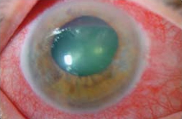
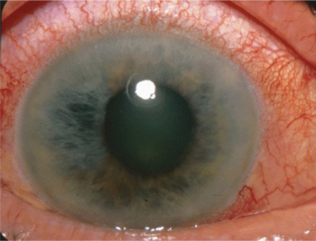
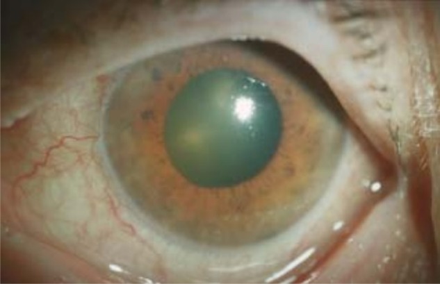
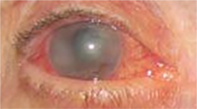
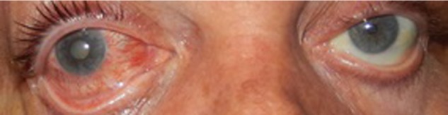
Glaucoma involves a characteristic optic neuropathy, often with elevated intraocular pressure. Before 1850, poor vision with a normal eye appearance, as occurs in primary open-angle glaucoma, was termed amaurosis, gutta serena, or black cataract. Few observers noted palpable hardness of the eye in amaurosis. On the other hand, angle-closure glaucoma can produce a green or gray pupil, and therefore was called, variously, glaucoma (derived from the Greek for glaucous, a nonspecific term connoting blue, green, or light gray) and viriditate oculi. Angle closure, with palpable hardness of the eye, mydriasis, and anterior prominence of the lens, was described in greater detail in the 18th and 19th centuries. The introduction of the ophthalmoscope in 1850 permitted the visualization of the excavated optic neuropathy in eyes with a normal or with a dilated greenish-gray pupil. Physicians developed a better appreciation of the role of intraocular pressure in both conditions, which became subsumed under the rubric "glaucoma".

Keywords: angle-closure glaucoma; glaucoma; history of ophthalmology; open-angle glaucoma.


Figures

Figure 1
Figure 1
A green mid-dilated pupil and an intraocular pressure of 50 mmHg, seen in bilateral angle-closure glaucoma and choroiditis due to Hodgkin lymphoma in a 27-year-old male. Courtesy of Wolters Kluwer Health (Baillif et al, 2011). Promotional and commercial use of the material in print, digital or mobile device format is prohibited without the permission of the publisher Wolters Klower Health.
Figure 2
Figure 2
A green, dilated pupil in acute angle-closure glaucoma. No fluorescein was instilled before the photograph. Courtesy of and personal communication (2014) Paulo Pierre-Filho, MD.
Figure 3
Figure 3
A green, fixed, mid-dilated left pupil in a 70-year-old woman with 3 days of left eye pain and a left afferent pupillary defect. Visual acuity was hand motions in the left eye. The left eye had an intraocular pressure of 46 mmHg, and a narrow anterior chamber angle by gonioscopy. The diagnosis of acute angle-closure glaucoma was made. Medical treatment ended the attack. Cataract surgery of the left eye was performed. No fluorescein was instilled before the photograph.
Figure 4
Figure 4
A grayish-green, mid-dilated pupil in acute angle-closure glaucoma. Courtesy of Jonathan Trobe, MD, and the University of Michigan Kellogg Eye Center.
Figure 5
Figure 5
A green, dilated pupil in a 70-year-old woman with acute angle-closure glaucoma and an intraocular pressure of 62 mmHg, secondary to intraocular hemorrhage from macular degeneration while on anticoagulants. Courtesy of Springer Science and Business Media (Schlote et al, 2005).
Figure 6
Figure 6
A greenish-gray, dilated pupil due to acute angle-closure attack in the right eye with an intraocular pressure over 70 mmHg. Courtesy of Andrew Doan, MD, PhD.
Figure 7
Figure 7
A gray pupil is seen in some instances of angle-closure glaucoma. A 54-year-old male with 2 days of right eye pain. The right eye intraocular pressure was 60 mmHg. The visual acuity in the right eye was hand motions, and had been poor for 1 year. The right pupil was fixed and dilated. The attack ended following medical treatment and iridotomy of the right eye. Right eye cataract surgery was performed. The examiner is holding the eyelids open during the photograph. No fluorescein was instilled before the photograph.
Figure 8
Figure 8
Cross-sectional diagram of the glaucomatous eye (top) and normal eye (bottom) as illustrated in the 19th century. Glaucoma was ascribed to anterior displacement of the lens, pressing against the iris, causing mydriasis and a narrow anterior chamber, well before the development of gonioscopy and cross-sectional imaging.

References

    1. Frezzotti R. The glaucoma mystery from ancient times to the 21st century. The glaucoma mystery: ancient concepts. Acta Ophthalmol Scand. 2000;78(S232):14–8. - PubMed
    1. Tsatsos M, Broadway D. Controversies in the history of glaucoma: is it all a load of old Greek? Br J Ophthalmol. 2007;91(11):1561–2. - PMC - PubMed
    1. Grzybowski A. Controversies in the history of glaucoma. Acta Ophthalmol. 2013;91(s252)
    1. Leffler CT, Schwartz SG, Hadi TM, Salman A, Vasuki V. The early history of glaucoma: the glaucous eye (800 BC to 1050 AD) Clin Ophthalmol. 2015;9:207–15. - PMC - PubMed
    1. Eye Diseases Prevalence Research Group Prevalence of open-angle glaucoma among adults in the United States. Arch Ophthalmol. 2004;122:532–8. - PMC - PubMed

LinkOut - more resources