. 2015 Oct 2:9:383.
doi: 10.3389/fncel.2015.00383. eCollection 2015.

Meninges harbor cells expressing neural precursor markers during development and adulthood

Affiliations

Meninges harbor cells expressing neural precursor markers during development and adulthood

Francesco Bifari et al. Front Cell Neurosci. .

Abstract

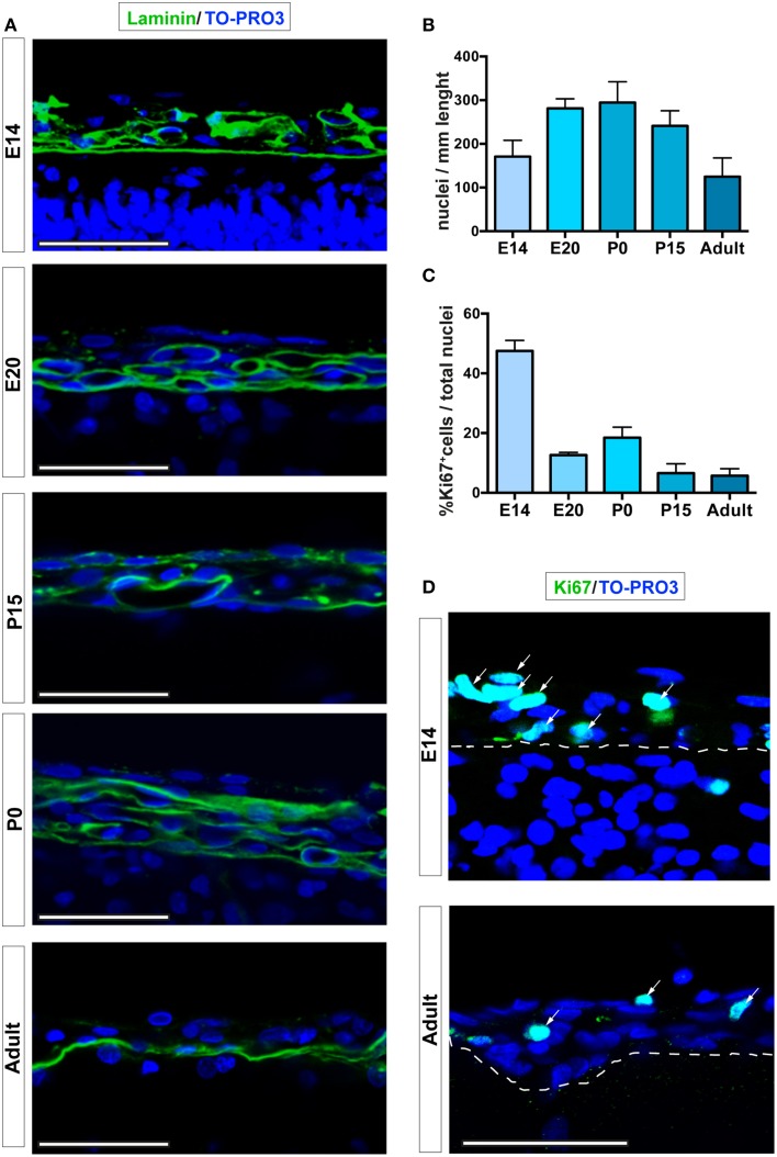
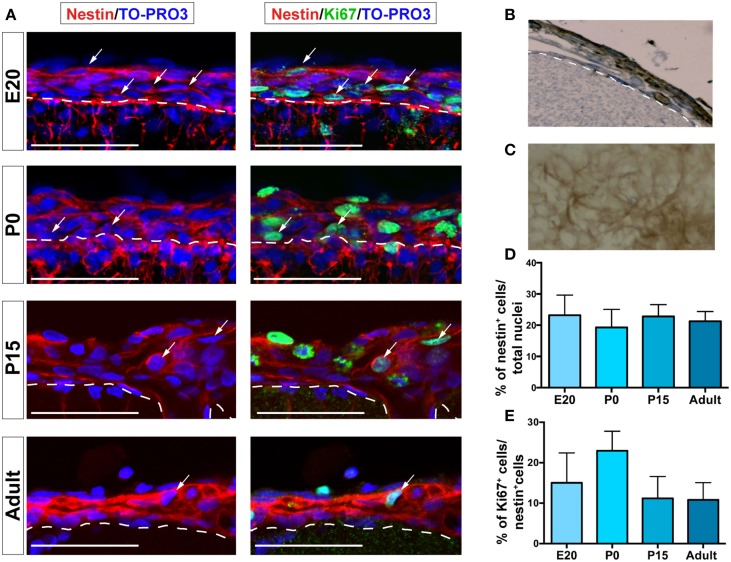
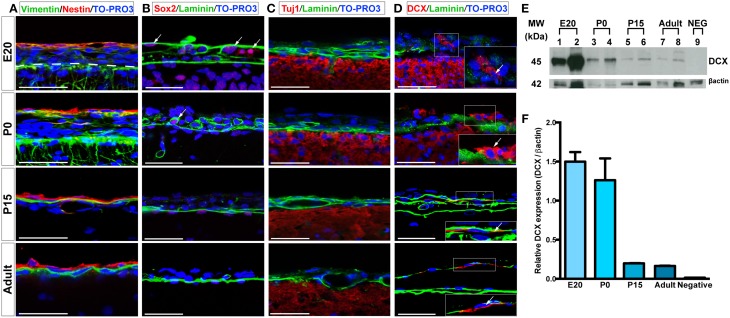
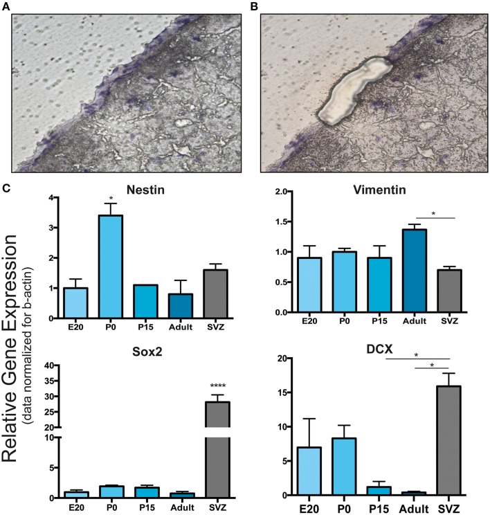
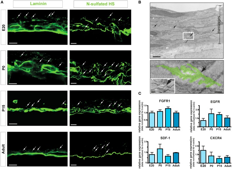
Brain and skull developments are tightly synchronized, allowing the cranial bones to dynamically adapt to the brain shape. At the brain-skull interface, meninges produce the trophic signals necessary for normal corticogenesis and bone development. Meninges harbor different cell populations, including cells forming the endosteum of the cranial vault. Recently, we and other groups have described the presence in meninges of a cell population endowed with neural differentiation potential in vitro and, after transplantation, in vivo. However, whether meninges may be a niche for neural progenitor cells during embryonic development and in adulthood remains to be determined. In this work we provide the first description of the distribution of neural precursor markers in rat meninges during development up to adulthood. We conclude that meninges share common properties with the classical neural stem cell niche, as they: (i) are a highly proliferating tissue; (ii) host cells expressing neural precursor markers such as nestin, vimentin, Sox2 and doublecortin; and (iii) are enriched in extracellular matrix components (e.g., fractones) known to bind and concentrate growth factors. This study underlines the importance of meninges as a potential niche for endogenous precursor cells during development and in adulthood.

Keywords: brain development; fractones; meninges; nestin; neural precursor cells; neural stem cell niche; proliferation.


Figures

Figure 1
Figure 1
Leptomeningeal cells at different developmental stages. (A) Confocal microscopy analysis of leptomeninges at different stages of development; from top to bottom: E14, E20, P0, P15, adult. Basal lamina of the pia mater was visualized by laminin immunoreactivity (green). (B) Quantification of number of meningeal cell nuclei present along 1 mm of brain sections; number of nuclei peaked at P0. (C) Percentage of meningeal cells positive for the proliferation marker Ki67. The number of Ki67+ cells is maximum at E14 and decreases going to adulthood. The number of rats analyzed in (B,C) was n = 3 at E14, n = 6 at E20, n = 3 at P0, n = 5 at P15, and n = 4 at adulthood; values represent mean ± SD (D). Confocal microscopy representative images of Ki67+ cells (green) of E14 and 6–8 weeks adult rat brain leptomeninges. Arrows indicate Ki67+ cells, the white dashed line highlights the border between neural parenchyma and meninges. Nuclei are stained with TO-PRO3 (blue). Scale bar: 50 μm.
Figure 2
Figure 2
Nestin+ and Ki67+ cells are present in leptomeninges. (A) Immunostaining of nestin (red, left column) and nestin (red)/Ki67 (green, right column) at different stages of development; from top to bottom: E20, P0, P15, adult. Confocal microscopy analysis revealed that nestin+ cells (red) are present in the leptomeninges from embryonic stage E20 up to adulthood. Nuclei are stained with TO-PRO3 (blue). Scale bar: 50 μm. (B,C) Immunoperoxidase staining (brown) with anti-nestin antibody of brain sections. (B) Coronal section; the white dashed line highlights the border between neural parenchyma and meninges. (C) En face view showing nestin+ cells as an intricate net covering the brain. (D) Quantification of nestin+ cells normalized for the total number of nuclei in meninges. (E) Percentage of nestin+/Ki67+ cells; the values are normalized for the number of nestin+ cells. In (D,E), values are mean ± SD.
Figure 3
Figure 3
Expression of neural progenitors markers in the leptomeninges. (A–D) Confocal microscopy images of leptomeninges in brain coronal sections stained with vimentin (green)/nestin (red) (A), Sox2 (red)/laminin (green) (B), Tuj1 (red)/laminin (green) (C), and DCX (red)/laminin (green) (D) at different stages of development; from top to bottom: E20, P0, P15, adult. Arrows in (B,D) point to Sox2+ and DCX+ cells respectively. Nuclei are stained with TO-PRO3 (blue). Scale bar: 50 μm. (E) Western Blot of meninges lysates. 7 μg of total protein lysate were loaded in lanes 1, 3, 5, 7, 9 and 10 μg in lanes 2, 4, 6, 8. Lanes 1–2: lysates from E20 meninges. Lanes 3–4: lysates from P0 meninges. Lanes 5–6: lysates from P15 meninges. Lanes 7–8: lysates from adult meninges. Lane 9: lysates from P0 meninges as negative control for the secondary antibody. Numbers on the left indicate molecular masses in kilodaltons (kDa). (F) Densitometric analysis of relative protein levels shown in (E). DCX expression was normalized for β-actin expression. DCX relative expression is high in E20 and P0 meningeal lysates and persists in P15 and adult meningeal lysates.
Figure 4
Figure 4
Laser capture microdissection and gene expression analysis of leptomeningeal cells. (A,B) Laser capture microdissection (LCM) was performed to distinguish leptomeningeal from parenchymal gene expression. (A) Shows a coronal brain section with the entire meningeal layer before LCM. (B) Shows the same section after meningeal dissection. From each stage of development (E20, P0, P15, adult), at least 1000 cells were collected from meningeal tissue (B). (C) qRT-PCR on collected samples was performed for gene expression analysis. As expected from immunofluorescence and WB analysis, we detected expression of nestin, vimentin, Sox2 and DCX genes. Expression of all these neural precursor-related genes persisted up to adulthood. SVZ samples from 6 to 8 weeks adult rats were used as positive control. *p < 0.05; ****p < 0.0001. Values are mean ± SEM of 3 replicates.
Figure 5
Figure 5
ECM components and fractones. (A) Confocal images of meninges in brain coronal sections showing the presence of immunoreactivities for laminin and N-sulfated HS. Dot-like aggregates (arrows) suggest organization of fractones in the leptomeninges. Scale bar: 10 μm. (B) Transmission electron microscopy representative image of P15 rat meninges. The white rectangle in the upper picture is enlarged in the lower frame; colored area highlights a fractone. Scale bar: 5 μm upper panel; 0.5 μm bottom panel and 1 μm in the insert. (C) Relative gene expression of FGFR1, EGFR, SDF1, and CXCR4 of rat leptomeninges at E20, P0, P15, and 6–8 weeks adult. Values are mean ± SEM of 3 replicates.

References

    1. Adeeb N., Mortazavi M. M., Tubbs R. S., Cohen-Gadol A. A. (2012). The cranial dura mater: a review of its history, embryology, and anatomy. Child Nerv. Syst. 28, 827–837. 10.1007/s00381-012-1744-6 - DOI - PubMed
    1. Angelov D. N., Vasilev V. A. (1989). Morphogenesis of rat cranial meninges. A light- and electron-microscopic study. Cell Tissue Res. 257, 207–216. 10.1007/BF00221652 - DOI - PubMed
    1. Beggs H. E., Schahin-Reed D., Zang K., Goebbels S., Nave K. A., Gorski J., et al. . (2003). FAK deficiency in cells contributing to the basal lamina results in cortical abnormalities resembling congenital muscular dystrophies. Neuron 40, 501–514. 10.1016/S0896-6273(03)00666-4 - DOI - PMC - PubMed
    1. Belmadani A., Ren D., Bhattacharyya B. J., Hope T. J., Perlman H., Miller R. J. (2015). Identification of a sustained neurogenic zone at the dorsal surface of the adult mouse hippocampus and its regulation by the chemokine SDF-1. Hippocampus. 10.1002/hipo.22428. [Epub ahead of print]. - DOI - PMC - PubMed
    1. Bifari F., Decimo I., Chiamulera C., Bersan E., Malpeli G., Johansson J., et al. . (2009). Novel stem/progenitor cells with neuronal differentiation potential reside in the leptomeningeal niche. J. Cell Mol. Med. 13, 3195–3208. 10.1111/j.1582-4934.2009.00706.x - DOI - PMC - PubMed