. 2015 Oct 1:6:1037.
doi: 10.3389/fmicb.2015.01037. eCollection 2015.

Lassa virus isolates from Mali and the Ivory Coast represent an emerging fifth lineage

Affiliations

Lassa virus isolates from Mali and the Ivory Coast represent an emerging fifth lineage

John T Manning et al. Front Microbiol. .

Abstract

Previous imported cases of Lassa fever (LF) into the United Kingdom from the Ivory Coast and Mali, as well as the detection of Lassa virus (LASV) among the Mastomys natalensis population within Mali has led to the suggestion that the endemic area for LF is expanding. Initial phylogenetic analyses arrange isolates from Mali and the Ivory Coast separately from the classical lineage IV isolates taken from Sierra Leone, Guinea, and Liberia. The availability of full genome sequences continues to increase, allowing for a more complete phylogenetic comparison of the isolates from Mali and the Ivory Coast to the other existing isolates. In this study, we utilized a Bayesian approach to infer the demographic histories of each LASV isolate for which the full sequence was available. Our results indicate that the isolates from Mali and the Ivory Coast group separately from the isolates of lineage IV, comprising a distinct fifth lineage. The split between lineages IV and V is estimated to have occurred around 200-300 years ago, which coincides with the colonial period of West Africa.

Keywords: Lassa fever; Lassa virus; genetic diversity; lineage; phylogenetics.


Figures
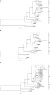
Figure 1
Figure 1
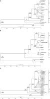
Bayesian analysis of complete LP, NP, and GPC nucleotide sequences. Complete open reading frames were aligned using MUSCLE, and analyzed using the Bayesian MCMC approach. The resulting trees for LP (A), NP (B), and GPC (C) were visualized using FigTree v1.4.2 and rooted using two representative Mopeia virus isolates. To better visualize distinct lineages, several Sierra Leonian, and Nigerian isolates were collapsed. These sections of the trees are provided in Figures S1–S6. Posterior probabilities are represented as node labels for the main clades, with 1 being 100% probability. The isolates are grouped by their respective lineages, as represented by the bars to the right of the trees. The scale represents nucleotide changes per site per year.
Figure 2
Figure 2
BEAST analysis of complete LP, NP, and GPC nucleotide sequences. Complete open reading frames were aligned using MUSCLE, and analyzed using the Bayesian MCMC approach. The resulting trees for LP (A), NP (B), and GPC (C) were visualized using FigTree v1.4.2 and rooted using two representative Mopeia virus isolates. To better visualize distinct lineages, several Sierra Leonian and Nigerian isolates were collapsed, and these sections of the trees are provided in Figures S7–S12. The node ages, in years, are included on the major nodes, with the 95% confidence ranges displayed in parentheses below the median node ages. The isolates are grouped by their lineages, as represented by the bars to the right of the trees. The reverse axis represents the age, in years, from the most recent isolate.
Figure 3
Figure 3
Map of LASV movement across West Africa. Based on the phylogenetic data, LASV has gradually spread west, beginning in Eastern Nigeria. Ehichioya et al. has previously illustrated the movement of LASV within Nigeria (Ehichioya et al., 2011). The isolates of lineage V and the BA366 Liberian isolate share a more direct common ancestor with lineage III than the Sierra Leone Isolates share with lineage III isolates. This suggests that the virus was likely present in Mali, the Ivory Coast, and Liberia prior to establishing itself in Sierra Leone. The areas from which the isolates were collected are shaded in gray.

References

    1. Andersen K. G., Shapiro B. J., Matranga C. B., Sealfon R., Lin A. E., Moses L. M., et al. . (2015). (2015). Clinical sequencing uncovers origins and evolution of Lassa Virus. Cell 162, 738–750. 10.1016/j.cell.2015.07.020 - DOI - PMC - PubMed
    1. Atkin S., Anaraki S., Gothard P., Walsh A., Brown D., Gopal R., et al. . (2009). The first case of Lassa fever imported from Mali to the United Kingdom, February 2009. Euro Surveill. 14:19145. Available online at: http://www.eurosurveillance.org/ViewArticle.aspx?ArticleId=19145 - PubMed
    1. Bowen M. D., Rollin P. E., Ksiazek T. G., Hustad H. L., Bausch D. G., Demby A. H., et al. . (2000). Genetic diversity among Lassa virus strains. J. Virol. 74, 6992. 10.1128/JVI.74.15.6992-7004.2000 - DOI - PMC - PubMed
    1. Demby A. H., Inapogui A., Kargbo K., Koninga J., Kourouma K., Kanu J., et al. . (2001). Lassa fever in Guinea: II. Distribution and prevalence of Lassa virus infection in small mammals. Vector Borne Zoonotic Dis. 1, 283–297. 10.1089/15303660160025912 - DOI - PubMed
    1. Drummond A. J., Ho S. Y., Phillips M. J., Rambaut A. (2006). Relaxed phylogenetics and dating with confidence. PLoS Biol. 4:e88. 10.1371/journal.pbio.0040088 - DOI - PMC - PubMed