Review
. 2015 Sep 30:6:1049.
doi: 10.3389/fmicb.2015.01049. eCollection 2015.

Choosing a suitable method for the identification of replication origins in microbial genomes

Affiliations
Review

Choosing a suitable method for the identification of replication origins in microbial genomes

Chengcheng Song et al. Front Microbiol. .

Abstract

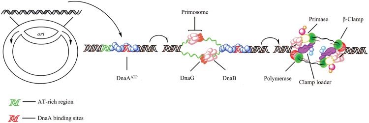
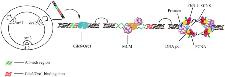
As the replication of genomic DNA is arguably the most important task performed by a cell and given that it is controlled at the initiation stage, the events that occur at the replication origin play a central role in the cell cycle. Making sense of DNA replication origins is important for improving our capacity to study cellular processes and functions in the regulation of gene expression, genome integrity in much finer detail. Thus, clearly comprehending the positions and sequences of replication origins which are fundamental to chromosome organization and duplication is the first priority of all. In view of such important roles of replication origins, tremendous work has been aimed at identifying and testing the specificity of replication origins. A number of computational tools based on various skew types have been developed to predict replication origins. Using various in silico approaches such as Ori-Finder, and databases such as DoriC, researchers have predicted the locations of replication origins sites for thousands of bacterial chromosomes and archaeal genomes. Based on the predicted results, we should choose an effective method for identifying and confirming the interactions at origins of replication. Here we describe the main existing experimental methods that aimed to determine the replication origin regions and list some of the many the practical applications of these methods.

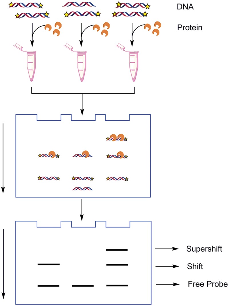
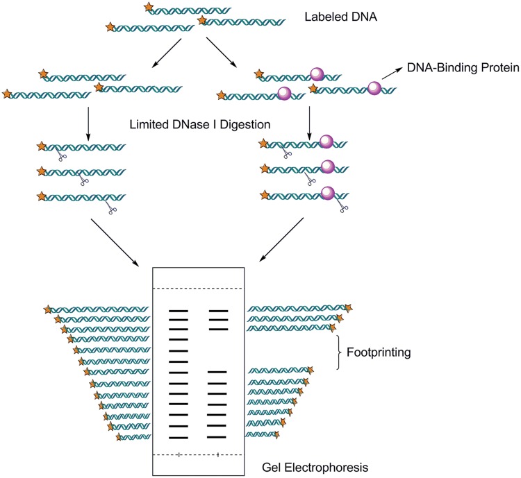
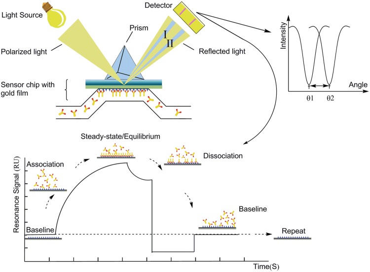
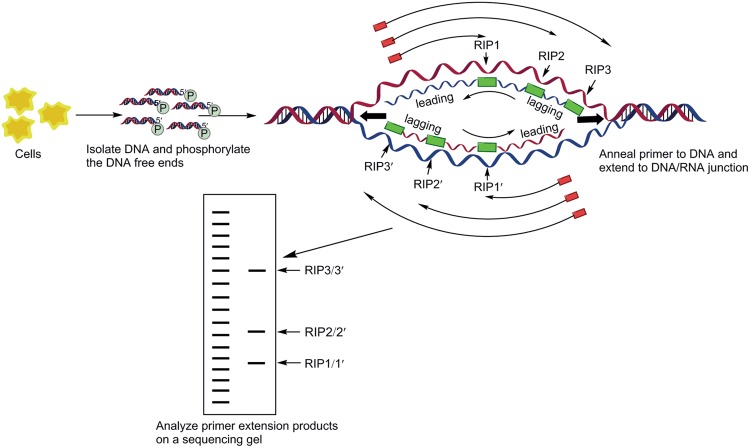
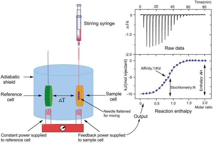
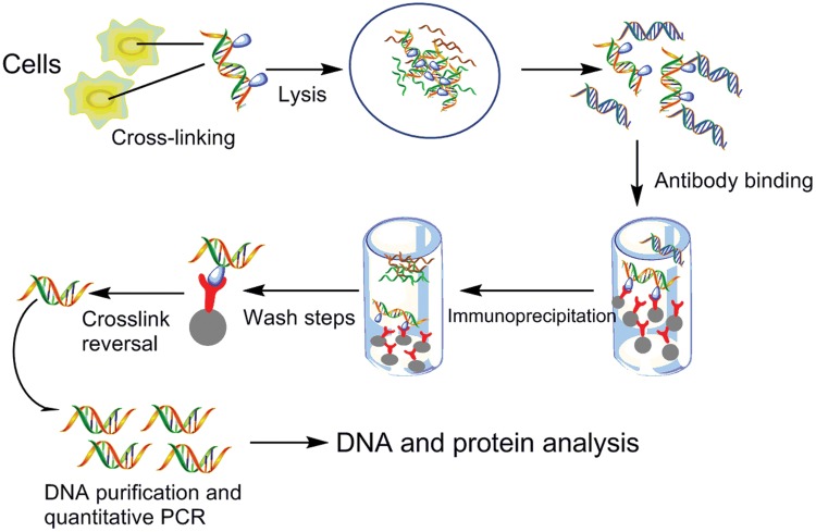
Keywords: ChIP; ChIP-seq; Dnase I footprinting; EMSA; ITC; RIP mapping; SPR; replication origin.


Figures

FIGURE 1
FIGURE 1
Initiation of replication in bacteria. In Escherichia coli, replication initiation requires binding of the DNA-binding protein DnaA to DnaA-boxes at the chromosome origin oriC which is regulated by SeqA (Dame et al., 2011). Then, with the activation of ATP, two DnaB hexamers and the helicase loader DnaC, one double hexamer for each replication direction, are positioned by DnaA into the loop (Wahle et al., 1989; Skarstad and Katayama, 2013). Primase (DnaG) which can enter the complex and synthesize two leading strand primers, stimulates release of the regulatory protein DnaC from DnaB after transiently binding to the DnaB replicative helicase (Arias-Palomo et al., 2013). Also, DnaB binds to the sliding clamp loader, a ring-shaped dimer of the β-subunit which in turn binds the DNA polymerases III (Kelman and O’Donnell, 1995; O’Donnell et al., 2013).
FIGURE 2
FIGURE 2
Initiation of replication in archaea. Archaeal circular chromosome can contain a single or multiple origins (oriC). Archaea have the AAA+ Orc1/Cdc6 origin-binding protein, which shares extensive sequence homology with eukaryotic ORC proteins (Zhang et al., 2009). Minichromosome maintenance (MCM) proteins bind preferentially to the oriC region. ATP bound Cdc6/Orc1 associates with the Cdc6/Orc1-origin complex and with the MCM helicase. Following ATP hydrolysis the Cdc6/Orc1 protein releases the helicase, and the primase replaces the Cdc6/Orc1 protein binding to MCM. MCM interacts with the archaeal GINS (go, ichi, nii, san [five, one, two, three in Japanese]) complex (Marinsek et al., 2006) which is additionally capable of binding primase. Each DNA Pol interacts with a trimer of PCNA (proliferating cell nuclear antigen). The flap endonuclease FEN1 and DNA ligase I are only assembled to PCNA clamp of similar structure to E. coli β (Michel and Bernander, 2014).
FIGURE 3
FIGURE 3
The schematic illustration of electrophoretic mobility shift assays (EMSA). Typically one compound is labeled to follow its mobility during electrophoresis. In general, a single protein binds to a single site. Once the length of the nucleotides is sufficient for the binding of two or more proteins, the protein–DNA complexes migrate as distinct bands, usually referred to as a super shift. If the labeled nucleotides are bound by the proteins, then the mobility of the labeled nucleotides through the electrophoretic medium will be retarded.
FIGURE 4
FIGURE 4
Dnase I footprinting. This Analysis involves endonuclease treatment of an end labeled DNA fragment bound to a protein. This technique relies on the fact that fragments of DNA that have DNA-binding proteins bound will move more slowly through an acrlyamide gel. The enzyme DNaseI will only cut exposed DNA. Limited digestion yields fragments terminating everywhere except in the footprint region, which is protected from digestion.
FIGURE 5
FIGURE 5
The schematic illustration of surface plasmon resonance (SPR) system. SPR detects changes in the refractive index in the immediate vicinity of the surface layer of a sensor chip. The sensor surface is gold with antibodies attached to it. During the measurement, the chip is irradiated from the bottom with a beam of a wide angle range within that of total internal reflection. The SPR angle shifts (from I to II in the diagram) when biomolecules binding events cause changes in the refractive index at the surface layer. The detector will determine the angle of the intensity decrease. This change in resonant angle can be monitored non-invasively in real time as a plot of resonance signal (proportional to mass change) versus time (Sawhney and Singh, 2000; Cooper, 2002; Pattnaik, 2005; Sahai, 2011; Hou and Cronin, 2013).
FIGURE 6
FIGURE 6
Replication initiation point (RIP) mapping. The replication bubble of semi-discontinuous replication is diagrammed here. After phosphorylation of any 5′-OH ends with polynucleotide kinase, replication intermediates enriched on the BND-cellulose column are treated with λ-exonuclease to digest nicked DNA. Digestion is confirmed on the agarose gel before proceeding to the primer extension reaction. The primer extension products are showed as arrows outside the replication bubble, stopping at DNA/RNA junctions on the DNA. Extension stops at the points labeled RIP1, RIP2, RIP3. Green rectangles depict the RNA primers of nascent strands. PCR products are purified and analyzed on a denaturing polyacrylamide gel. Due to asynchrony, the replication bubble can be of various sizes, resulting in various length, is the transition point from discontinuous to continunous synthesis. Sequencing and RIP reactions were analyzed side by side on the same gel (Gerbi and Bielinsky, 1997; Matsunaga et al., 2003; Lee and Romero, 2012).
FIGURE 7
FIGURE 7
Basic principle of isothermal titration calorimetry. Schematic representation of the isothermal titration calorimeter (left) and a characteristic titration experiment (upper right) with its evaluation (lower right). In (upper right) picture, the titration thermogram is represented as heat per unit of time released after each injection of the ligand into the protein (black), as well as the dilution of ligand into buffer (blue). In (lower right) picture, the dependence of released heat in each injection versus the ratio between total ligand concentration and total protein concentration is represented. Circles represent experimental data and the line corresponds to the best fitting to a model considering n identical and independent sites. The syringe is inserted in the sample cell and a series of injections are made (Freyer and Lewis, 2008; Martinez et al., 2013).
FIGURE 8
FIGURE 8
Chromatin immunoprecipitation. Protein and associated chromatin in living cells or tissues are temporarily bonded, the DNA–protein complexes (chromatin-protein) are then sheared into ∼500 bp DNA fragments using either enzymatic digestion or physical shearing by sonication. Cross-linked DNA fragments associated with the protein(s) of interest using formaldehyde are selectively immunoprecipitated from the cell debris using appropriate protein-specific antibody. After the cross-links are reversed, the associated DNA fragments are purified and their sequence is determined. These DNA sequences are supposed to be associated with the protein of interest in vivo. The DNA undergoes PCR amplification using primers targeting a particular genomic locus. These DNA sequences can be subjected to a number of downstream analysis techniques, including targeted approaches, like semiquantitative PCR and quantitative PCR, and genome-wide analyses using microarrays (ChIP–chip) and deep sequencing (ChIP-seq), ChIP-on-chip (Shah, 2009; Vinckevicius and Chakravarti, 2012).

Similar articles

Cited by

References

    1. Abe Y., Jo T., Matsuda Y., Matsunaga C., Katayama T., Ueda T. (2007). Structure and function of DnaA N-terminal Domains: specific sites and mechanisms in inter-DnaA interaction and in DnaB helicase loading on oriC. J. Biol. Chem. 282 17816–17827. 10.1074/jbc.M701841200 - DOI - PubMed
    1. Alves C., Cunha C. (2012). Electrophoretic mobility shift assay: analyzing protein–nucleic acid interactions. Nat. Protoc. 2 1849–1861. 10.1038/nprot.2007.249 - DOI - PMC - PubMed
    1. Arias-Palomo E., O’shea V. L., Hood I. V., Berger J. M. (2013). The bacterial DnaC helicase loader is a DnaB ring breaker. Cell 153 438–448. 10.1016/j.cell.2013.03.006 - DOI - PMC - PubMed
    1. Aves S. J. (2009). “DNA replication initiation,” in DNA Replication eds Vengrova S., Dalgaard J. Z. (New York, NY: Humana Press; ) 1–16. 10.1093/jexbot/52.355.193 - DOI
    1. Baird C. L., Myszka D. G. (2001). Current and emerging commercial optical biosensors. J. Mol. Recognit. 14 261–268. 10.1002/jmr.544 - DOI - PubMed