

Eumycetoma Osteomyelitis of the Calcaneus in a Child: A Radiologic-Pathologic Correlation following Total Calcanectomy
- PMID: 26483983
- PMCID: PMC4592886
- DOI: 10.1155/2015/129020
Eumycetoma Osteomyelitis of the Calcaneus in a Child: A Radiologic-Pathologic Correlation following Total Calcanectomy
Abstract
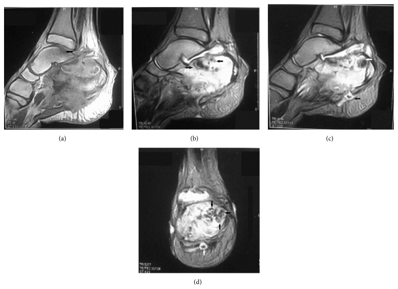
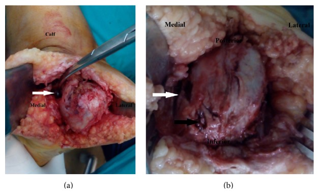
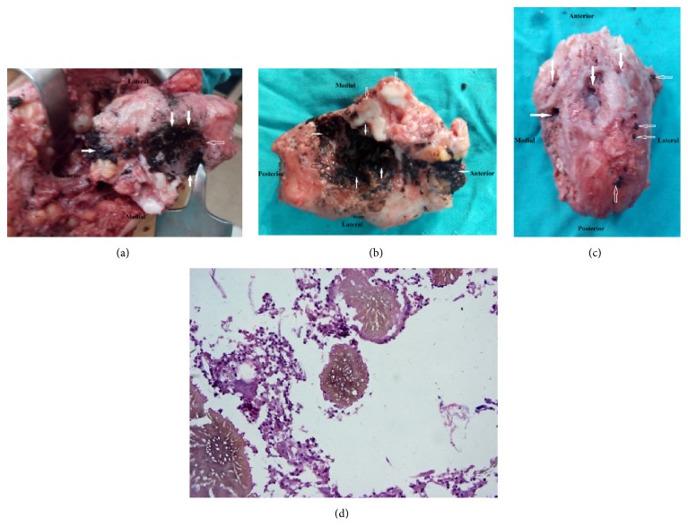
Fungi are unusual causes of pedal osteomyelitis in children and adolescents. Eumycetoma is a chronic cutaneous and subcutaneous infection caused by various genera of fungi. A provisional diagnosis of foot mycetoma is made after clinical assessment. Radiologic-pathologic correlation is an essential supplement for the accurate diagnosis of osteoarticular infections. This paper aims to sensitize orthopedic surgeons, radiologists, and pathologists to the importance of correlative imaging findings in relation to surgical and microscopic pathology in osteoarticular infections, specifically eumycetoma osteomyelitis of the foot. From our review of the published data, the present case is the first report of radiologic-pathologic correlation in eumycetoma osteomyelitis of the calcaneus. This paper describes a case of eumycetoma osteomyelitis of the calcaneus in a child in which diagnostic X-rays and magnetic resonance imaging (MRI) were correlated with the surgical and microscopic pathologic features, for establishing an appropriate diagnosis and treatment. We conclude that there is a significant agreement between radiologic and pathologic evaluation for assessment of eumycetoma osteomyelitis of the calcaneus. Radiologic-pathologic correlation amplified our interpretation of imaging information available on plain radiographs and MRI and augmented diagnostic confidence. Similarly, anatomic-histopathological correlations consolidated diagnostic accuracy.
Figures
Similar articles
-
Primary mycetoma osteomyelitis of the calcaneus with active subcutaneous nodules.J Foot Ankle Surg. 2007 Jul-Aug;46(4):302-6. doi: 10.1053/j.jfas.2007.02.005. J Foot Ankle Surg. 2007. PMID: 17586446
-
Eumycetoma Osteomyelitis Calcaneus in Adolescent; report of case and literature review.BMC Infect Dis. 2021 Sep 23;21(1):995. doi: 10.1186/s12879-021-06695-3. BMC Infect Dis. 2021. PMID: 34556037 Free PMC article. Review.
-
Total calcanectomy for the treatment of chronic calcaneal osteomyelitis.Foot Ankle Int. 1998 Dec;19(12):849-55. doi: 10.1177/107110079801901210. Foot Ankle Int. 1998. PMID: 9872473
-
[Hematogenous osteomyelitis of the calcaneus in children: 26 cases].Rev Chir Orthop Reparatrice Appar Mot. 2008 Sep;94(5):434-42. doi: 10.1016/j.rco.2008.02.004. Epub 2008 May 2. Rev Chir Orthop Reparatrice Appar Mot. 2008. PMID: 18774017 French.
-
Oral eumycetoma of infancy: a rare presentation and a brief review.J Craniomaxillofac Surg. 2014 Jan;42(1):35-40. doi: 10.1016/j.jcms.2012.12.012. Epub 2013 Mar 21. J Craniomaxillofac Surg. 2014. PMID: 23522832 Review.
Cited by
-
Surgical management of eumycetoma: experience from Gezira Mycetoma Center, Sudan.Trop Med Health. 2019 Jan 14;47:6. doi: 10.1186/s41182-018-0129-2. eCollection 2019. Trop Med Health. 2019. PMID: 30675125 Free PMC article.
-
The Surgical Treatment of Mycetoma.PLoS Negl Trop Dis. 2016 Jun 23;10(6):e0004690. doi: 10.1371/journal.pntd.0004690. eCollection 2016 Jun. PLoS Negl Trop Dis. 2016. PMID: 27336736 Free PMC article. Review.
-
Acute osteoarticular infections in children are frequently forgotten multidiscipline emergencies: beyond the technical skills.EFORT Open Rev. 2021 Jul 8;6(7):584-592. doi: 10.1302/2058-5241.6.200155. eCollection 2021 Jul. EFORT Open Rev. 2021. PMID: 34377550 Free PMC article. Review.
-
A scoping review of mycetoma profile in Egypt: revisiting the global endemicity map.Trans R Soc Trop Med Hyg. 2023 Jan 3;117(1):1-11. doi: 10.1093/trstmh/trac085. Trans R Soc Trop Med Hyg. 2023. PMID: 36084235 Free PMC article.
-
Pediatric calcaneal osteomyelitis: an analysis of literature-reported 128 cases.BMC Infect Dis. 2024 Sep 18;24(1):998. doi: 10.1186/s12879-024-09887-9. BMC Infect Dis. 2024. PMID: 39294568 Free PMC article.
References
-
- Develoux M., Dieng M. T., Kane A., Ndiaye B. Management of mycetoma in West-Africa. Bulletin de la Societe de Pathologie Exotique. 2004;96(5):376–382. - PubMed
-
- Kumar J., Kumar A., Sethy P., Gupta S. The dot-in-circle sign of mycetoma on MRI. Diagnostic and Interventional Radiology. 2007;13(4):193–195. - PubMed
-
- Khan S. H. M., Bloem J. L. Radiologic-pathologic correlations of bone infections. In: Gourtsoyiannis N. C., Ros P. R., editors. Radiologic-Pathologic Correlations from Head to Toe: Understanding the Manifestations of Disease. Berlin, Germany: Springer; 2005. pp. 647–659.
LinkOut - more resources
Full Text Sources
Other Literature Sources
