Review
. 2015 Aug 22;5(4):e29716.
doi: 10.5812/aapm.29716. eCollection 2015 Aug.

Interventional Therapies for Chronic Low Back Pain: A Focused Review (Efficacy and Outcomes)

Affiliations
Review

Interventional Therapies for Chronic Low Back Pain: A Focused Review (Efficacy and Outcomes)

Vikram B Patel et al. Anesth Pain Med. .

Abstract

Context: Lower back pain is considered to be one of the most common complaints that brings a patient to a pain specialist. Several modalities in interventional pain management are known to be helpful to a patient with chronic low back pain. Proper diagnosis is required for appropriate intervention to provide optimal benefits. From simple trigger point injections for muscular pain to a highly complex intervention such as a spinal cord stimulator are very effective if chosen properly. The aim of this article is to provide the reader with a comprehensive reading for treatment of lower back pain using interventional modalities.

Evidence acquisition: Extensive search for published literature was carried out online using PubMed, Cochrane database and Embase for the material used in this manuscript. This article describes the most common modalities available to an interventional pain physician along with the most relevant current and past references for the treatment of lower back pain. All the graphics and images were prepared by and belong to the author.

Results: This review article describes the most common modalities available to an interventional pain physician along with the most relevant current and past references for the treatment of lower back pain. All the graphics and images belong to the author. Although it is beyond the scope of this review article to include a very detailed description of each procedure along with complete references, a sincere attempt has been made to comprehensively cover this very complex and perplexing topic.

Conclusion: Lower back pain is a major healthcare issue and this review article will help educate the pain practitioners about the current evidence based treatment options.

Keywords: Decompression; Discography; Facet Joint; Interventional Therapies; Intradiscal Procedures, Disc; Low Back Pain; Procedures; Sacroiliac joint; Spinal Cord Stimulation.


Figures
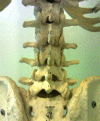
Figure 1.
Figure 1.. Lumbar Spine Anatomy
Figure 2.
Figure 2.. A Cross Section of the Lumbar Vertebral Level Showing the Different Sections of the Spine
Figure 3.
Figure 3.. Lumbar Interlaminar Epidural Injection AP and Lateral Views
Figure 4.
Figure 4.. Caudal Epidural Injection AP and Lateral Views
Figure 5.
Figure 5.. Transforaminal Injection AP and Lateral Views; Infra-Pedicular Approach
Figure 6.
Figure 6.. Transforaminal Injection AP and Lateral Views; Supra-Pedicular Approach
Figure 7.
Figure 7.. Lumbar Spine Anatomy Showing the Medial Branches
Figure 8.
Figure 8.. Classic Oblique View Optimized for the “Scotty Dog” Image Depicting the “Eye”
Note the tangential placement of the needle for medial branch neurotomy.
Figure 9.
Figure 9.. Contrast Spread for Medial Branch Block
Figure 10.
Figure 10.. A, B: Lumbar Intra-Articular Facet Joint Injections
Figure 11.
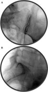
Figure 11.. A, B, C and D, Radiofrequency of L5-S1 Level, AP and Lateral Views
Note the tangential angles of the needles and the L5 medial branch (dorsal ramus) ablation over the ala of the sacrum.
Figure 12.
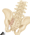
Figure 12.. A and B, Anatomy of the SI Joint (Courtesy Gray’s Anatomy Online)
The SI joint articular surface is highlighted in the left image.
Figure 13.
Figure 13.. A and B: Sacroiliac Joint Intra-Articular Injection Antero-Posterior and Lateral Views
Note the angle of the needle in lateral view, parallel to the joint.
Figure 14.
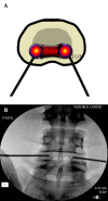
Figure 14.. The Image Showing the Spherical Lesions Created by Cooled RF Lesioning and the Targeted Areas Around the Posterior Sacral Foramina as Well as the L5 Lesion Targets
Figure 15.
Figure 15.. Antero-Posterior and Lateral Views of the RF Lesioning for the SI Joint at the Edge of the Medial Joint Border of the Joint Using the “Leap Frog” Technique
Figure 16.
Figure 16.. Simplicity® Probe for SI Joint RF Lesioning (Courtesy Neurotherm)
Figure 17.
Figure 17.. Entry Point for the Discogram With Optimized View for the L4-5 Disc
The articular elements of the inferior level bisect the end-plate of the level above in an ideal view.
Figure 18.
Figure 18.. Dual Needle Set for Diagnostic Discography
Note the curved inner needle. This curve helps negotiate the needle within the nucleus so that the tip is positioned centrally within the disc. The curvature is reduced significantly as the needle exits the introducer.
Figure 19.
Figure 19.. A and B, Dual Needle Discography
Note that even though the introducer needle is inferior to the center of the disc, the inner needle can achieve a central location of the tip due to the curvature, thus eliminating the need for re-introduction of the needle.
Figure 20.
Figure 20.. A and B, Printed Data From a Discography Procedure Showing Graphical as Well as Numeric Readings
The leads are confirmed to be in the posterior epidural space with a lateral view.
Figure 21.
Figure 21.. A and B, Post Discogram CT Scan Image Showing Annular Disruption
Figure 22.
Figure 22.. A and B, Intradiscal Electrothermal Treatment (IDET)
Compare to the fluoroscopic image on the right taken during the procedure.
Figure 23.
Figure 23.. A and B, Biacuplasty® Procedure With Cooled Radiofrequency
The entry point is at the posterolateral annulus and the thermal element is guided through the introducer at the annular-nuclear junction with the final position in the posterior aspect of the disc.
Figure 24.
Figure 24.. A, B, C and D: Spinal Cord stimulator Lead Placement for Low Back Pain as Well as Radicular Pain in a Post-Laminectomy Pain Patient Using a Dual Lead Configuration
The lesion is generated between the tips of the probes and provides a near complete coverage of the posterior annulus.

References

    1. Williams KA, Gonzalez-Fernandez M, Hamzehzadeh S, Wilkinson I, Erdek MA, Plunkett A, et al. A multi-center analysis evaluating factors associated with spinal cord stimulation outcome in chronic pain patients. Pain Med. 2011;12(8):1142–53. doi: 10.1111/j.1526-4637.2011.01184.x. - DOI - PubMed
    1. van Dulmen S, Sluijs E, van Dijk L, de Ridder D, Heerdink R, Bensing J. Patient adherence to medical treatment: a review of reviews. BMC Health Serv Res. 2007;7:55. doi: 10.1186/1472-6963-7-55. - DOI - PMC - PubMed
    1. Manchikanti L, Cash KA, Pampati V, McManus CD, Damron KS. Evaluation of fluoroscopically guided caudal epidural injections. Pain Physician. 2004;7(1):81–92. - PubMed
    1. Hansen HC. Is fluoroscopy necessary for sacroiliac joint injections? Pain Physician. 2003;6(2):155–8. - PubMed
    1. Jasper JF. Role of digital subtraction fluoroscopic imaging in detecting intravascular injections. Pain Physician. 2003;6(3):369–72. - PubMed

LinkOut - more resources