

4-hydroxynonenal regulates mitochondrial function in human small airway epithelial cells
- PMID: 26484418
- PMCID: PMC4747170
- DOI: 10.18632/oncotarget.6131
4-hydroxynonenal regulates mitochondrial function in human small airway epithelial cells
Abstract
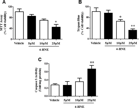
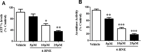
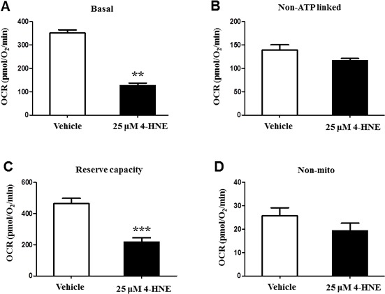
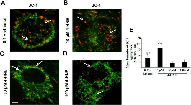
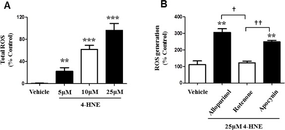
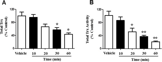
Prolonged exposure to oxidative stress causes Acute Lung Injury (ALI) and significantly impairs pulmonary function. Previously we have demonstrated that mitochondrial dysfunction is a key pathological factor in hyperoxic ALI. While it is known that hyperoxia induces the production of stable, but toxic 4-hydroxynonenal (4-HNE) molecule, it is unknown how the reactive aldehyde disrupts mitochondrial function. Our previous in vivo study indicated that exposure to hyperoxia significantly increases 4-HNE-Protein adducts, as well as levels of MDA in total lung homogenates. Based on the in vivo studies, we explored the effects of 4-HNE in human small airway epithelial cells (SAECs). Human SAECs treated with 25 μM of 4-HNE showed a significant decrease in cellular viability and increased caspase-3 activity. Moreover, 4-HNE treated SAECs showed impaired mitochondrial function and energy production indicated by reduced ATP levels, mitochondrial membrane potential, and aconitase activity. This was followed by a significant decrease in mitochondrial oxygen consumption and depletion of the reserve capacity. The direct effect of 4-HNE on the mitochondrial respiratory chain was confirmed using Rotenone. Furthermore, SAECs treated with 25 μM 4-HNE showed a time-dependent depletion of total Thioredoxin (Trx) proteins and Trx activity. Taken together, our results indicate that 4-HNE induces cellular and mitochondrial dysfunction in human SAECs, leading to an impaired endogenous antioxidant response.
Keywords: 4-HNE; Immune response; Immunity; Immunology and Microbiology Section; ROS; acute lung injury; hyperoxia; mitochondrial dysfunction.
Conflict of interest statement
No potential conflicts of interest were disclosed.
Figures

Similar articles
-
Thioredoxin-deficient mice, a novel phenotype sensitive to ambient air and hypersensitive to hyperoxia-induced lung injury.Am J Physiol Lung Cell Mol Physiol. 2015 Mar 1;308(5):L429-42. doi: 10.1152/ajplung.00285.2014. Epub 2014 Dec 24. Am J Physiol Lung Cell Mol Physiol. 2015. PMID: 25539854 Free PMC article.
-
4-Hydroxynonenal induces mitochondrial-mediated apoptosis and oxidative stress in SH-SY5Y human neuronal cells.Basic Clin Pharmacol Toxicol. 2012 May;110(5):441-8. doi: 10.1111/j.1742-7843.2011.00834.x. Epub 2011 Dec 20. Basic Clin Pharmacol Toxicol. 2012. PMID: 22118713
-
Alterations in mitochondrial respiratory functions, redox metabolism and apoptosis by oxidant 4-hydroxynonenal and antioxidants curcumin and melatonin in PC12 cells.Toxicol Appl Pharmacol. 2008 Jan 15;226(2):161-8. doi: 10.1016/j.taap.2007.09.002. Epub 2007 Sep 11. Toxicol Appl Pharmacol. 2008. PMID: 17935746
-
Electrophilic Aldehyde 4-Hydroxy-2-Nonenal Mediated Signaling and Mitochondrial Dysfunction.Biomolecules. 2022 Oct 25;12(11):1555. doi: 10.3390/biom12111555. Biomolecules. 2022. PMID: 36358905 Free PMC article. Review.
-
Mitochondrial Dysfunction, Altered Mitochondrial Oxygen, and Energy Metabolism Associated with the Pathogenesis of Schizophrenia.Int J Mol Sci. 2023 Apr 28;24(9):7991. doi: 10.3390/ijms24097991. Int J Mol Sci. 2023. PMID: 37175697 Free PMC article. Review.
Cited by
-
Adjunct treatment with ozone to enhance therapy of knee osteoarthritis: preliminary results.Clin Rheumatol. 2024 Jun;43(6):2093-2101. doi: 10.1007/s10067-024-06972-x. Epub 2024 Apr 26. Clin Rheumatol. 2024. PMID: 38671261
-
Mitochondrial dysfunction in alveolar and white matter developmental failure in premature infants.Pediatr Res. 2017 Feb;81(2):286-292. doi: 10.1038/pr.2016.216. Epub 2016 Nov 3. Pediatr Res. 2017. PMID: 27901512 Free PMC article. Review.
-
Involvement of 4-hydroxy-2-nonenal in the pathogenesis of pulmonary fibrosis.Mol Cell Biochem. 2021 Dec;476(12):4405-4419. doi: 10.1007/s11010-021-04244-9. Epub 2021 Aug 31. Mol Cell Biochem. 2021. PMID: 34463938 Review.
-
The Protective Roles of ROS-Mediated Mitophagy on 125I Seeds Radiation Induced Cell Death in HCT116 Cells.Oxid Med Cell Longev. 2016;2016:9460462. doi: 10.1155/2016/9460462. Epub 2016 Dec 29. Oxid Med Cell Longev. 2016. PMID: 28119765 Free PMC article.
-
Impact of Lysine Succinylation on the Biology of Fungi.Curr Issues Mol Biol. 2024 Jan 23;46(2):1020-1046. doi: 10.3390/cimb46020065. Curr Issues Mol Biol. 2024. PMID: 38392183 Free PMC article. Review.
References
-
- Crapo JD. Morphologic changes in pulmonary oxygen toxicity. Annual review of physiology. 1986;48:721–731. - PubMed
-
- Chaudhary P, Sharma R, Sahu M, Vishwanatha JK, Awasthi S, Awasthi YC. 4-Hydroxynonenal induces G2/M phase cell cycle arrest by activation of the ataxia telangiectasia mutated and Rad3-related protein (ATR)/checkpoint kinase 1 (Chk1) signaling pathway. The Journal of biological chemistry. 2013;288:20532–20546. - PMC - PubMed
Publication types
MeSH terms
Substances
Grants and funding
LinkOut - more resources
Full Text Sources
Other Literature Sources
Research Materials
