. 2015 Oct 20;10(10):e0141040.
doi: 10.1371/journal.pone.0141040. eCollection 2015.

The Direct Interaction between Two Morphogenetic Proteins Is Essential for Spore Coat Formation in Bacillus subtilis

Affiliations

The Direct Interaction between Two Morphogenetic Proteins Is Essential for Spore Coat Formation in Bacillus subtilis

Rachele Isticato et al. PLoS One. .

Abstract

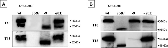
In Bacillus subtilis the protective layers that surround the mature spore are formed by over seventy different proteins. Some of those proteins have a regulatory role on the assembly of other coat proteins and are referred to as morphogenetic factors. CotE is a major morphogenetic factor, known to form a ring around the forming spore and organize the deposition of the outer surface layers. CotH is a CotE-dependent protein known to control the assembly of at least nine other coat proteins. We report that CotH also controls the assembly of CotE and that this mutual dependency is due to a direct interaction between the two proteins. The C-terminal end of CotE is essential for this direct interaction and CotH cannot bind to mutant CotE deleted of six or nine C-terminal amino acids. However, addition of a negatively charged amino acid to those deleted versions of CotE rescues the interaction.


Conflict of interest statement

Competing Interests: The authors have declared that no competing interests exist.

Figures

Fig 1
Fig 1. CotE in vivo assembly in mature spores and during sporulation.
Proteins for western blot analysis were extracted from: (A) mature spores of a wild type strain (wt), or isogenic strains lacking CotH (cotH; ER220) or CotE (cotE; RH211) or over-producing CotH (P A cotH; RG24) (24); (B) mother cell or forespore compartments of sporulating cells of a wild type strain (wt), or an isogenic strain lacking CotH (cotH; ER220), 3 (T3), 7 (T7), and 9 (T9) hours after the initiation of sporulation; (C) mature spores of a wild type strain (wt), or isogenic strains lacking CotH (cotH; ER220), or CotG and CotH (cotG cotH; AZ603). Proteins were fractionated on 15% SDS-PAGE, electrotransfered on a membrane and reacted with anti-CotE antibody.
Fig 2
Fig 2. Immunoprecipitation analysis of the in vitro CotH-CotE interactions.
CotH-His was bound to a Ni-NTA column and the flowthrough (FTCotH) and washes (W1-W3, here only W3 is shown) were collected. Untagged CotE was then added, and flow through (FTCotE), washed (W1—W8, here only W8 is shown), and eluted (E1—E4) proteins collected as described in Materials and Methods. Proteins were fractionated on 12.5% polyacrylamide gels, electrotransferred to membranes, and reacted with anti-CotH (A), anti-CotE (B) antibodies. The same experiment was also performed without CotH-His (C).
Fig 3
Fig 3. Effects of deletions in CotE on in vivo CotH assembly.
(A) CotE and various deletion mutant versions of the protein are indicated. To the left of each construct is the strain name and the deleted amino acids. Coat proteins were extracted from mature spores of wild type and mutants, and analyzed by western blot with anti-CotE (B) or anti-CotH (C) antibodies. White triangles indicate likely CotE degradation products. Molecular masses are indicated in kilodaltons.
Fig 4
Fig 4. Effects of C-terminal deletions of CotE on in vivo CotH assembly.
(A) The amino acid sequences of the C termini of CotE and various mutant versions. To the left of each sequence are the strain name and the number of deleted amino acids. Negatively charged amino acids when present at the C terminus are indicated in bold. Western blot with anti-CotE antibody (B), SDS-PAGE (C) and western blot with anti-CotH- antibody (D) of coat proteins extracted from spores of the strain indicated in panel A. Triangles in panel C indicate the predicted CotE bands. Proteins were fractionated on 12.5% polyacrylamide gels, electrotransferred to membranes, and reacted with anti-CotH and anti-CotE antibodies.
Fig 5
Fig 5. Rescue of in vivo CotH assembly by addition of negatively charged residues.
Strains bearing versions of CotE lacking nine (A) or six (B) C-terminal amino acids were modified by adding the indicated C terminal residue and analyzed by western blot. Proteins were fractionated on 12.5% polyacrylamide gels, electrotransferred to membranes, and reacted with anti-CotH antibody.
Fig 6
Fig 6. Time course of in vivo CotE-CotH interaction and in vitro pull down experiments.
(A) Western blot of proteins extracted from the forespore compartment of sporulating cells of a wild type strain (wt), or isogenic strains lacking CotH (cotH; ER220), carrying a deleted version of CotE (-9; TB124) or carrying a deleted version with a negatively charged C terminus (-9EE). Proteins were extracted 6 (T6), 10 (T10), and 18 (T18) hours after the initiation of sporulation. (B) Untagged CotE -9 and -9EE versions were independently added to a Ni-NTA column bound to CotH-His as in Fig 2A. Flow through (FTCotE), washed (W1—W8, here only W8 is shown), and eluted (E1—E4) proteins collected as described in Materials and Methods. Proteins were fractionated on 12.5% polyacrylamide gels, electrotransferred to membranes, and reacted with anti-CotH and anti-CotE antibodies.
Fig 7
Fig 7. Effects of CotE mutations on CotB and CotG assembly during sporulation.
Western blot with anti-CotG (A) and anti-CotB (B) antibodies of proteins extracted from mature spores of a wild type strain (wt) or of isogenic strains lacking CotH (cotH; ER220) or carrying modified version of CotE (-9; TB124, -9EE; RH401). Proteins were fractionated on 12.5% polyacrylamide gels, electrotransferred to membranes, and reacted with the anti-CotG (A) and anti-CotB (B) antibodies.
Fig 8
Fig 8. Effects of CotE mutations on assembly of CotC and CotU during sporulation.
Western blot with anti-CotC antibody of proteins extracted from mature spores of a wild type strain (wt), or isogenic strains carrying either deleted version of CotE (-6; SL483, -9; TB124). The various forms of CotC and CotU are indicated. CotC** indicates a post-translationally modified form of CotC [31]. Proteins were fractionated on 12.5% polyacrylamide gels, electrotransferred to membranes, and reacted with the indicated antibodies.

Similar articles

Cited by

References

    1. Losick R, Youngman P, Piggot PJ. Genetics of endospore formation in Bacillus subtilis . Ann. Rev. Genet. 1986; 20:625–669. - PubMed
    1. Stragier P, Losick R. Molecular genetics of sporulation in Bacillus subtilis . Ann. Rev. Genet. 1996; 30:297–241. - PubMed
    1. Nicholson WL, Munakata N, Horneck G, Melosh HJ, Setlow P. Resistance of Bacillus subtilis endospores to extreme terrestrial and extraterrestrial environments. Microbiol. Molec. Biol. Rev. 2000; 64:548–572. - PMC - PubMed
    1. Setlow P. Spores of Bacillus subtilis: their resistance to and killing by radiation, heat and chemicals. J. Appl. Microbiol. 2006; 101:514–525. - PubMed
    1. Dworkin J, Shah IM. Exit from dormancy in microbial organisms. Nat. Rev. Microbiol. 2010; 8:890–896. 10.1038/nrmicro2453 - DOI - PubMed

Publication types

MeSH terms

Substances

LinkOut - more resources