. 2015 Oct 20;11(10):e1005223.
doi: 10.1371/journal.ppat.1005223. eCollection 2015 Oct.

Rescue of a Plant Negative-Strand RNA Virus from Cloned cDNA: Insights into Enveloped Plant Virus Movement and Morphogenesis

Affiliations

Rescue of a Plant Negative-Strand RNA Virus from Cloned cDNA: Insights into Enveloped Plant Virus Movement and Morphogenesis

Qiang Wang et al. PLoS Pathog. .

Abstract

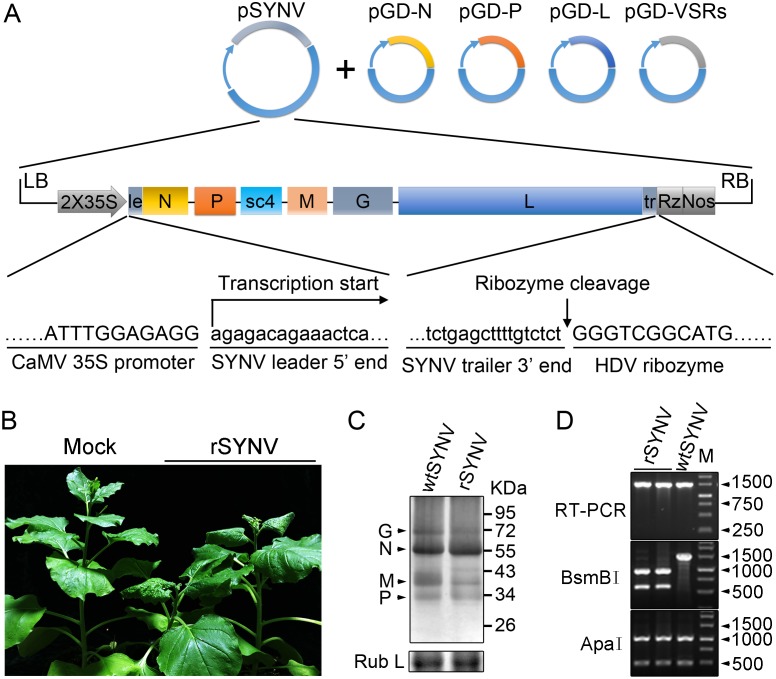
Reverse genetics systems have been established for all major groups of plant DNA and positive-strand RNA viruses, and our understanding of their infection cycles and pathogenesis has benefitted enormously from use of these approaches. However, technical difficulties have heretofore hampered applications of reverse genetics to plant negative-strand RNA (NSR) viruses. Here, we report recovery of infectious virus from cloned cDNAs of a model plant NSR, Sonchus yellow net rhabdovirus (SYNV). The procedure involves Agrobacterium-mediated transcription of full-length SYNV antigenomic RNA and co-expression of the nucleoprotein (N), phosphoprotein (P), large polymerase core proteins and viral suppressors of RNA silencing in Nicotiana benthamiana plants. Optimization of core protein expression resulted in up to 26% recombinant SYNV (rSYNV) infections of agroinfiltrated plants. A reporter virus, rSYNV-GFP, engineered by inserting a green fluorescence protein (GFP) gene between the N and P genes was able to express GFP during systemic infections and after repeated plant-to-plant mechanical passages. Deletion analyses with rSYNV-GFP demonstrated that SYNV cell-to-cell movement requires the sc4 protein and suggested that uncoiled nucleocapsids are infectious movement entities. Deletion analyses also showed that the glycoprotein is not required for systemic infection, although the glycoprotein mutant was defective in virion morphogenesis. Taken together, we have developed a robust reverse genetics system for SYNV that provides key insights into morphogenesis and movement of an enveloped plant virus. Our study also provides a template for developing analogous systems for reverse genetic analysis of other plant NSR viruses.


Conflict of interest statement

The authors have declared that no competing interests exist.

Figures

Fig 1
Fig 1. Recovery of recombinant SYNV in N. benthamiana.
(A) Schematic representation of the SYNV infectious cDNA clone and supporting plasmids. The pSYNV plasmid is designed for transcription to yield the SYNV antigenome (ag) RNA and contains the full-length SYNV cDNA positioned between a truncated CaMV double 35S promoter (2X35S) and the HDV ribozyme sequence in the pCB301 plasmid. Note that the SYNV gene order is shown in the antigenome sense. The pGD-N, pGD-P and pGD-L supporting plasmids encode the viral N, P and L core NC proteins, respectively, and the pGD-VSRs encode the TBSV p19, BSMV γB, and TEV P1/HC-Pro suppressors of RNA silencing (VSRs). le: leader; tr: trailer; Rz: ribozyme; LB: left border sequence; RB: right border sequence; Nos: nopaline synthase terminator. (B) Symptoms of N. benthamiana plants systemically infected with rSYNV. Agrobacterium cultures containing the pSYNV, pGD-N, pGD-P, pGD-L and the three VSRs plasmids were mixed and infiltrated into N. benthamiana leaves. Infected plants showing stunting and typical vein clearing symptoms were photographed at 35 days post infiltration (dpi) along with a mock control (uninfected healthy plant). (C) Detection of viral structural proteins in wtSYNV- and rSYNV-infected N. benthamiana plants. Total protein samples were analyzed by Western blotting with anti-SYNV virion antibody. The Coomassie blue stained Rubisco large subunit (Rub L) was used as a loading control. The positions of the SYNV G, N, M and P proteins are indicated along the left side of the gel and the numbers along the right side are protein size markers in KDa. (D) Restriction enzyme site identification of wtSYNV and rSYNV cDNAs. Total RNAs were extracted from wtSYNV- and rSYNV-infected plants and used as templates for reverse transcription PCR (RT-PCR). The wtSYNV and rSYNV RT-PCR products (∼1500 bp) were digested with BsmBI or ApaI and analyzed on 1.5% agarose gels. Positions of DNA size markers (M) are shown in base pairs.
Fig 2
Fig 2. Improvement of SYNV minireplicon (MR) expression by optimizing the core protein ratios.
(A) Schematic representation of the SYNV MR-GFP-RFP containing GFP and RFP reporter genes substituted for the N and P ORFs. The SYNV MR-GFP-RFP antigenomic RNA was transcribed from a CaMV double 35S promoter (2X35S), and flanked by a Hammerhead ribozyme (HH Rz) and HDV ribozyme (HDV Rz) sequence to produce exact 5′- and 3′- ends. le: leader; tr: trailer; LB: left border sequence; RB: right border sequence; Nos: nopaline synthase terminator; N/P GJ: N/P gene junction. (B) Visualization of plaques expressing GFP reporter protein in infiltrated plants. Equal volumes of 0.8 OD Agrobacterium cultures harboring the SYNV MR-GFP-RFP, pGD-NPL and the three VSRs plasmids were mixed and infiltrated into N. benthamiana leaves. Additional volumes of bacterial cultures containing the pGD-N (upper panels), pGD-P (middle panels) or pGD-L plasmids (bottom panels) at 0.2, 0.4 or 0.8 OD as indicated on the top of panels, were also included in the mixture to test their effects on reporter expression. Infiltrated leaves were photographed at 9 dpi with a fluorescence microscope under the GFP channel. Scale bar, 200 μm. (c) Detection of GFP and RFP protein levels in agroinfiltrated leaves by Western blotting using GFP- and RFP-specific antibodies. The Coomassie blue-stained Rubisco large subunit (Rub L) serves as a total protein loading control.
Fig 3
Fig 3. Expression of GFP engineered into rSYNV.
(A) Diagram showing insertion of the GFP gene between the N and P genes of the rSYNV antigenome to produce rSYNV-GFP. The recombinant vector transcribes GFP mRNA under the control of a duplicated N/P gene junction sequence. (B) Time course of GFP foci appearing in N. benthamiana leaves after agroinfiltration with rSYNV-GFP derivatives. N. benthamiana leaves were agroinfiltrated with bacterial mixtures containing plasmids encoding the rSYNV-GFP agRNA, the N, P, L proteins and the VSR suppressor proteins, and photographed by fluorescence microscopy at 6, 9, 12 and 15 dpi. The white arrow indicates GFP entering a leaf vein. Scale bar, 200 μm. (C) Symptoms of rSYNV- and rSYNV-GFP-infected plants. Photographs were taken at 30 dpi under white light (upper panels) and ultraviolet (UV) light (bottom panels). (D) Immunoblot analysis of SYNV proteins and GFP expression in plants systemically infected with rSYNV-GFP. Total proteins extracted from rSYNV (lane 1) and rSYNV-GFP (lane 2) infected plants were analyzed by Western blotting with antibodies against disrupted SYNV virions (α:SYNV) or GFP (α:GFP). (E) Stable maintenance of GFP during serial passages of rSYNV-GFP in N. benthamiana. rSYNV-GFP passaged to healthy plants by mechanical inoculation, and after 5 serial passages, the infected plants were photographed at 16 dpi under white (upper panels) and UV light (bottom panels). (F) Total RNAs and proteins were extracted from upper infected leaves of each plant passage and analyzed by RT-PCR using GFP-specific primers or by immmunoblotting (WB) with αSYNV and αGFP antibodies. Numbers 1 to 5 on the top of the panel represent passages No. 1 to 5, respectively. Coomassie blue stained Rubisco large subunit (Rub L) was used as a loading control.
Fig 4
Fig 4. Cell-to-cell movement analysis of rSYNV sc4, M and G deletion mutants.
(A) Schematic representation of rSYNV-GFP and rSYNV-GFP mutants with Δsc4, ΔM or ΔG deletions. Note that the SYNV gene order is shown in the antigenome sense. (B) Cell-to-cell movement of rSYNV-GFP and deletion derivatives. N. benthamiana leaves were agroinfiltrated with the rSYNV-GFP plasmid or the indicated mutant rSYNV-GFP plasmids along with supporting plasmids for expression of the N, P, L core proteins and the VSRs. Leaves were photographed with a fluorescence microscope at 8 and 14 dpi. Scale bar, 100 μm. (C) Complementation of rSYNV-eGFP-Δsc4 cell-to-cell movement by the sc4 protein expressed in trans from the MR-sc4-RFP minireplicon. Agrobacterium strains harboring the rSYNV-eGFP-Δsc4 plasmid, the MR-sc4-RFP plasmid, along with the supporting plasmids indicated in the panel B legend, were mixed and infiltrated into N. benthamiana leaves. Fluorescence images for GFP, RFP and the overlaid images are shown at 8 and 14 dpi. Scale bar, 100 μm.
Fig 5
Fig 5. Role of the SYNV glycoprotein in systemic infection and virion maturation.
(A) Symptoms of N. benthamiana plants after agroinfiltration with rSYNV-GFP and rSYNV-GFP-ΔG infectious clones. Leaves were photographed under white light (WL) or UV light at 30 dpi for rSYNV-GFP (upper panels) and at 35 dpi for rSYNV-GFP-ΔG (lower panels). Leaf tissue was also photographed with a fluorescence microscope (FM) to show GFP expression patterns in the mesophyll tissue. Scale bar, 200 μm. (B) Total proteins extracted from newly emerged leaf tissues were detected by immunoblotting with antibodies raised against SYNV virions (αSYNV), G protein (αG) or GFP (αGFP). The bottom panel is a loading control showing the Coomassie blue stained Rubisco large subunit (Rub L). (C and D) Electron micrographs of thin sections of N. benthamiana plants infected with rSYNV-GFP (C) and rSYNV-GFP-ΔG (D). The boxed sectors in the panels are magnified (40,000 X) to highlight the appearance of the virions and nucleocapsid cores. The red arrows in (C) show cross sections of the invaginated inner nuclear envelopes surrounding the enveloped virions. V: virion; C: core; Vp: viroplasm; Im: inner nuclear membranes; Ch: chloroplast; M: mitochondria.

Similar articles

Cited by

References

    1. King AMQ, Adams MJ, Carstens EB, Lefkowitz EJ. Virus Taxonomy: Ninth Report of the International Committee on Taxonomy of Viruses. London, UK: Elsevier Academic Press; 2011.
    1. Mann KS, Dietzgen RG. Plant rhabdoviruses: new insights and research needs in the interplay of negative-strand RNA viruses with plant and insect hosts. Arch Virol. 2014; 159: 1889–1900. 10.1007/s00705-014-2029-z - DOI - PubMed
    1. Kormelink R, Garcia ML, Goodin M, Sasaya T, Haenni AL. Negative-strand RNA viruses: the plant-infecting counterparts. Virus Res. 2011; 162: 184–202. 10.1016/j.virusres.2011.09.028 - DOI - PubMed
    1. Ammar el D, Tsai CW, Whitfield AE, Redinbaugh MG, Hogenhout SA. Cellular and molecular aspects of rhabdovirus interactions with insect and plant hosts. Annu Rev Entomol. 2009; 54: 447–468. 10.1146/annurev.ento.54.110807.090454 - DOI - PubMed
    1. Falk BW, Tsai JH. Biology and molecular biology of viruses in the genus Tenuivirus. Annu Rev Phytopathol. 1998; 36: 139–163. - PubMed

Publication types

MeSH terms