. 2015 Oct 20;11(10):e1005610.
doi: 10.1371/journal.pgen.1005610. eCollection 2015 Oct.

Canonical Poly(A) Polymerase Activity Promotes the Decay of a Wide Variety of Mammalian Nuclear RNAs

Affiliations

Canonical Poly(A) Polymerase Activity Promotes the Decay of a Wide Variety of Mammalian Nuclear RNAs

Stefan M Bresson et al. PLoS Genet. .

Abstract

The human nuclear poly(A)-binding protein PABPN1 has been implicated in the decay of nuclear noncoding RNAs (ncRNAs). In addition, PABPN1 promotes hyperadenylation by stimulating poly(A)-polymerases (PAPα/γ), but this activity has not previously been linked to the decay of endogenous transcripts. Moreover, the mechanisms underlying target specificity have remained elusive. Here, we inactivated PAP-dependent hyperadenylation in cells by two independent mechanisms and used an RNA-seq approach to identify endogenous targets. We observed the upregulation of various ncRNAs, including snoRNA host genes, primary miRNA transcripts, and promoter upstream antisense RNAs, confirming that hyperadenylation is broadly required for the degradation of PABPN1-targets. In addition, we found that mRNAs with retained introns are susceptible to PABPN1 and PAPα/γ-mediated decay (PPD). Transcripts are targeted for degradation due to inefficient export, which is a consequence of reduced intron number or incomplete splicing. Additional investigation showed that a genetically-encoded poly(A) tail is sufficient to drive decay, suggesting that degradation occurs independently of the canonical cleavage and polyadenylation reaction. Surprisingly, treatment with transcription inhibitors uncouples polyadenylation from decay, leading to runaway hyperadenylation of nuclear decay targets. We conclude that PPD is an important mammalian nuclear RNA decay pathway for the removal of poorly spliced and nuclear-retained transcripts.


Conflict of interest statement

The authors have declared that no competing interests exist.

Figures
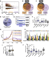
Fig 1
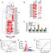
Fig 1. Global analysis of PPD targets.
(A) Scatter plot of DEGs from each of the three datasets tested. The log2 fold change (FC) is relative to the untreated total or nuclear RNA as appropriate. The x-axis is an average FPKM of the control samples for the two biological replicates. (B) Venn diagram of the upregulated DEGs identified in each of the three samples. (C) Venn diagram of the downregulated DEGs identified in each of the three samples. (D) Pie chart of the annotations assigned to the 353 high-confidence upregulated DEGs. (E) Strand-specific sequence traces from the RNF139 locus. The plus strand is in black; the minus strand is in blue. (F) Box-and-whiskers graph of the fold change of the siPAP total samples for each of the high-confidence target categories. For all box-and-whisker plots, the box corresponds to the 25th through the 75th percentile, the horizontal line is the median and the whiskers represent the upper and lower 25 percent. In this graph, asterisks indicate a p-value <0.0001 (Mann-Whitney test). Thirteen RNAs were categorized into two groups. (G) Composite RNA profiles comparing the fold changes from the high-confidence PROMPTs (solid lines) or the entire genome (dotted lines). The data are from one biological replicate; the other replicate is shown in S1B Fig. (H) Bar graph of results from qRT-PCR of six PROMPTs under five PPD inactivation conditions as listed. The values are averages and the error bars are standard deviation (n = 3). (I) Box-and-whisker plots of the number of exons for the up- or downregulated DEGs or all expressed genes. Expressed genes were defined as those with FPKM>1 (n = 13044; asterisk, p-value <0.0001; Mann-Whitney test; “ns”, not significant). (J) Box-and-whiskers plot of the number of exons for each category of high-confidence targets. (asterisk, p-value <0.0001; Mann-Whitney test).
Fig 2
Fig 2. NcSNHGs are degraded by PPD or NMD.
(A) Heat map showing the changes in spliced ncSNHG levels following RRP6 and DIS3 knockdown (“siExo”), LALA expression, PAP knockdown, cordycepin treatment, PABPN1 knockdown, cycloheximide (CHX) treatment, or UPF1 knockdown. Log2 fold change (FC) values were determined by qRT-PCR (n = 3). 7SK RNA was used as a loading control for the cycloheximide experiment, while β-actin or GAPDH was used for all other samples. (B) Same as in (A), but the relative changes in spliced (left) and unspliced (right) transcripts are shown. The left panel is reproduced from (A). (C) Bar graphs of qRT-PCR data comparing the average relative levels of six ncSNHGs following cycloheximide, siPAP or both treatments. The error bars are standard deviation (n = 3). (D) Correlation between intron number and the fold change in transcript levels following LALA expression. Expression values are derived from the experiments in (A); the red line is a linear regression. (E) Nuclear enrichment scores calculated from each expressed gene (FPKM>0.5, n = 13,114) were placed into 32 bins and color-coded from red to blue. (F) Each ncSNHG was plotted by the average log2(FC) values from (A) and color-coded by its NES as determined in (F).
Fig 3
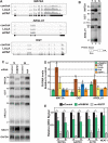
Fig 3. RI-RNAs are subject to PPD.
(A) Nuclear sequence traces of three RI-RNAs. The RI is shown as a gray box in the gene diagrams. (B) Northern blot using total (T), cytoplasmic (C), or nuclear (N) fractions and exon probes that hybridize to both RI and mRNA isoforms. The ARGLU1 probe cross-hybridizes with 28S rRNA; pre-rRNA and rRNA control for fractionation (C) Northern blot of specific RNAs from cells treated with siControl, siPABPN1, or siPAP. (D) Quantification of the RI isoforms from northern blots (siPABPN1, siPAP, CHX), or qRT-PCR (siUPF1, cordycepin). Each value is normalized to GAPDH or ACTB and expressed relative to the matched control. Error bars are standard deviation from the mean (asterisk, p-value <0.05; unpaired Students t-test; n = 3). (E) NRO assays using cells treated with control or PABPN1 siRNAs. All values are relative to the control. The no 4SUTP is a negative control in which UTP was substituted for 4SUTP. Two amplicons (labeled “A” and “B”) were used for each gene except NEAT1. Error bars are standard deviation from the mean (asterisk, p-value <0.01; unpaired Students t-test; n = 4).
Fig 4
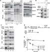
Fig 4. RI-RNAs are hyperadenylated upon ActD treatment.
(A) Northern blot of RNAs from cells treated with ActD and deadenylated with RNase H and oligo(dT) as indicated. The probes hybridized to exons (lanes 1–6, 13–18) or the RI (lanes 7–12). The asterisk marks RNAs that are fully spliced but hyperadenylated. (B) RNAs were cleaved ~500 nt from their poly(A) addition site and examined by northern blot with a probe to the 3´ cleavage product. ActD treatment was for 6 hrs. RNA markers (kb) are shown on the right. (C) Northern blots for specific transcripts using RNA from cells transfected with control or PABPN1 siRNAs +/- 6-hr ActD treatment. The bottom panel is an RNase H assay as in (B). (D) Scheme of the 4SU pulse-chase and ActD time courses. For the 4SU experiments, cells were washed and grown in label-free media for an additional hour prior to beginning the time course. This step was necessary to allow unincorporated 4SU in the cell to be depleted. (E) Decay profiles of SNHG19 as determined by 4SU or ActD.
Fig 5
Fig 5. A large fraction of nuclear RNA is hyperadenylated upon ActD treatment.
(A) Total cellular poly(A) tails were examined by northern blot after ActD treatment. Mobility of molecular weight markers (kb) is shown. (B) Bulk poly(A) tails were examined from untreated or ActD treated cells from whole cells (T), cytoplasmic (C) or nuclear (N) fractions. (C) Northern blot for bulk poly(A) tails using RNA from cells treated with a control siRNA or siRNAs targeting PABPN1 +/-6-hr ActD treatment. (D) Scheme for metabolic labeling approach to examine bulk poly(A) tail dynamics. (E) Results of a metabolic labeling assay examining the bulk soluble and insoluble poly(A) tails in cells +/- ActD treatment. Lane 1 is a negative control from cells without EU treatment.
Fig 6
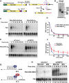
Fig 6. Role of hyperadenylation in PPD.
(A) Left, Cartoons of the PANΔENE-AAUAAA and PANΔENE-A35 plasmids depicting the TetRP (green), PAN RNA sequence (yellow), PAN RNA polyadenylation signals (black), A35 stretch (red), MALAT1 3´-end cleavage sequence (purple), and the position of the ENE deletion (Δ); the diagrams are not to scale. Right, Scheme of the production of PANΔENE-A35 by RNase P cleavage in cells. The color scheme is the same as the DNA diagrams; the cap is shown as a gray circle. The MALAT1 mascRNA sequence is represented by the cloverleaf structure. (B) Poly(A) tail length analysis of PANΔENE-A35. RNA was harvested and treated with RNase H in the presence or absence of oligo(dT). (C) Representative transcription pulse-chase assay with the indicated constructs. The “-” samples were harvested prior to the two-hour transcription pulse. 7SK RNA was used as a loading control. (D) Quantification of the pulse-chase assays; error bars show the standard deviation of the mean (n = 3). (E and F) Representative transcription pulse chase and quantification of PANΔENE-A35 following treatment with the indicated siRNAs (n = 3) (G) Illustration of the PAP-tethering approach. (H) Transcription pulse-chase analysis of TetRP-driven PANΔENE containing six MS2 binding sites. Cells were treated with either control (left) or PABPN1 (right) siRNAs. Cells were co-transfected with PANΔENE-6MS2 and either MS2-PAP or MS2 expression constructs.

Similar articles

Cited by

References

    1. Jensen TH, Jacquier A, Libri D. Dealing with Pervasive Transcription. Mol Cell. Elsevier Inc; 2013;52: 473–484. 10.1016/j.molcel.2013.10.032 - DOI - PubMed
    1. Fasken MB, Corbett AH. Mechanisms of nuclear mRNA quality control. RNA Biol. 2009;6: 237–241. - PubMed
    1. Kervestin S, Jacobson A. NMD: a multifaceted response to premature translational termination. Nat Rev Mol Cell Biol. Nature Publishing Group; 2012;13: 700–712. 10.1038/nrm3454 - DOI - PMC - PubMed
    1. Beaulieu YB, Kleinman CL, Landry-Voyer A-M, Majewski J, Bachand F. Polyadenylation-Dependent Control of Long Noncoding RNA Expression by the Poly(A)-Binding Protein Nuclear 1. Lee JT, editor. PLoS Genet. 2012;8: e1003078 10.1371/journal.pgen.1003078 - DOI - PMC - PubMed
    1. Bresson SM, Conrad NK. The human nuclear poly (a)-binding protein promotes RNA hyperadenylation and decay. PLoS Genet. 2013. - PMC - PubMed

Publication types