Review
. 2015 Dec:6:524-551.
doi: 10.1016/j.redox.2015.08.020. Epub 2015 Oct 8.

Reperfusion injury and reactive oxygen species: The evolution of a concept

Affiliations
Review

Reperfusion injury and reactive oxygen species: The evolution of a concept

D Neil Granger et al. Redox Biol. 2015 Dec.

Abstract

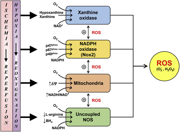
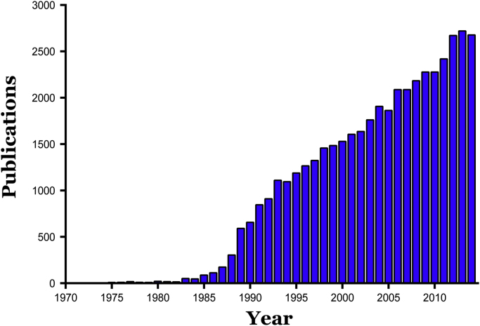
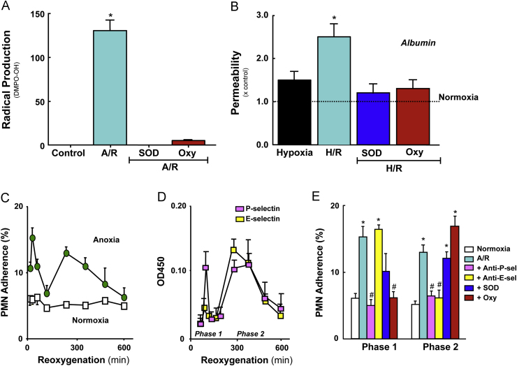
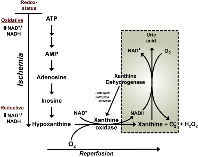
Reperfusion injury, the paradoxical tissue response that is manifested by blood flow-deprived and oxygen-starved organs following the restoration of blood flow and tissue oxygenation, has been a focus of basic and clinical research for over 4-decades. While a variety of molecular mechanisms have been proposed to explain this phenomenon, excess production of reactive oxygen species (ROS) continues to receive much attention as a critical factor in the genesis of reperfusion injury. As a consequence, considerable effort has been devoted to identifying the dominant cellular and enzymatic sources of excess ROS production following ischemia-reperfusion (I/R). Of the potential ROS sources described to date, xanthine oxidase, NADPH oxidase (Nox), mitochondria, and uncoupled nitric oxide synthase have gained a status as the most likely contributors to reperfusion-induced oxidative stress and represent priority targets for therapeutic intervention against reperfusion-induced organ dysfunction and tissue damage. Although all four enzymatic sources are present in most tissues and are likely to play some role in reperfusion injury, priority and emphasis has been given to specific ROS sources that are enriched in certain tissues, such as xanthine oxidase in the gastrointestinal tract and mitochondria in the metabolically active heart and brain. The possibility that multiple ROS sources contribute to reperfusion injury in most tissues is supported by evidence demonstrating that redox-signaling enables ROS produced by one enzymatic source (e.g., Nox) to activate and enhance ROS production by a second source (e.g., mitochondria). This review provides a synopsis of the evidence implicating ROS in reperfusion injury, the clinical implications of this phenomenon, and summarizes current understanding of the four most frequently invoked enzymatic sources of ROS production in post-ischemic tissue.

Keywords: Hypoxia-reoxygenation; Ischemia-reperfusion; Mitochondria; NADPH oxidase; Uncoupled nitric oxide synthase; Xanthine oxidase.


Figures

fx1
Graphical abstract
Fig. 1
Fig. 1
Publication frequency of articles dealing with ischemia-reperfusion injury from 1970 through 2014. Based on PubMed search (June, 2015) using search term “ischemia-reperfusion injury” or “reperfusion injury”.
Fig. 2
Fig. 2
Responses of vascular endothelial cells (EC) to anoxia/reoxygenation (A/R) or hypoxia/reoxygenation (H/R). (Panel A) Radical production by human aortic EC exposed to 60 min anoxia and 10 min reoxygenation. Measurements derived from electron paramagnetic resonance using the spin trap 5,5-dimethyl-1-pyrroline N-oxide (DMPO). The large signal detected after reoxygenation was not evident when the EC were treated with either superoxide dismutase (SOD) or oxypurinol (Oxy). Data from Zweier et al. . (Panel B) Albumin permeability across monolayers of bovine pulmonary artery EC exposed to 90 min hypoxia, with or without 90 reoxygenation. While hypoxia per se did not increase monolayer permeability, H/R elicited a significant increase. The H/R-induced permeability response was prevented by pretreatment with either SOD or oxypurinol (Oxy). Data from Inauen et al. . (Panel C) Neutrophil (PMN) adhesion response on human umbilical vein endothelial cells (HUVEC) exposed to 60 min anoxia, followed by 30–600 min reoxygenation. A biphasic adhesion response was noted with an initial peak (phase 1) at 30 min and a later peak (phase 2) at 240 min. (Panel D) Biphasic increases in endothelial expression of P- and E-selectin on HUVEC exposed to A/R (as described for Panel C). (Panel E) PMN adhesion on HUVEC monolayers exposed to A/R (as per Panel C) following treatment with a blocking antibody to either P-selectin (anti-P-sel) or E-selectin (anti-E-sel), SOD or Oxy. Data in Panels C–E from Ichikawa et al. .
Fig. 3
Fig. 3
Potential mechanisms of ROS production by XOR in tissues exposed to ischemia and reperfusion. In the setting of ischemia, ATP is catabolized to hypoxanthine and the dehydrogenase form of XOR (XDH) is converted, via limited proteolysis and sulfhydryl oxidation, to the oxidase form (XO). Upon reperfusion, the restored tissue O2 reacts with hypoxanthine (or xanthine) and XO to generate both superoxide (O2-) and hydrogen peroxide (H2O2), which can consequently interact to yield more reactive secondary species , . The conversion of XDH to XO may not be required for ROS production following reperfusion (see boxed area of figure). During ischemia, the redox status of the tissue is altered from an oxidative state (higher level of NAD+ relative to NADH) to a reductive state (higher NADH relative to NAD+). This altered redox state has been shown to enhance the generation of O2- from XDH in the presence of xanthine .
Fig. 4
Fig. 4
Evidence implicating leukocyte-associated Nox in the lung injury response to 1 h ischemia and 2 h reperfusion. Data are presented for postischemic lungs from bone marrow chimeras produced by the transplantation of marrow from wild type (WT) mice into p47phox−/− recipients or vice versa (donor→recipient). (Panel A) Malondialdehyde (MDA) level in bronchioalveolar lavage fluid after reperfusion. (Panel B) Lung vascular permeability estimated from Evans blue accumulation in lung tissue. (Panel C) Pulmonary edema measured by wet/dry weight ratio after reperfusion. (Panel D) Lung airway resistance after reperfusion. Data derived from Yang et al. .
Fig. 5
Fig. 5
Stimuli that may act on Nox-2 positive cells following ischemia and reperfusion to elicit activation and/or induction of the enzyme. Hypoxia-inducible factor-1α (HIF-1α), leukotriene B4 (LTB4), platelet activating factor (PAF), complement component-5a (C5a), tumor necrosis factor-α (TNF-α), interleukin-1β (IL-1β), and interferon-Υ (IFN-γ).
Fig. 6
Fig. 6
ROS production by the electron transport chain (ETC): role of mitochondrial proton motive force (∆ψ). Electron carriers, NADH and FADH2 feed electrons (e) into complex I and complex II of the ETC, respectively. As e traverse through the ETC they release some of their energy to pump H+ into the intermembrane space generating a ∆ψ. The ∆ψ is used to move H+ through ATP synthase and phosphorylate ADP to ATP. At several points along the ETC (esp. complex I and III) there is a potential for e to leak and reduce O2 to superoxide (O2·-). The rate of O2·- production is directly related to the level of ∆ψ; ∆ψ was varied using an uncoupling agent or inhibitors of ETC (inset; from Korshunov et al. [318]). A feedback mechanism exists to limit O2·- production by dissipating the ∆ψ. In this scenario, excessive O2·- production activates the uncoupling protein (UCP) allowing H+ to diffuse back into the matrix bypassing the ATP synthase. The increased efflux of H+ lowers the ∆ψ and reduces O2·- production. Red arrows, forward e flow; red dashed arrow, reverse electron flow; Q, Coenzyme Q; Cyt, cytochrome c. (modified from Krauss et al. and Chen and Zweier [298]).
Fig. 7
Fig. 7
Mitochondrial anti-oxidant system. Superoxide (O2·-) generated by the ETC (see Fig. 6 ) is converted to H2O2 by superoxide dismutase (SOD); MnSOD in the matrix and CuZnSOD in the intermembrane space, respectively. Catalase, glutathione peroxidase (GPx), and peroxiredoxin (Prx) convert H2O2 to H2O. GPx and Prx require glutathione (GSH) and thioredoxin (TrxSH) for their reduction, respectively. Glutathione disulfide (GSSG) is reduced back to GSH by glutathione reductase (GR), while TrxS is reduced back to TrxSH by thioredoxin reductase (TrxR). Both GR and TrxR depend on reducing equivalents from NADPH generated by transhydrogenase (NNT) (modified from Murphy and Kowaltowski et al. [304]).
Fig. 8
Fig. 8
Hypoxia/reoxygenation (H/R): mitochondrial ROS-induced ROS release leading to cell death. (Panel A) H/R-induced increase in ROS and decrease in membrane potential (∆ψ) of a single mitochondria of a cardiomyocyte. Immediately upon reoxygenation there is an increase ∆ψ and gradual increase in ROS. Once a threshold level of ROS is achieved, ROS generation increases sharply and ∆ψ falls precipitously. (Panel B) H/R-induced loss of ∆ψ and ROS in a network of mitochondria within a cardiomyocyte. Numerous mitochondria that are completely depolarized (dark areas in upper panel) are associated with ROS production (green areas in lower panel). (Panel C) H/R-induced cell death. Several hours after reoxygenation about 2/3 of the cardiomyocytes are dead. The number of cells dying after H/R is reduced by an inhibitor of MTP (cyclosporine A; CSA). Adapted from Juhaszova et al. .
Fig. 9
Fig. 9
Monomeric and dimeric forms of NOS. (Panel A) NOS monomers can transfer electrons from NADPH to the flavins in the reductase domain where limited amounts of O2·- can be formed. NOS monomers cannot bind the cofactor (BH4) or the substrate (arginine) and cannot generate NO. (Panel B) Formation of a NOS dimer requires heme, which allows for the transfer of electrons from the reductase domain of one monomer to the oxidase domain of the adjacent monomer. When sufficient arginine and BH4 are present, NOS dimers couple their heme, allowing for O2 reduction and the synthesis of NO. From Forstermann and Sessa where the structure and catalytic function of NOS are discussed in greater detail.
Fig. 10
Fig. 10
Role of BH4 in NOS uncoupling in endothelial cells during H/R in vitro (Panels A–C) and I/R-induced cardiac injury in vivo (Panel D). (Panel A) Intracellular O2·- production (DHE-derived fluorescence intensity) is increased in endothelial cells subjected to H/R. *p<0.002 vs control (normoxia). (Panel B) BH4 decreases while BH2 increases in endothelial cells subjected to H/R. *p<0.004 vs control (normoxia). (Panel C) NO production by endothelial cells is decreased during H/R and is partially rescued by BH4 supplementation. *p<0.002 vs control (normoxia) #p<0.002 vs H/R. (Panel D) Liposomal BH4 (6R BH4) reduces myocardial infarction (as percent of area at risk; AAR) in rat hearts subjected to I/R. Control represents I/R and vehicle represents empty liposome. *p<0.001 vs control. Panels A–C modified from De Pascali et al. while panel D modified from Xie et al. .

Similar articles

Cited by

References

    1. Carden D.L., Granger D.N. Pathophysiology of ischaemia-reperfusion injury. J. Pathol. 2000;190:255–266. - PubMed
    1. Raedschelders K., Ansley D.M., Chen D.D. The cellular and molecular origin of reactive oxygen species generation during myocardial ischemia and reperfusion. Pharmacol. Ther. 2012;133:230–255. - PubMed
    1. Costantino C., Corday E., Lang T.W., Meerbaum S., Brasch J., Kaplan L., Rubins S., Gold H., Osher J. Revascularization after 3 h of coronary arterial occlusion: effects on regional cardiac metabolic function and infarct size. Am. J. Cardiol. 1975;36:368–384. - PubMed
    1. Kloner R.A., Ganote C.E., Whalen D.A., Jr., Jennings R.B. Effect of a transient period of ischemia on myocardial cells. II. Fine structure during the first few minutes of reflow. Am. J. Pathol. 1974;74:399–422. - PMC - PubMed
    1. Reimer K.A., Lowe J.E., Rasmussen M.M., Jennings R.B. The wavefront phenomenon of ischemic cell death. 1. Myocardial infarct size vs duration of coronary occlusion in dogs. Circulation. 1977;56:786–794. - PubMed

Publication types

Substances