. 2015 Oct 20;10(10):e0140685.
doi: 10.1371/journal.pone.0140685. eCollection 2015.

Peroxisomal Import Reduces the Proapoptotic Activity of Deubiquitinating Enzyme USP2

Affiliations

Peroxisomal Import Reduces the Proapoptotic Activity of Deubiquitinating Enzyme USP2

Katharina Reglinski et al. PLoS One. .

Abstract

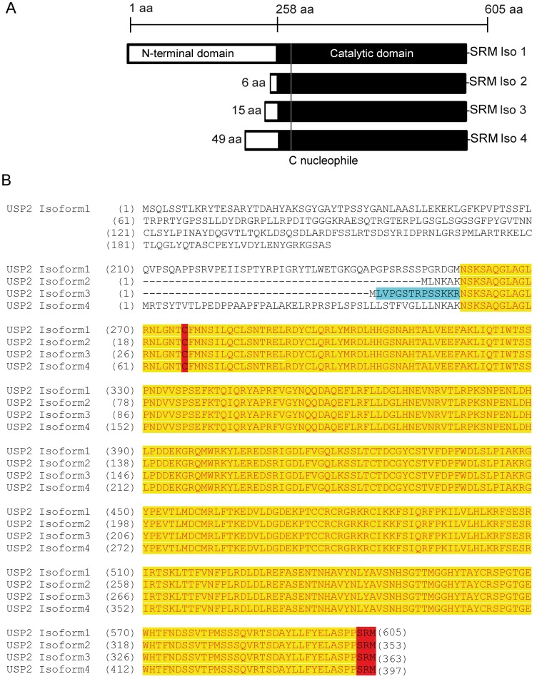
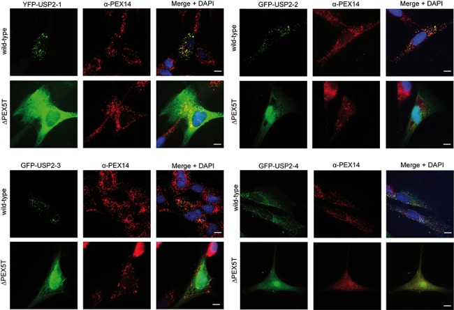
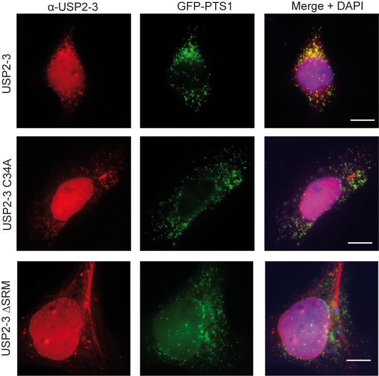
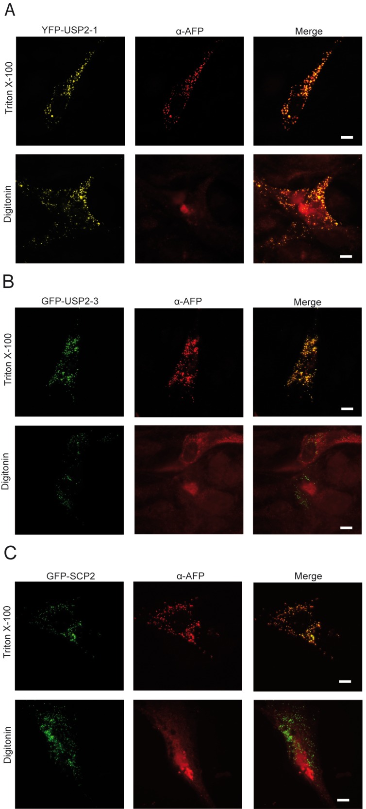
The human deubiquitinating enzyme ubiquitin-specific protease 2 (USP2) regulates multiple cellular pathways, including cell proliferation and apoptosis. As a result of alternative splicing four USP2 isoenzymes are expressed in human cells of which all contain a weak peroxisome targeting signal of type 1 (PTS1) at their C-termini. Here, we systematically analyzed apoptotic effects induced by overexpression and intracellular localization for each isoform. All isoforms exhibit proapoptotic activity and are post-translationally imported into the matrix of peroxisomes in a PEX5-dependent manner. However, a significant fraction of the USP2 pool resides in the cytosol due to a weaker PTS1 and thus low affinity to the PTS receptor PEX5. Blocking of peroxisomal import did not interfere with the proapoptotic activity of USP2, suggesting that the enzyme performs its critical function outside of this compartment. Instead, increase of the efficiency of USP2 import into peroxisomes either by optimization of its peroxisomal targeting signal or by overexpression of the PTS1 receptor did result in a reduction of the apoptotic rate of transfected cells. Our studies suggest that peroxisomal import of USP2 provides additional control over the proapoptotic activity of cytosolic USP2 by spatial separation of the deubiquitinating enzymes from their interaction partners in the cytosol and nucleus.


Conflict of interest statement

Competing Interests: The authors have declared that no competing interests exist.

Figures

Fig 1
Fig 1. Sequence alignment of the four different isoforms of USP2.
A: Schematic overview of four isoforms of human USP2. The sequence of the C-terminal region containing the catalytic domain (in black) and the peroxisomal targeting signal (SRM) is identical for all four isoforms. The isoforms differ in length and composition of the N-terminal domain (in white). B: Sequence alignment of the four USP2 isoforms. During this study, antibodies have been raised against a 13mer peptide representing the N-terminal amino acids of isoform 3 (blue). The catalytic domain is highlighted in yellow. The catalytically critical cysteine residue as well as the C-terminal peroxisomal targeting sequence–SRM are shown in red.
Fig 2
Fig 2. All USP2 isoforms localize to peroxisomes.
Fluorescence microscopy images from wild-type fibroblasts and PEX5-deficient fibroblasts, transfected with YFP-USP2-1, GFP-USP2-2, GFP-USP2-3 and GFP-USP2-4 (green). Congruent fluorescent pattern of the labelled USP2 isoforms and PEX14 indicate peroxisomal localization. All four USP2 isoforms are associated with peroxisomes, when expressed in wild-type fibroblasts. The cytosolic and nuclear fluorescent pattern, which is not congruent with the PEX14 staining, indicates a mistargeting as seen in the PEX5 deficient fibroblasts. Cells were counterstained with DAPI to mark the nucleus. Scale bar: 10 μm.
Fig 3
Fig 3. Untagged USP2-3 shows a PTS1-dependent peroxisomal localisation.
Immunofluorescence microscopy images of HEK-293 cells, which were co-transfected with the peroxisomal marker protein GFP-PTS1 and either USP2-3, the catalytically inactive variant (USP2-3 C34A), or the PTS1 deficient variant (USP2-3 ∆SRM) as indicated. The overexpressed USP2-3 variants were detected with a peptide antibody raised against the N-terminal isoform-specific region of USP2-3 (red). USP2-3 and the catalytically inactive variant are partially localised to peroxisomes, cytosol and nucleus, while USP2-3 (ΔSRM) is found only in cytosol and nucleus. Peroxisomes are labelled by the GFP-PTS1 (green). Scale bar: 10 μm.
Fig 4
Fig 4. USP2 is a peroxisomal matrix protein.
Immunofluorescence microscopy images of wild-type fibroblasts, containing YFP-USP2-1 (A), or GFP-USP2-3 (B) or GFP-SCP2, a peroxisomal matrix protein (C). Immunofluorescence microscopy was performed using antibodies against fluorescent proteins (AFP). Cells were incubated either with 1% Triton X-100, permeabilising all cellular membranes, or Digitonin (25 μg/ml), permeabilizing only the plasma membrane. Using Triton X-100, peroxisomal matrix proteins become accessible for the antibodies, as indicated by the congruent staining of the fluorophore (green) and the AFP antibody (red). Using Digitonin, only proteins facing the cytosolic compartment can interact with the antibodies, leading to a diffuse staining of the AFP antibody, when the fluorescent protein is compartmentalized into cell organelles. YFP-USP2-1 and GFP-USP2-3 could only be detected by the AFP antibody after permeabilization with Triton X-100, indicating that these proteins enter the peroxisomal lumen. Scale bar: 10 μm.
Fig 5
Fig 5. In vitro deubiquitination assay.
Purified USP2-3, its catalytically inactive variant (C34A) and a variant lacking the PTS-1 (∆SRM) were tested for their ability to deubiquitinate Ub-PEX5 (A) and Ub-GST (B) in vitro. Equal amounts of USP2 and the substrate (200 μg) were incubated for 1h at 37°C followed by SDS-PAGE and Coomassie staining. A: A recombinant Ub-PEX5 fusion construct was designed to mimic monoubiquitinated PEX5. Here the first 12 amino acids of PEX5 are replaced by Ubiquitin. PEX5 is monoubiquitinated at the cysteine in position 11. The thioester bond of the original monoubiquitinated PEX5 is replaced by a peptide bond in the Ub-PEX5 model substrate. The recombinant Ub-PEX5 appears as a double band. The upper band of purified Ub-PEX5, represents Ub-PEX5 and the lower one supposedly deubiquitinated PEX5. Ub-PEX5 shifted to the lower mass upon incubation with USP2-3 and USP2-3 ∆SRM, indicative for the deubiquitination of these constructs. This shift was not seen with the catalytic inactive USP2-3 C34A, indicating that this construct was not capable to deubiquitinate Ub-PEX5. The deubiquitination could be inhibited by N-ethylmaleimide (NEM). B: Ubiquitin fused to GST was used as a control substrate. The upper band represents the entire fusion construct, while the lower band represents deubiquitinated GST. As Ub-PEX5, also Ub-GST shifted to the lower mass upon incubation with USP2-3 and USP2-3 ∆SRM, indicative for the deubiquitination of these constructs. Again, the catalytic inactive USP2-3 C34A, was not capable to deubiquitinate Ub-GST and the deubiqutination could be inhibited by N-ethylmaleimide (NEM).
Fig 6
Fig 6. The efficiency of the peroxisomal import of USP2-3 is increased by exchange of its targeting signal.
A: Mammalian Two-Hybrid analysis of the interaction of PEX5 and USP2-3 variants with different C-terminal sequences. HEK-293 cells were transfected with PEX5, fused to the activation domain, and indicated variants of USP2-3, fused to the binding domain. The interaction between both proteins was monitored by expression of the reporter protein chloramphenicol transferase (CAT). Expression was quantified by a CAT-Elisa assay. The results show that USP2 interacts with PEX5. The PTS1 has a critical role for the PEX5 interaction, as there is no CAT-expression detected when the PTS1-signal is deleted (ΔSRM). In contrast, there is an increased interaction when the PTS1 of USP2 is replaced by the more common PTS1 signal SKL. The statistical significance of the differences between the analyzed USP2-3 variants in their ability to interact with PEX5 was found using a one-way ANOVA test (p<0.0001) and pairwise comparisons performed using the Tukey’s multiple comparison test. The Tukey’s comparison test revealed a significant increase in the expression of the reporter protein after replacement of the PTS1-signal (USP2-3 SRM>SKL) when compared to USP2-3 (***p<0.005). Mean values are shown with standard deviation from three independent experiments which were performed in triplicate for each USP2 variant. B: USP2-3 SRM>SKL is predominantly localized in peroxisomes. Immunofluorescence microscopy images of HEK-293 cells, transfected with USP2-3, USP2-3 ΔSRM or USP2-3 SRM>SKL as indicated. USP2 variants were visualized with an isoform specific antibody (red). Most of USP2-3 and USP2-3 ΔSRM localized to the cytosol and nucleus while the predominant peroxisomal localization of USP2-3 SRM>SKL is indicated by its clear colocalization with the peroxisomal marker GFP-PTS1 (green). Statistical analysis of peroxisomal localization of the different USP2-3 variants by fluorescence intensity measurement in the areas positive for peroxisome staining (bottom panel). For the statistical analysis, nine cells of each condition were analysed. The intensity of the red fluorescence was measured in regions where the green fluorescence was above a threshold (peroxisomes, indicated by the GFP-PTS1 marker protein). The intensity of the red fluorescence in the area of peroxisomes is presented in a boxplot, IQR 25–75 percentile, Whiskas 10–90 percentile. The peroxisomal localization of USP2-3 SRM>SKL, indicated by a higher intensity of the red fluorescence in the region of peroxisomes, is significantly higher than for USP2-3, as indicated by an one way ANOVA analysis and a multiple comparison (Tukey’s) test, (*p<0.05, **p<0.01, ***p<0.005). Scale bar: 10 μm. C: Localization of USP2 variants by subcellular fractionation studies. HEK-293 cells, transiently transfected with the indicated USP2-3 variants were subjected to subcellular fractionation studies by differential centrifugation. After permeabilization of the plasma membrane with 25μg/ml Digitonin, the cell organelles were sedimented by centrifugation. The resulting supernatant contained released cytosolic proteins, while permeabilized cells including organelles are present in the sediments. The results show that USP2-3 and USP2-3 ∆SRM are mostly localized in the cytosol (C) with only a small portion associated with the sedimented organelles (P). The exchange of the PTS1 signal results in an increased organelle localization of USP2-3 SRM>SKL, indicated by its predominant presence in the sediment. Catalase was used as peroxisomal marker. Immunodetection was carried out with antibodies against USP2, catalase and PEX5.
Fig 7
Fig 7. All isoforms of USP2 exhibit proapoptotic activity.
A: The different isoforms of USP2 and their catalytic mutants were overexpressed in HEK-293 cells. Expression was analyzed by immunoblotting using USP2-X antibodies. The molecular masses of the isoforms are: USP2-1: 68 kDa; USP2-2:40 kDa, USP2-3: 41 kDa; USP2-4: 45 kDa. Each full lengths isoform is denoted by an arrow. The asterisk indicates a degradation product of around 37 kDa, which is common for all isoforms. Prohibitin was used as a loading control. B: All Wild-type USP2 isoforms but not their catalytically inactive mutants induce cell death. HEK-293 cells were transfected with the control plasmid (pcDNA3.1) or plasmids expressing the indicated USP2 variants. The induction of apoptosis was monitored by a caspase activity assay, using a luminescent substrate for Caspase 3 and 7. In comparison with the vector control, the caspase activity was significantly increased after expression of each of the USP2 isoforms. Mean values are shown with standard deviation from three independent experiment, which were performed in triplicate for each construct. Two way ANOVA analysis showed that the proapoptotic effect of USP2 depends on the isoform as well as on the activity of the enzyme (p<0.0001). (RUL = Relative Light Unit).
Fig 8
Fig 8. The proapoptotic effect of USP2-3 overexpression is reduced by optimization of the peroxisomal import.
A: Activity of caspase 3/7 in HEK-293 cells after expression of different USP2-3 variants. Cells were transfected with the empty control plasmid pcDNA3.1 or plasmids, encoding the indicated USP2-3 variants. 24 h after transfection caspase activity was measured by a luminescence assay. Expression of USP2-3 did result in an increase of the caspase activity, while caspase acitivity upon expression of the inactive USP2-3 C34A variant was only slightly above the control. The apoptotic effect of the different USP2-3 variants differ significantly, which was shown by one way ANOVA analysis (p<0.0001). However, in comparison to the unmodified protein, the caspase activity was significantly decreased upon expression USP2-3 SRM>SKL, which was shown by a Tukey’s multiple comparisons test (*p<0.05). Mean values are shown with standard deviation from three independent experiments, which were performed in triplicate for each USP2 variant. B: Activity of Caspase 3/7 of FlpUSP2-3 cells after transfection with the empty vector or plasmids coding for PEX5 or mutated PEX5 C11A, which does not support peroxisomal protein import. 48 h after transfection, caspase activity was monitored by a luminescence assay (upper panel). The caspase activity significantly differs upon the expressed PEX5 variants (one way ANOVA analysis; p<0.001). Tukey’s multiple comparisons test revealed that in comparison to the control (empty vector), transfection with the import defective PEX5 C11A mutant had no significant effect on the caspase activity, but was significantly decreased after transfection of wild-type PEX5 (***p<0.005). Shown are mean values with standard deviation from three independent experiments, which were performed in triplicate for each construct. Expression of the different PEX5 variants was analyzed by immunoblotting (lower panel). Prohibitin was used as a loading control. PEX5, USP2-3 and Prohibitin were detected with specific antibodies. (RUL = Relative Light Unit).

Similar articles

Cited by

References

    1. Chau V, Tobias JW, Bachmair A, Marriott D, Ecker DJ, Gonda DK, et al. A multiubiquitin chain is confined to specific lysine in a targeted short-lived protein. Science. 1989;243:1576–83. - PubMed
    1. Bhattacharya S, Ghosh MK. Cell death and deubiquitinases: perspectives in cancer. Biomed Res Int. 2014;2014:435197 10.1155/2014/435197 - DOI - PMC - PubMed
    1. Allende-Vega N, Sparks A, Lane DP, Saville MK. MdmX is a substrate for the deubiquitinating enzyme USP2a. Oncogene. 2010;29(3):432–41. 10.1038/onc.2009.330 - DOI - PubMed
    1. Stevenson LF, Sparks A, Allende-Vega N, Xirodimas DP, Lane DP, Saville MK. The deubiquitinating enzyme USP2a regulates the p53 pathway by targeting Mdm2. EMBO J. 2007;26(4):976–86. 10.1038/sj.emboj.7601567 - DOI - PMC - PubMed
    1. Shi Y, Solomon LR, Pereda-Lopez A, Giranda VL, Luo Y, Johnson EF, et al. Ubiquitin-specific cysteine protease 2a (USP2a) regulates the stability of Aurora-A. J Biol Chem. 2011;286(45):38960–8. 10.1074/jbc.M111.231498 - DOI - PMC - PubMed

Publication types