. 2015 Oct 20;10(10):e0140958.
doi: 10.1371/journal.pone.0140958. eCollection 2015.

MiR-486 and miR-92a Identified in Circulating HDL Discriminate between Stable and Vulnerable Coronary Artery Disease Patients

Affiliations

MiR-486 and miR-92a Identified in Circulating HDL Discriminate between Stable and Vulnerable Coronary Artery Disease Patients

Loredan S Niculescu et al. PLoS One. .

Abstract

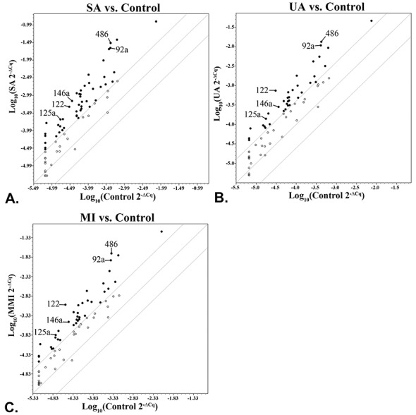
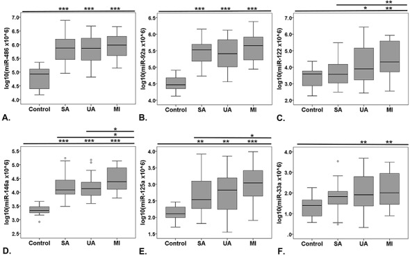
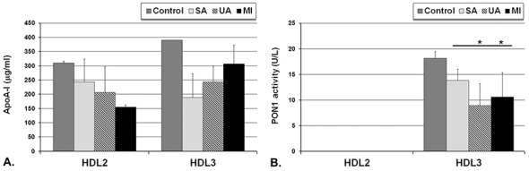
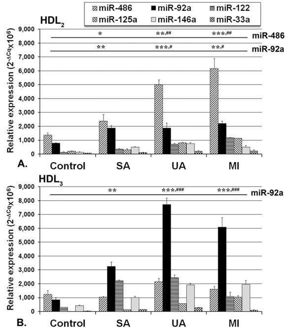
Small non-coding microRNAs (miRNAs) are implicated in gene regulation, including those involved in coronary artery disease (CAD). Our aim was to identify whether specific serum miRNAs present in the circulating lipoproteins (Lp) are associated with stable or vulnerable CAD patients. A cardiovascular disease-focused screening array was used to assess miRNAs distribution in sera collected from 95 CAD patients: 30 with stable angina (SA), 39 with unstable angina (UA), 26 at one month after myocardial infarction (MI) and 16 healthy control subjects. We found that miR-486, miR-92a and miR-122 presented the highest expression in CAD sera. These miRNA together with miR-125a, miR-146a and miR-33a were further individually analyzed by TaqMan assays. The results were consistent with PCR-array screening data that all of these miRNAs were significantly increased in CAD patients compared to controls. Using a binary logistic regression model, we established that miR-486 and miR-92a in association with some high-density lipoprotein (HDL) components can designate vulnerable CAD patients. Further, all classes of Lp were isolated from sera by density gradient ultracentrifugation. Analysis of the selected miRNAs in each Lp class showed that they were associated mainly with HDL, miR-486 and miR-92a having the highest levels. In UA and MI patients, miR-486 prevailed in HDL2, while miR-92a prevailed in HDL3, and their levels discriminate between stable and vulnerable CAD patients. We identified two circulating miRNAs that in association with some lipid metabolism biomarkers can be used as an additional tool to designate vulnerable CAD patients.


Conflict of interest statement

Competing Interests: The authors have declared that no competing interests exist.

Figures

Fig 1
Fig 1. Distribution of circulating miRNAs profiled in the pooled sera from coronary artery disease (CAD) patients: with stable angina (SA) (A), unstable angina (UA) (B), and patients at one month after myocardial infarction (MI) (C), illustrated as scatter plots of the fold regulation values expressed relative to control group (boundary = 4).
The miRNAs selected for individual validation are depicted by arrows. Black dots = up-regulated miRNA (fold regulation over 4), empty dots = unchanged miRNA (fold regulation between -4 and 4, between the boundaries).
Fig 2
Fig 2. Boxplot distribution of the highest ranked miRNAs after miRNAs screening analysis.
Levels of miR-486 (A), miR-92a (B), miR-122 (C), miR-125a (D), miR-146a (E) and miR-33a (F) measured in the sera from control subjects and coronary artery disease (CAD) patients with stable angina (SA), unstable angina (UA) and patients at one month after myocardial infarction (MI). Data are expressed as log-transformed individual 2-ΔCq values. Note that significantly increased serum miRNAs levels were measured in all CAD patients vs control sera, but they do not differ statistically between the CAD groups (*p<0.05, **p<0.01, ***p<0.001 vs. control group, Oneway ANOVA analysis with LSD Posthoc test).
Fig 3
Fig 3. Levels of apolipoprotein A-I (apoA-I) (A) and paraoxonase-1 (PON1) activity (B) in HDL2 and HDL3 fractions isolated by isopycnic density gradient ultracentrifugation from pooled sera of control subjects and the coronary artery disease (CAD) patients with stable angina (SA), unstable angina (UA) and patients at one month after myocardial infarction (MI); the procedure was performed on 3 independent pools of sera from each group.
Data are expressed as means ± standard deviation and analyzed with Oneway ANOVA analysis with LSD Posthoc test; * p<0.05 vs. SA group.
Fig 4
Fig 4. Distribution of selected miRNAs in HDL populations: HDL2 (A) and HDL3 (B) isolated by density gradient ultracentrifugation from pooled sera obtained from coronary artery disease (CAD) patients with stable angina (SA), unstable angina (UA) and patients at one month after myocardial infarction (MI) and control; the procedure was performed on 3 pools of sera from each group.
Data are expressed as mean 2-ΔCq values multiplied with the same coefficient as for serum (*p<0.05, **p<0.01, ***p<0.001 vs. control group; ##p<0.01, ###p<0.001 vs. SA group, Oneway ANOVA test with LSD Posthoc analysis).

References

    1. Lusis AJ. Atherosclerosis. Nature. 2000;407(6801):233–41. Epub 2000/09/23. 10.1038/35025203 - DOI - PMC - PubMed
    1. Hansson GK, Hermansson A. The immune system in atherosclerosis. Nat Immunol. 2011;12(3):204–12. Epub 2011/02/16. 10.1038/ni.2001 . - DOI - PubMed
    1. Bartel DP. MicroRNAs: genomics, biogenesis, mechanism, and function. Cell. 2004;116(2):281–97. Epub 2004/01/28. 10.1016/S0092-8674(04)00045-5 . - DOI - PubMed
    1. Ono K, Kuwabara Y, Han J. MicroRNAs and cardiovascular diseases. FEBS J. 2011;278(10):1619–33. Epub 2011/03/15. 10.1111/j.1742-4658.2011.08090.x - DOI - PMC - PubMed
    1. Kroh EM, Parkin RK, Mitchell PS, Tewari M. Analysis of circulating microRNA biomarkers in plasma and serum using quantitative reverse transcription-PCR (qRT-PCR). Methods. 2010;50(4):298–301. Epub 2010/02/12. 10.1016/j.ymeth.2010.01.032 . - DOI - PMC - PubMed

Publication types

MeSH terms