. 2016 Jan;37(1):99-108.
doi: 10.1097/MAO.0000000000000879.

Anti CD163+, Iba1+, and CD68+ Cells in the Adult Human Inner Ear: Normal Distribution of an Unappreciated Class of Macrophages/Microglia and Implications for Inflammatory Otopathology in Humans

Anti CD163+, Iba1+, and CD68+ Cells in the Adult Human Inner Ear: Normal Distribution of an Unappreciated Class of Macrophages/Microglia and Implications for Inflammatory Otopathology in Humans

Jennifer T O'Malley et al. Otol Neurotol. 2016 Jan.

Abstract

Hypothesis: Identification, characterization, and location of cells involved in the innate immune defense system of the human inner ear may lead to a better understanding of many otologic diseases and new treatments for hearing and balance-related disorders.

Background: Many otologic disorders are thought to have, as part of their disease process, an immune component. Although resident macrophages are known to exist in the mouse inner ear, the innate immune cells in the human inner ear are, to date, unknown.

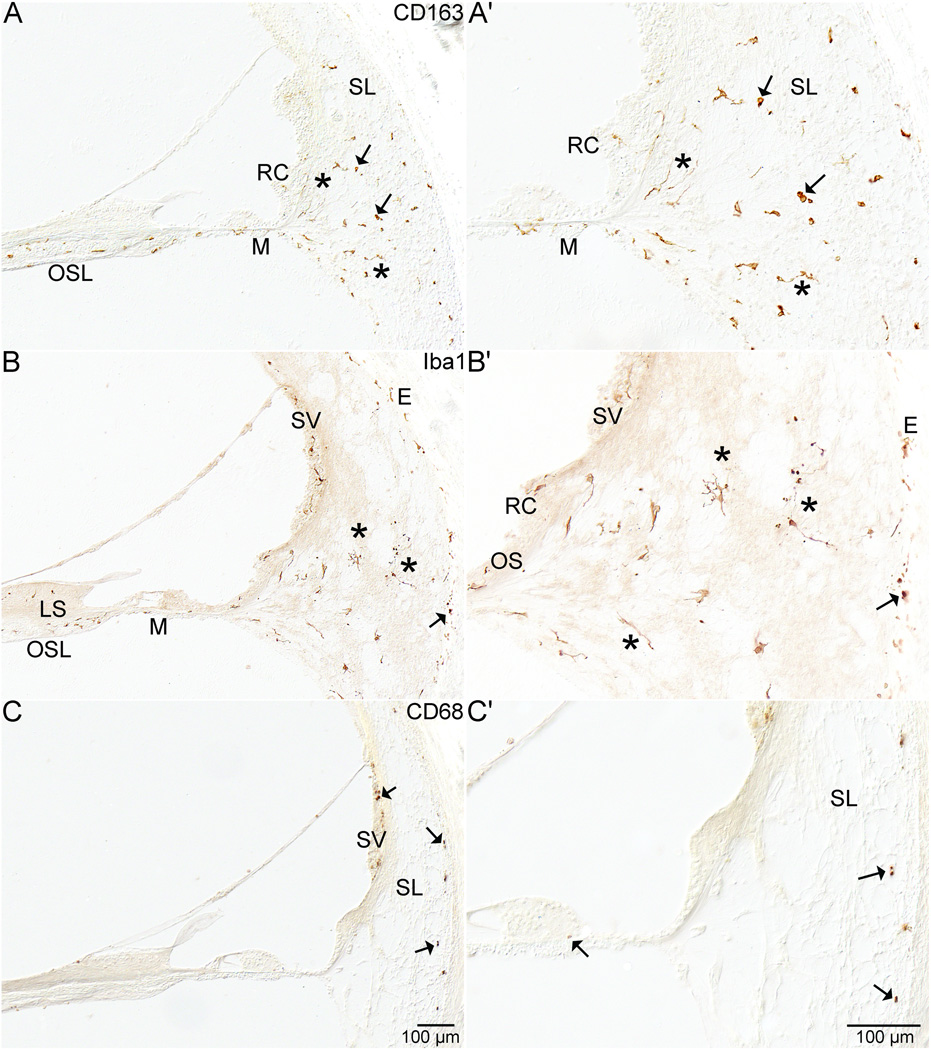
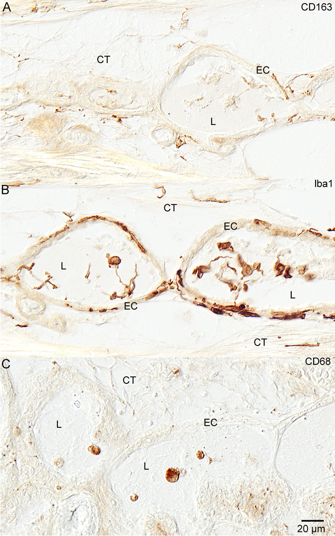
Methods: Primary antibodies against CD163, Iba1, and CD68 (markers known to be specific for macrophages/microglia) were used to immunohistochemically stain celloidin embedded archival temporal bone tissue of normal individuals with no known otologic disorders other than changes associated with age.

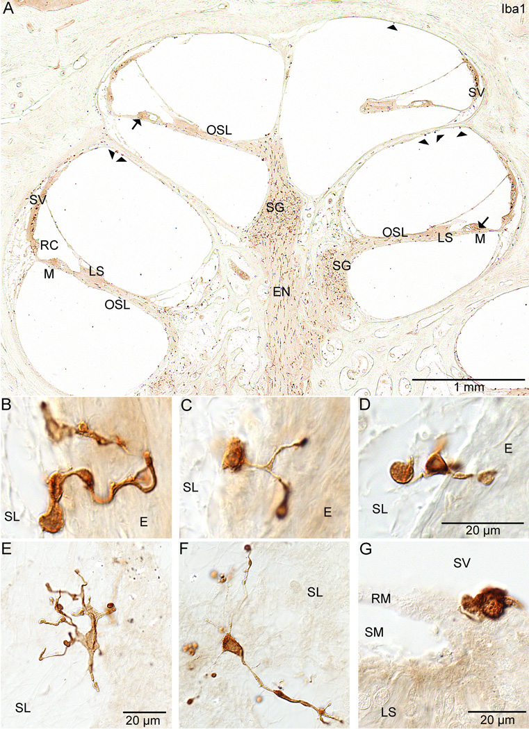
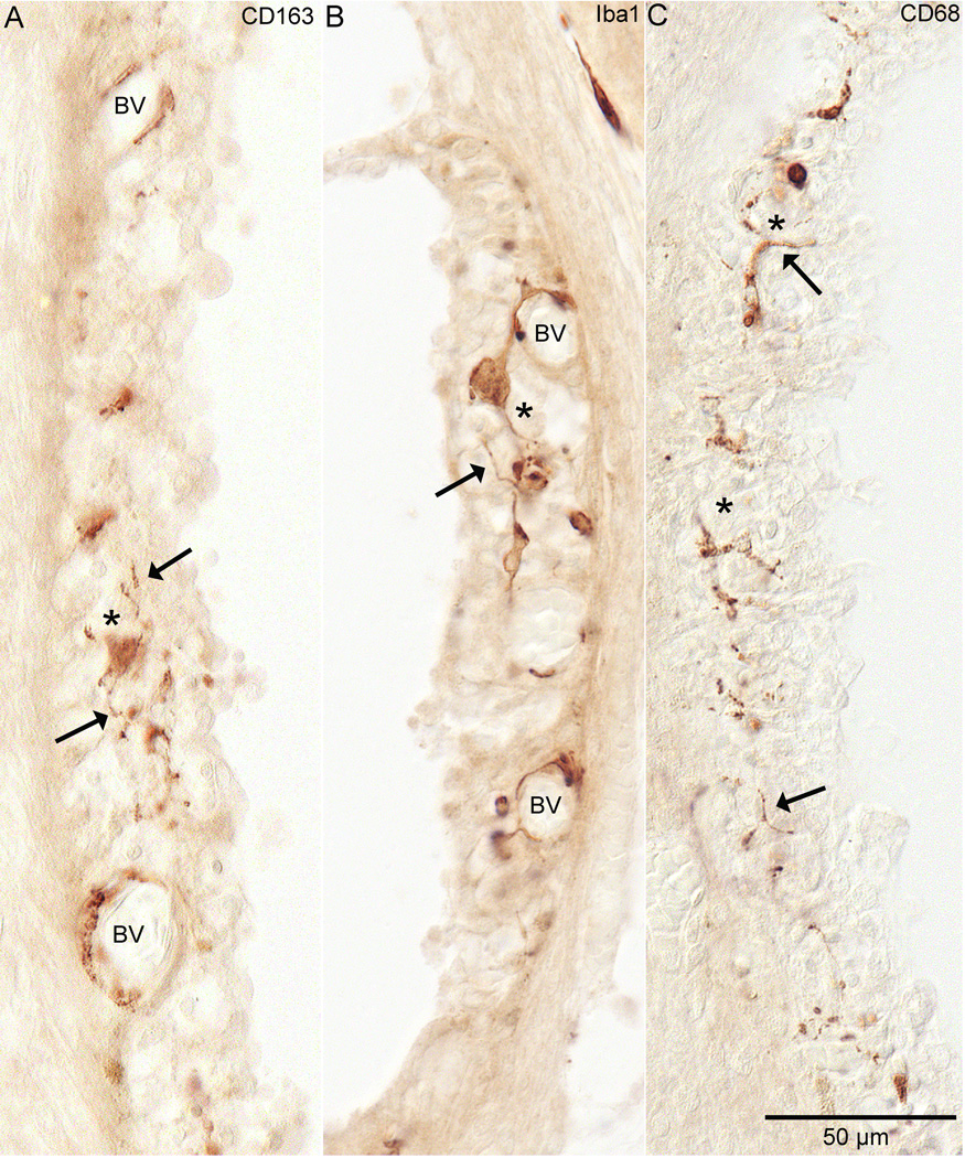
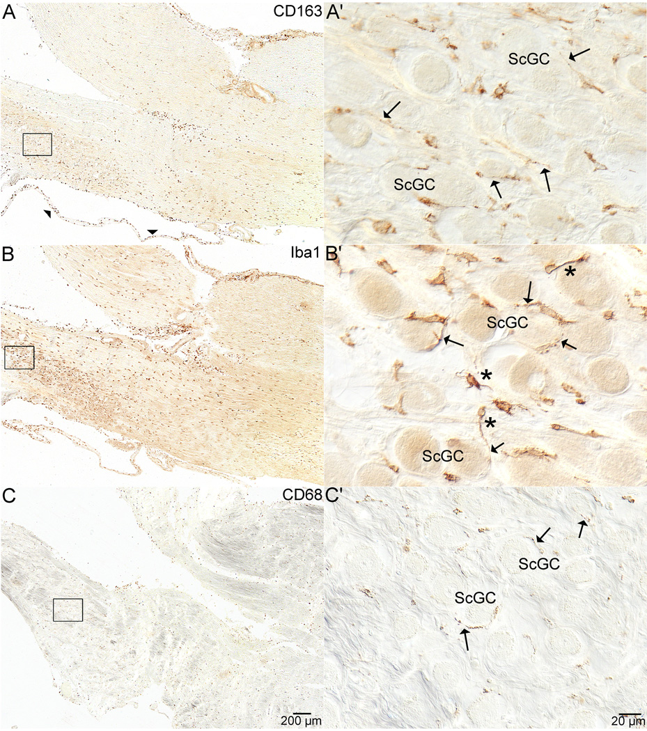
Results: Cells were positively stained throughout the temporal bone within the connective tissue and supporting cells with all three markers. They were often associated with neurons and on occasion entered the sensory cell areas of the auditory and vestibular epithelium.

Conclusions: We have immunohistochemically identified an unappreciated class of cells in the normal adult inner ear consistent in staining characteristics and morphology with macrophages/microglia. As in other organ systems, it is likely these cells play an essential role in organ homeostasis that has not yet been elucidated within the ear.


Conflict of interest statement

There are no declared conflicts of interest.

Figures

Figure 1
Figure 1. Cochlea
Figure 1 demonstrates staining in the cochlea with all three antibodies. Figure 1A and 1A' show the anti-CD163 in the lower basal turn of the cochlea in specimen # 1. Both ramified (*) and amoeboid cells (arrow) are demonstrated. Positive staining cells are evident in the osseous spiral lamina (OSL), within the mesenchymal cell layer (M) below the basilar membrane, in the root cells (RC), and throughout the spiral ligament (SL). Figure 1B and 1B' show anti-Iba1 positive staining in the lower basal turn of specimen # 6. Both ramified (*) and amoeboid cells (arrow) are stained. Staining is evident in the osseous spiral lamina (OSL), within the limbus spiralis (LS), in the mesenchymal cell layer (M) below the basilar membrane, in the outer sulcus (OS) and root cell area (RC), within the stria vascularis (SV), and entering the endochondral (E) layer of bone within the otic capsule. Figure 1C and C' show the anti-CD68 staining from the upper basal turn of specimen #7. There was less staining in the spiral ligament with this antibody than with the other two antibodies. Mostly amoeboid staining (arrows) was evident in the spiral ligament (SL) and in the stria vascularis (SV).
Figure 2
Figure 2. Cochlear staining with anti-Iba1
Figure 2 shows staining in the cochlea with the antibody against Iba1 only. Figure 2A shows a midmodiolar section through the cochlea of specimen #6. The abundance of staining within the cochlea can be appreciated. Staining can be seen along the eighth nerve (EN) within the modiolus, in Rosenthal's canal within the spiral ganglion (SG), and along the osseous spiral lamina (OSL). Staining is evident in the limbus spiralis (LS) and within the mesenchymal cell layer below the basilar membrane (M). Staining was also present below the cells of Hensen (arrows) in the organ of Corti. Positive staining cells were seen inserting into the outer sulcus and root cell areas (RC). The stria vascularis (SV) also demonstrated positive staining cells within the intermediate cell layer. Positive stained cells (arrowheads) are present in the scala vestibuli along the bone. Figures 2B, 2C (specimen #2), and 2D (specimen #6) show ramified cells in specimens from the spiral ligament area. These cells are partly embedded in the spiral ligament (SL) and partly in the endochondral (E) bone of the otic capsule. Figures 2E and 2F show stained macrophages/microglia from the spiral ligament (SL) of specimen #6 (also visible in figures 1B and 1B'). These cells demonstrate many ramified processes. Figure 2G is also from specimen #6 and demonstrates the amoeboid form of an Iba1 positive staining cell on Reissner's membrane (RM) at its junction with the limbus spiralis (LS). The scala media (SM) and scala vestibuli (SV) are also seen.
Figure 3
Figure 3. Stria Vascularis
Staining by all three antibodies was seen in the stria vascularis. In figure 3A, anti-CD163 staining in specimen #6 from the hook region of the cochlea is demonstrated. Note the staining around the blood vessels (BV) and in the middle of the stria (*) in the layer that would contain the intermediate cells. These cells have ramified cell processes (arrow). Figure 3B shows anti-Iba1 staining from the upper middle turn of specimen #2. Very similar staining to CD163 is evident with this antibody. Note the staining around blood vessels (BV) and the staining within the middle of the stria in the intermediate cell layer (*). These cells, like the CD163+ cells, also demonstrate ramified processes (arrow). Figure 3C shows anti-CD68 staining in specimens #2 from the hook region of the cochlea. Note that it is also within the intermediate cell layer. Also note that most of it is occurring in what are cell processes (arrow).
Figure 4
Figure 4. Scarpa’s Ganglion Cells
Figure 4 shows staining with all three antibodies in the internal auditory canal where the vestibular and auditory portions of the eighth nerve separate from one another near Scarpa’s ganglion. In figure 4A, staining of anti-CD163 from specimen #3 can be seen throughout the eighth nerve and in the peri-neural membrane that surrounds the nerve (arrowheads). Figure 4A' shows higher magnification of boxed area in 4A. The staining is spread throughout the ganglion and occurs in ramified processes (arrows) around Scarpa’s ganglion cells (ScGC). Figure 4B, also from specimen #3, demonstrates anti-Iba1 staining in the same area as in 4A. 4B' shows higher magnification of the anti-Iba1 staining and ramified cells are visible (*). Additionally, stained cell processes of the macrophages\microglia (arrows) are seen encircling the ganglion cells (ScGC). Figure 4C shows staining with the anti-CD68 antibody from specimen #2. The staining in the nerve and around Scarpa’s ganglion was not as prominent as with the other two antibodies, but was still present throughout the eighth nerve (4C). Stained cell processes of the macrophages\microglia (arrows) are seen encircling the ganglion cells (ScGC in 4 C').
Figure 5
Figure 5. Endolymphatic Duct
Figure 5 shows staining with all three antibodies in the distal endolymphatic duct. Figure 5A shows anti-CD163 staining in the endolymphatic duct of specimen #6. Positive staining was seen in the connective tissue (CT) around the duct as well as in the epithelial cells (EC) of the duct and in the duct lumen (L) as well. There were both round and ramified cells that stained. Figure 4B shows anti-Iba1 staining in the endolymphatic duct of specimen #6. There is plentiful staining in all of the same locations as with the anti-CD163. Staining was seen in the connective tissue (CT), epithelial cells (EC), and within the lumen (L). Figure 5C demonstrates anti-CD68 staining in the endolymphatic duct of specimen #2. The most prominent staining here was in round cells within the lumen (L) of the duct.

Similar articles

Cited by

References

    1. Lim DJ. Functional morphology of the mucosa of the middle ear and eustachian tube. Ann Otol Rhinol Laryngol. 1976;85(2 Suppl 25 Pt 2):36–43. - PubMed
    1. Masuda M, Yamazaki K, Kanzaki J, Hosoda Y. Immunohistochemical and ultrastructural investigation of the human vestibular dark cell area: roles of subepithelial capillaries and T lymphocyte-melanophage interaction in an immune surveillance system. Anat Rec. 1997;249(2):153–162. - PubMed
    1. Altermatt HJ, Gebbers JO, Muller C, Arnold W, Laissue JA. Human endolymphatic sac: evidence for a role in inner ear immune defence. ORL J Otorhinolaryngol Relat Spec. 1990;52(3):143–148. - PubMed
    1. Rask-Andersen H, Danckwardt-Lillieström N, Friberg U, House W. Lymphocyte-macrophage activity in the human endolymphatic sac. Acta Otolaryngol Suppl. 1991;485:15–17. - PubMed
    1. Jansson B, Rask-Andersen H. Osmotically induced macrophage activity in the endolymphatic sac. On the possible interaction between periaqueductal bone marrow cells and the endolymphatic sac. ORL J Otorhinolaryngol Relat Spec. 1992;54(4):191–197. - PubMed

Publication types

MeSH terms