. 2015 Dec;12(6):377-86.
doi: 10.1089/zeb.2015.1086. Epub 2015 Oct 20.

Intersectional Gene Expression in Zebrafish Using the Split KalTA4 System

Affiliations

Intersectional Gene Expression in Zebrafish Using the Split KalTA4 System

Rafael Gois Almeida et al. Zebrafish. 2015 Dec.

Abstract

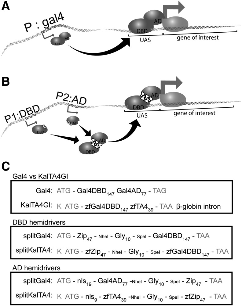
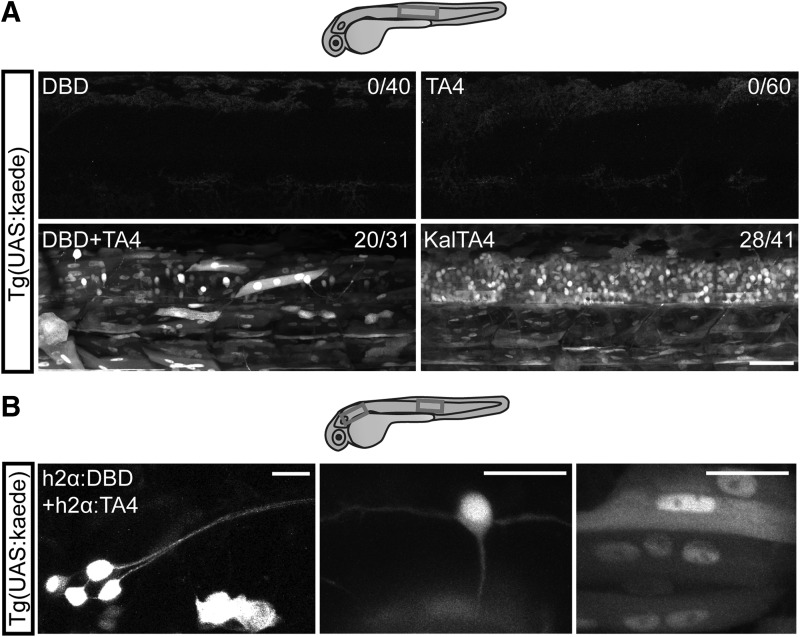
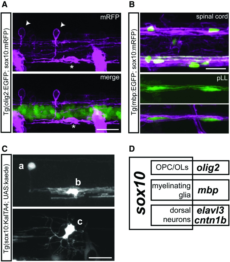
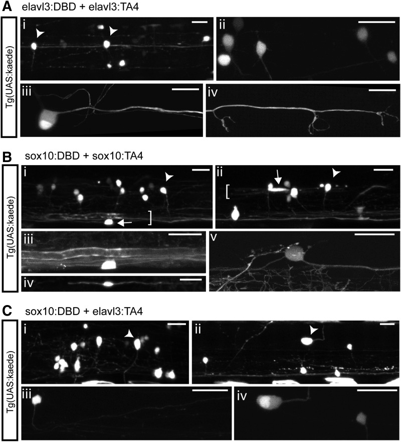
In this study, we describe the adaptation of the split Gal4 system for zebrafish. The Gal4-UAS system is widely used for expression of genes-of-interest by crossing driver lines expressing the transcription factor Gal4 (under the control of the promoter of interest) with reporter lines where upstream activating sequence (UAS) repeats (recognized by Gal4) drive expression of the genes-of-interest. In the Split Gal4 system, hemi-drivers separately encode the DNA-binding domain (DBD) and the activation domain (AD) of Gal4. When encoded under two different promoters, only those cells in the intersection of the promoters' expression pattern and in which both promoters are active reconstitute a functional Gal4 and activate expression from a UAS-driven transgene. We split the zebrafish-optimized version of Gal4, KalTA4, and generated a hemi-driver encoding the KalTA4 DBD and a hemi-driver encoding KalTA4's AD. We show that split KalTA4 domains can assemble in vivo and transactivate a UAS reporter transgene and that each hemi-driver alone cannot transactivate the reporter. Also, transactivation can happen in several cell types, with similar efficiency to intact KalTA4. Finally, in transient mosaic expression assays, we show that when hemi-drivers are preceded by two distinct promoters, they restrict the expression of an UAS-driven reporter from a broader pattern (sox10) to its constituent smaller neuronal pattern. The Split KalTA4 system should be useful for expression of genes-of-interest in an intersectional manner, allowing for more refined manipulations of cell populations in zebrafish.


Figures

<b>FIG. 1.</b>
FIG. 1.
Overview of Gal4, Split Gal4, KalTA4, and Split KalTA4 systems. (A) Gal4/UAS system: Gal4 driver lines contain a transgene encoding Gal4 (DBD and AD in a single open-reading frame) under a specific promoter (P). In a cell where P is active, Gal4 protein is expressed and can bind UAS in effector lines, activating transcription of the downstream gene-of-interest (gray arrow). (B) Split Gal4 system: Gal4 DBD hemi-driver is encoded under one promoter (P1) and Gal4 AD hemi-driver in a separate open-reading frame under a different promoter (P2). In a cell where both promoters are active, the two domains assemble and reconstitute Gal4 function, leading to expression of genes-of-interest only in the intersection of two expression domains. (C) Structure of Gal4, KalTA4, split Gal4, and split KalTA4 coding sequences. Compared to Gal4, KalTA4 includes a stronger Kozak sequence (K), zebrafish-optimized codon usage (zf), the core repeats of the VP16 AD (TA4), and a β-globin intron; these features were used to generate the split KalTA4 hemi-drivers. ATG: start codon, TAG/TAA: stop codon, Gly: decaglycine linker, Zip: heterodimerizing leucine zippers, nls: nuclear localization signal. Numbers indicate length of feature in amino acid residues. NheI and SpeI restriction sites from the original split Gal4 constructs were maintained in split KalTA4. AD, activation domain; DBD, DNA-binding domain; UAS, upstream activating sequence.
<b>FIG. 2.</b>
FIG. 2.
Split KalTA4 hemi-drivers reconstitute functional KalTA4 in several cell types. (A) Lateral view of the trunk region of 2 dpf Tg(UAS:kaede) larvae injected with split KalTA4 hemi-driver mRNA or control KalTA4 mRNA. Either DBD or TA4 mRNA alone does not activate kaede expression. DBD and TA4 mRNA in combination, or intact KalTA4 mRNA, is sufficient to activate the reporter UAS:kaede. Since heterozygous parent UAS:kaede fish were outcrossed, the subset of nonfluorescent embryos are likely nontransgenic offspring. Numbers on top right indicate number of kaede+ embryos/number of total embryos. Boxes indicate approximate imaged regions. Scale bar: 50 μm. (B) Lateral view of the pLL ganglion (left) or trunk region (middle and right) of 2 dpf Tg(UAS:kaede) larvae injected with h2α:DBD and h2α:TA4. h2α-driven split KalTA4 expression results in activation of the reporter UAS:kaede in a variety of cell types, including peripheral neurons in the pLL ganglion (left), central neurons in the spinal cord such as a commissural primary ascending interneuron (middle) and muscle cells (right). Boxes indicate areas of imaged cells. Scale bars: 20 μm. pLL, posterior lateral line.
<b>FIG. 3.</b>
FIG. 3.
Reporter transgenes indicate expression of sox10 in several cell types. (A) Lateral view of the spinal cord of 2 dpf Tg(olig2:EGFP; sox10:mRFP) larva. Arrowheads: examples of sox10:mRFP+ dorsal neurons; asterisk: example of olig2:GFP+ sox10:mRFP+ OPC. (B) Lateral view of the spinal cord (top) and pLL (bottom) of 4 dpf Tg(mbp:EGFP; sox10:mRFP) larva. Many OPCs become mbp:EGFP+ mature OLs by 4 dpf in the spinal cord, and are associated with sox10:mRFP myelin sheaths, as are mbp:EGFP+ Schwann cells in the pLL. (C) Lateral view of the spinal cord of 3 dpf Tg(sox10:KalTA4; UAS:kaede) larva. Cell a: dorsal neuron; cell b: OL; cell c: OPC. (D) Venn Diagram of the expression patterns of sox10, olig2, mbp, elavl3, and cntn1b, indicating some of the cell types found in the intersection of each subdomain. All scale bars: 20 μm. OL, oligodendrocyte; OPC, oligodendrocyte precursor cell. Color images available online at www.liebertpub.com/zeb
<b>FIG. 4.</b>
FIG. 4.
Split KalTA4 system restricts sox10 expression pattern to each component cell type. (A) Coinjection of elavl3:DBD and elavl3:TA4 activate reporter expression in neurons. (i) Overview of two spinal cord segments showing kaede+ neurons (arrowheads indicate examples of neuron somas). (ii) High-resolution view of spinal interneurons. (iii–iv) A pLL neuron and its axon innervating neuromasts. (B) Coinjection of sox10:DBD and sox10:TA4 activate reporter expression in neurons and glia. (i, ii) Spinal cord overviews showing kaede+ neurons (arrowheads) and OLs (arrows) forming myelin sheaths (brackets). (iii) High-resolution view of ventral OL in (i). (iv) A Schwann cell in the pLL. (v) High-resolution view of a Rohon-Beard cell in the dorsal spinal cord. (C) Coinjection of sox10:DBD and elavl3:TA4 activate reporter expression only in neurons. (i, ii) Spinal cord overviews showing kaede+ neurons (arrowheads). (iii) High-resolution view of a circumferential descending interneuron. (iv) High-resolution view of Rohon-Beard neuron and of a dorsal interneuron in (ii). All panels are lateral views at 4 dpf; all scale bars: 20 μm.

Similar articles

Cited by

References

    1. Zhang J, et al. . Creating new fluorescent probes for cell biology. Nat Rev Mol Cell Biol 2002;3:906–918 - PubMed
    1. Fenno L, Yizhar O, Deisseroth K. The development and application of optogenetics. Annu Rev Neurosci 2011;34:389–412 - PMC - PubMed
    1. Elliott DA, Brand AH. The GAL4 system: a versatile system for the expression of genes. Methods Mol Biol 2008;420:79–95 - PubMed
    1. Kettleborough RN, et al. . A systematic genome-wide analysis of zebrafish protein-coding gene function. Nature 2013;496:494–497 - PMC - PubMed
    1. Stuart GW, McMurray JV, Westerfield M. Replication, integration and stable germ-line transmission of foreign sequences injected into early zebrafish embryos. Development 1988;103:403–412 - PubMed

Publication types

MeSH terms

LinkOut - more resources