. 2015 Oct 20;10(10):e0140645.
doi: 10.1371/journal.pone.0140645. eCollection 2015.

Singlet Oxygen-Mediated Oxidation during UVA Radiation Alters the Dynamic of Genomic DNA Replication

Affiliations

Singlet Oxygen-Mediated Oxidation during UVA Radiation Alters the Dynamic of Genomic DNA Replication

Dany Graindorge et al. PLoS One. .

Abstract

UVA radiation (320-400 nm) is a major environmental agent that can exert its deleterious action on living organisms through absorption of the UVA photons by endogenous or exogenous photosensitizers. This leads to the production of reactive oxygen species (ROS), such as singlet oxygen (1O2) and hydrogen peroxide (H2O2), which in turn can modify reversibly or irreversibly biomolecules, such as lipids, proteins and nucleic acids. We have previously reported that UVA-induced ROS strongly inhibit DNA replication in a dose-dependent manner, but independently of the cell cycle checkpoints activation. Here, we report that the production of 1O2 by UVA radiation leads to a transient inhibition of replication fork velocity, a transient decrease in the dNTP pool, a quickly reversible GSH-dependent oxidation of the RRM1 subunit of ribonucleotide reductase and sustained inhibition of origin firing. The time of recovery post irradiation for each of these events can last from few minutes (reduction of oxidized RRM1) to several hours (replication fork velocity and origin firing). The quenching of 1O2 by sodium azide prevents the delay of DNA replication, the decrease in the dNTP pool and the oxidation of RRM1, while inhibition of Chk1 does not prevent the inhibition of origin firing. Although the molecular mechanism remains elusive, our data demonstrate that the dynamic of replication is altered by UVA photosensitization of vitamins via the production of singlet oxygen.


Conflict of interest statement

Competing Interests: The authors have declared that no competing interests exist.

Figures

Fig 1
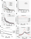
Fig 1. Singlet oxygen is generated by photosensitization of vitamins.
Absorption spectra between 300 and 500 nm of various dilutions in H2O of (A) vitamins (Vit) and (B) amino acids (AA). (C) Comparison between the absorption spectrum of a mixture of vitamins 1x and amino acids 1x diluted in H2O (Mix Vit1x + AA1x) and the absorption spectrum of MEMi. The stock solutions of vitamins and amino acids were 100x and 50x, respectively. Each spectrum is the average ± SD of 3 to 4 independent spectra. Solutions of AA (D) and Vit (E) were diluted in D2O and excited at 370 ± 7 nm. The 1O2 luminescence of excited solutions was recorded as described in Material and Methods. (F) The 1O2 luminescence of each dilution of Vit was normalized with the appropriate absorbance at 370 nm, and is compared to the 1O2 luminescence of a solution of Rose Bengal (RB).
Fig 2
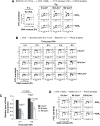
Fig 2. UVA radiation induces S-phase slowdown that is prevented by NaN3, a quencher of singlet oxygen.
(A) Transformed human fibroblasts (MRC5Vi) were pulse-labeled with 10 μM BrdU for 30 min (T½h) and exposed to UVA radiation in MEMi containing or not 10 mM NaN3 or 10 mM NAC. Thereafter, the cells were incubated at 37°C for 7.5 h and fixed in cold 70% EtOH (T8h). (B) The cells were exposed to UVA radiation in MEMi, incubated for different periods of time at 37°C, pulse-labeled with BrdU for 30 min, and fixed in cold 70% EtOH. (C) Histograms of the relative level of BrdU incorporation (FITC anti-BrdU axis) at T½h and T8h in non irradiated and irradiated cells displayed in panel B. The values represent the mean +/- SD of 3 to 5 independent experiments. Statistical significances were determined by Student t-test. (D) The cells were exposed to UVA radiation in MEMi containing or not 10 mM NaN3, immediately pulse-labeled with BrdU for 30 min, and fixed in cold 70% EtOH. The cells were analysed by bi-variable flow cytometry for BrdU incorporation (FITC anti-BrdU) and DNA content (propidium iodide, PI). The arrows highlight the delay of DNA replication (panels in A) and the dashed lines position the maximum level of BrdU incorporation in untreated cells (panels in B and D). It is to note that this level is lower in UVA-irradiated cells thus highlighting a defect of BrdU incorporation.
Fig 3
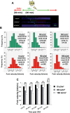
Fig 3. UVA-induced ROS impinge on the replication forks velocity.
(A) Scheme of the experimental protocol. MRC5Vi cells were exposed or not to UVA and then pulse-labeled for 30 min of IdU (green signal) followed by 30 min of CldU (red signal). DNA was extracted immediately after the 2nd pulse. Single stranded DNA (ssDNA) and incorporation of the thymidine analogs were detected as described in the Material and Methods. The picture is an example of the tracks length of IdU (green track), CldU (red track) and ssDNA (blue track). (B) Asynchronous MRC5Vi cells were untreated or exposed to 80 and 160 kJ/m2 of UVA radiation. IdU and CldU labeling was performed sequentially immediately after radiation. sd: standard deviation; med.: mediane; n: number of replication forks analysed. (C) The experiments were conducted as described in (B) with the exception that IdU (green) and CldU (red) labelings were performed at various time points (i.e. 0h, ½h, 1h, 2h and 4h) after UVA radiation. To determine the impact of UVA radiation on fork velocity, only the length of CldU tracks was scored. The values correspond to the mean of the forks velocity and are representatives of two experiments. A total of 500 to 3000 forks were analysed for these experiments. **P<0.001; *P<0.05 (two-tailed test).
Fig 4
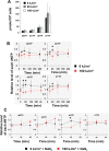
Fig 4. The transient drop of dNTP pool after photosensitization by UVA is prevented by NaN3.
MRC5Vi cells in mid S-phase (condition S4R, see Material and Methods) were untreated or exposed to 80 and 160 kJ/m2 of UVA radiation in MEMi. The intracellular pool of dNTPs was measured at various time points post UVA radiation. (A) Quantification of dGTP, dCTP, dATP and dTTP in non irradiated cells and immediately after UVA radiation. The values represent the mean +/- sd of 5 independent experiments. (B) The relative level of each dNTP was measured at the indicated time points post UVA. The values represent the mean +/- sd of 5 independent experiments and were normalized to the value at T0–0 kJ/m2. (C) The cells were exposed to 160 kJ/m2 of UVA radiation in MEMi containing 10 mM NaN3. The relative level of each dNTP was measured at the indicated time points. The values represent the mean +/- sd of 3 independent experiments. *P<0.05 (t-test)
Fig 5
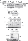
Fig 5. Reversible oxidation of the RRM1 subunit of RNR in response to UVA-induced ROS.
MRC5Vi cells were untreated or exposed to UVA radiation in the absence (A) or presence (B) of 10 mM NaN3. (C) MRC5Vi cells were transiently transfected with siCtr, siTrx1, or siGrx1 and 48 h post transfection exposed or not to 80 kJ/m2 of UVA. (D) Cells depleted (+ BSO) or not (- BSO) in intracellular GSH were exposed to 80 kJ/m2 of UVA and lysed either immediately or 5 and 10 min post UVA radiation. The expressions of RRM1, RRM2, p53R2, Trx1, Grx1, and proteins S-glutathionylated (prot-SSG) were detected by Western blotting in non reducing (- ß-mercaptoethanol, - BME) or reducing (+ ß-mercaptoethanol, +BME) conditions. Actin was used as a loading control. RRM1ox and RRM1red stand for the oxidized and reduced form of the RRM1 subunit of the RNR, respectively. Each blot is representative of 2 independent experiments. The vertical lines in panels A and C denote non-adjacent bands from the same blot. Numbers in brackets (panel A) indicate the apparent molecular weight of each subunit.
Fig 6
Fig 6. The firing of origins is reduced in response to UVA-induced ROS.
Asynchronous or S-phase synchronised (condition S4R, see Material and Methods) cells were exposed to 80 and 160 kJ/m2 of UVA in MEMi. IdU and CldU were then sequentially added to the medium 4 h (asynchronous cells) or 2 h (S-phase cells) after UVA radiation. The forks density is defined as the ratio between the number of CldU-IdU-CldU signals (Red-Green-Red signals) and the length of replicated DNA. The percentage of cells in S-phase at the labeling time was determined by flow cytometry and was found to be 47% for the asynchronous population and 80% for S-phase synchronised cells. The data represent the mean ± sd of 4 independent experiments for A and 3 independent experiments for B (excepted for the dose of 80 kJ/m2 of UVA for which only 2 data were available).
Fig 7
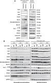
Fig 7. No relocalization or post-translational modifications of some components of the replication machinery after UVA radiation.
Asynchronous MRC5Vi cells or synchronized in early S-phase (condition S0R, see Material and Methods) are untreated or exposed to 80 and 160 kJ/m2 of UVA radiation. (A) Subcellular localization of Cdc6, Cdc7, Dbf4, Cdc45, Mcm2, Mcm10, Orc2, PCNA, RPA32 in asynchronous cells (Asyncro.) or cells synchronized in early S-phase (Synchro.). (B) The fractions of chromatin-bound proteins and total soluble proteins were recovered at various time points post UVA. The expression levels of Cdc6, Cdc7, Dbf4, Cdc45, Mcm2, p-Mcm2(Ser40/41), Mcm10, Orc2, PCNA, and RPA32 were detected by Western blotting in denaturing and reducing conditions. Lamin A/C and GAPDH were used as loading control for the chromatin-bound and soluble fractions, respectively. The apparent molecular weight of each protein is indicated on the right side of each blot (panel 7A). The stars (*) indicate the position of the non specific bands detected by Orc2 antibody (H-300, SCBT).

Similar articles

Cited by

References

    1. Madronich S, McKenzie RL, Bjorn LO, Caldwell MM (1998) Changes in biologically active ultraviolet radiation reaching the Earth's surface. J Photochem Photobiol B 46: 5–19. - PubMed
    1. Baier J, Maisch T, Maier M, Engel E, Landthaler M, Baumler W (2006) Singlet oxygen generation by UVA light exposure of endogenous photosensitizers. Biophys J 91: 1452–1459. - PMC - PubMed
    1. Wondrak GT, Jacobson MK, Jacobson EL (2006) Endogenous UVA-photosensitizers: mediators of skin photodamage and novel targets for skin photoprotection. Photochem Photobiol Sci 5: 215–237. - PubMed
    1. Baumler W, Regensburger J, Knak A, Felgentrager A, Maisch T (2012) UVA and endogenous photosensitizers—the detection of singlet oxygen by its luminescence. Photochem Photobiol Sci 11: 107–117. 10.1039/c1pp05142c - DOI - PubMed
    1. Bracchitta G, Catalfo A, Martineau S, Sage E, De Guidi G, Girard PM (2013) Investigation of the phototoxicity and cytotoxicity of naproxen, a non-steroidal anti-inflammatory drug, in human fibroblasts. Photochem Photobiol Sci 12: 911–922. 10.1039/c3pp25326k - DOI - PubMed

Publication types