. 2015 Dec 1;6(38):40734-46.
doi: 10.18632/oncotarget.5754.

Molecular mechanisms of cisplatin cytotoxicity in acute promyelocytic leukemia cells

Affiliations

Molecular mechanisms of cisplatin cytotoxicity in acute promyelocytic leukemia cells

Sanjay Kumar et al. Oncotarget. .

Abstract

Cis-diamminedichloroplatinum (II) (cisplatin) is a widely used anti-tumor drug for the treatment of a broad range of human malignancies with successful therapeutic outcomes for head and neck, ovarian, and testicular cancers. It has been found to inhibit cell cycle progression and to induce oxidative stress and apoptosis in acute promyelocytic leukemia (APL) cells. However, its molecular mechanisms of cytotoxic action are poorly understood. We hypothesized that cisplatin induces cytotoxicity through DNA adduct formation, oxidative stress, transcriptional factors (p53 and AP-1), cell cycle regulation, stress signaling and apoptosis in APL cells. We used the APL cell line as a model, and applied a variety of molecular tools to elucidate the cytotoxic mode of action of cisplatin. We found that cisplatin inhibited cell proliferation by a cytotoxicity, characterized by DNA damage and modulation of oxidative stress. Cisplatin also activated p53 and phosphorylated activator protein (AP-1) component, c-Jun at serine (63, 73) residue simultaneously leading to cell cycle arrest through stimulation of p21 and down regulation of cyclins and cyclin dependent kinases in APL cell lines. It strongly activated the intrinsic pathway of apoptosis through alteration of the mitochondrial membrane potential, release of cytochrome C, and up-regulation of caspase 3 activity. It also down regulated the p38MAPK pathway. Overall, this study highlights the molecular mechanisms that underline cisplatin toxicity to APL cells, and provides insights into selection of novel targets and/or design of therapeutic agents to treat APL.

Keywords: AP-1 and p53; APL cell line; cell cycle modulation; cisplatin.


Conflict of interest statement

CONFLICTS OF INTEREST

Authors declare no conflict of interest.

Figures
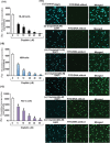
Figure 1
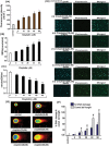
Figure 1. Cisplatin inhibits growth and induced formation of DNA-adduct in APL cells
APL cells (HL-60, NB4 and KG-1a) were exposed to various concentrations (0, 5, 10, 20, 40, and 80 μM) of cisplatin for 24 hours and further incubated for 24 hours with tritium labeled thymidine. After incubation, cells were harvested by centrifugation and counted using liquid scintillation analyzer as described in the material and method section. 3H-methyl thymidine incorporation was expressed as cpm/dish. Data represent the means of three independent experiments ± SDs. Highly statistically significant decreases (p < 0.01) in cell proliferation were observed in all cisplatin treated APL cells including HL-60 [1A], NB4 [1B] and Kg-1a [1C] cells. This reduction in cell growth was concentration-dependent. Cisplatin –induced formation of DNA adduct was assessed by immunocytochemistry and confocal microscopy analysis as described in the material and methods section. APL cells were exposed to various concentrations of cisplatin for 48 hours and performed immunocytochemistry as well as confocal microscopy using FITC filter to confirm DNA adduct formation. The results showed that cisplatin caused a significant concentration -dependent increase in DNA-adduct formation in APL cells [1D (i-vi)].
Figure 2
Figure 2. Cisplatin induces cytotoxicity in APL cells
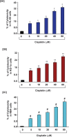
APL cells were exposed to various concentrations (0, 5, 10, 20, 40 & 80 μM) of cisplatin for 48 hours and LDH released in medium was measured using Promega non-radioactive cytotoxicity assay technical bulletin protocol. Then % cytotoxicity was calculated by dividing the levels of released LDH in treated cells over the total LDH released from control cells. Highly statistically significant increases (p < 0.01) in cytotoxicity were observed in all cisplatin treated APL cells including HL-60 [2A], KG-1a [2B] and NB4 [2C] cells in a concentration - dependent fashion.
Figure 3
Figure 3. Cisplatin induces oxidative stress and clastogenic effect in APL cells
Cisplatin induced oxidative stress and clastogenic effect in APL cells through generation of reactive oxygen species (ROS) and formation of DNA adduct. APL cells were exposed to various concentrations (0, 5, 10, 20, 40, and 80 μM) of cisplatin for 48 hours and further treated 30 min with dichlorofluorescein diacetate (DCFDA). After incubation, ROS released was measured through measuring DCF fluorescence intensity by spectrofluorementry. Data represent the means of three independent experiments ± SDs (*P < 0.01) [3A]. APL cells were exposed to various concentrations of cisplatin and lipid peroxidation one product, malondialdehyde (MDA) formation measured by spectrophotometry. Data represent the means of three independent experiments ± SDs (**P < 0.01) [3B]. APL cells were exposed to various concentrations of cisplatin and GSH level was measured by spectrophotometry. Data represent the means of three independent experiments ± SDs (#P < 0.05) [3C]. APL cells were exposed to various concentrations of cisplatin and DNA damage analyzed by TUNEL assay [3D (i-vi)]. APL cells were exposed to various concentrations of cisplatin and DNA damage was analyzed by alkaline gel electrophoresis (Comet) assay and characterized by comet tail length and % DNA damage. Data represent the means of three independent experiments ± SDs % DNA damage (*P < 0.01) and comet tail length ($, P < 0.01) [3E-3F].
Figure 4
Figure 4. Cisplatin modulates cell cycle regulation by transcriptional factors
Cisplatin induced cell cycle arrest at G1 checkpoint by stimulating transcription factors, p53 and phosphorylation of activator protein -1(AP-1) components in APL cells. APL cells were exposed to various concentrations (0, 5, 10, 20, 40 and 80 μM) of cisplatin for 48 hours and western blotting was performed for checking the expression level of cell cycle regulatory proteins, p53, p21, cdk6 & cyclin D3 [4A]. Transcriptional factor, AP-1 expression and their components, c-Jun phosphorylatin at ser 63 & Ser73 residue were checked by western blotting in both control and cisplatin treated APL cells [4B]. APL cells were exposed to various concentrations of cisplatin and the expression level of cell proliferation marker, Ki67 was assessed by immunocytochemistry and confocal imaging method. The results show a significant reduction in the expression level of Ki67 in cisplatin treated cells compared to control cells [4C (i-vi)].
Figure 5
Figure 5. Cisplatin induces intrinsic pathway of apoptosis
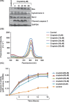
Cisplatin-induced cytotoxicity activated the intrinsic pathway of apoptosis in APL cells. APL cells were exposed to various concentrations (0, 5, 10, 20, 40 and 80 μM) of cisplatin for 48 hours and western blotting was performed to check the expression level of pro-apoptotic proteins (cytochrome C & Bax), caspase 3, and anti-apoptotic protein, Bcl-2 [5A]. APL cells were exposed to various concentrations of cisplatin and the mitochondria of all treated and control cells were isolated. Isolated mitochondria were treated with JC-1 dye for 10 min and mitochondrial membrane potential was assessed by spectrofluorometry [5B]. APL cells were exposed to various concentrations of cisplatin and caspase 3 activity was measured at different time points [5C].
Figure 6
Figure 6. Cisplatin induces apoptosis and modulates stress signaling
Cisplatin effect on JC-1 monomer expression and MAPK signaling pathway in APL cells was assessed. APL cells were incubated to various concentrations (0, 5, 10, 20, 40, and 80 μM) of cisplatin for 48 hours and mitochondria were isolated. Isolated mitochondria were treated with JC-1 dye 10 min, and immunocytochemistry and confocal imaging were performed to check the expression level of JC-1 monomer [6A (i-vi)]. APL cells were incubated to various concentrations of cisplatin and western blotting was performed to check the phosphorylation level of MAPK signaling molecules like JNK, p38 & Erk [6B].
Figure 7
Figure 7. Molecular mechanisms of Cisplatin-induced toxicity in APL cells
The overall molecular mechanisms of cisplatin cytotoxicity in APL cells involve many steps. In brief, cisplatin enters into the cell and interacts with DNA to form DNA adduct and also inhibits DNA synthesis. It produces ROS and creates oxidative stress inside the cell. DNA adduct formation and oxidative stress in APL cells cause the activation of p53 signaling and AP-1 phosphorylation leading to a change in mitochondrial membrane potential and cell cycle arrest in cells. This leads to cytochrome C release from the mitochondria, which activates caspase 9 and a series of complexes leading to activation of caspase 3 and apoptosis. Cisplatin cytotoxicity also modulates MAPK signaling pathway and overall leads to cancer cell death [7].

References

    1. Powell BL. Arsenic trioxide in acute promyelocytic leukemia: potion not poison. Expert Rev Anticancer Ther. 2011;11:1317–1319. - PubMed
    1. Kumar S, Yedjou CG, Tchounwou PB. Arsenic trioxide induces oxidative stress, DNA damage, and mitochondrial pathway of apoptosis in human leukemia (HL-60) cells. J Exp Clin Cancer Res. 2014;33:42. - PMC - PubMed
    1. Ghavamzadeh A, Alimoghaddam K, Ghaffari SH, Rostami S, Jahani M, Hosseini R, Mossavi A, Baybordi E, Khodabadeh A, Iravani M, Bahar B, Mortazavi Y, Totonchi M, Aghdami N. Treatment of acute promyelocytic leukemia with arsenic trioxide without ATRA and/or chemotherapy. Ann Oncol. 2006;17:131–134. - PubMed
    1. Ghavamzadeh A, Alimoghaddam K, Rostami S, Ghaffari SH, Jahani M, Iravani M, Mousavi SA, Bahar B, Jalili M. Phase II study of single-agent arsenic trioxide for the front-line therapy of acute promyelocytic leukemia. J Clin Oncol. 2011;29:2753–2757. - PubMed
    1. Tomita A, Kiyoi H, Naoe T. Mechanisms of action and resistance to all-trans retinoic acid (ATRA) and arsenic trioxide (As2O3) in acute promyelocytic leukemia. Int. J. Hematol. 2013;97:717–725. - PubMed

Publication types

MeSH terms