. 2015 Oct 19;18(1):20078.
doi: 10.7448/IAS.18.1.20078. eCollection 2015.

Tim-3 blocking rescue macrophage and T cell function against Mycobacterium tuberculosis infection in HIV+ patients

Affiliations

Tim-3 blocking rescue macrophage and T cell function against Mycobacterium tuberculosis infection in HIV+ patients

Isabel Sada-Ovalle et al. J Int AIDS Soc. .

Abstract

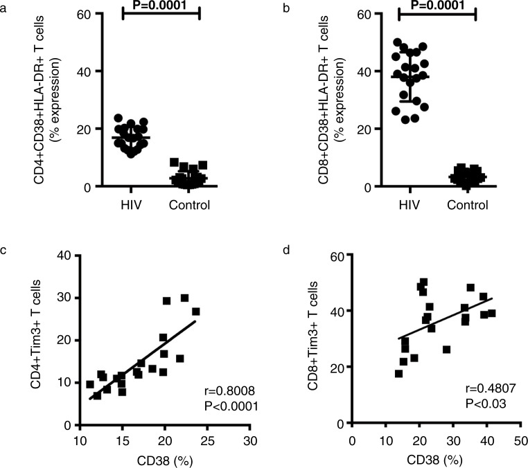
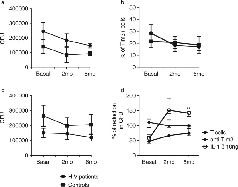
Introduction: T cell immunoglobulin and mucin domain (Tim) 3 and programmed death 1 (PD-1) are co-inhibitory receptors involved in the so-called T cell exhaustion, and in vivo blockade of these molecules restores T cell dysfunction. High expression of Tim-3 and PD-1 is induced after chronic antigen-specific stimulation of T cells during HIV infection. We have previously demonstrated that the interaction of Tim-3 with its ligand galectin-9 induces macrophage activation and killing of Mycobacterium tuberculosis. Our aim in this study was to analyze the Tim-3 expression profile before and after six months of antiretroviral therapy and the impact of Tim-3 and PD-1 blocking on immunity against M. tuberculosis.

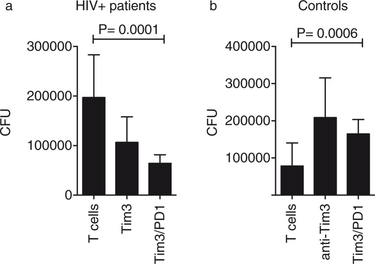
Materials and methods: HIV+ patients naïve to anti-retroviral therapy (ART) were followed up for six months. Peripheral immune-cell phenotype (CD38/HLA-DR/galectin-9/Tim-3 and PD-1) was assessed by flow cytometry. Supernatants were analyzed with a multiplex cytokine detection system (human Th1/Th2 cytokine Cytometric Bead Array) by flow cytometry. Control of bacterial growth was evaluated by using an in vitro experimental model in which virulent M. tuberculosis-infected macrophages were cultured with T cells in the presence or absence of Tim-3 and PD-1 blocking antibodies. Interleukin-1 beta treatment of infected macrophages was evaluated by enumerating colony-forming units.

Results: We showed that HIV+ patients had an increased expression of Tim-3 in T cells and were able to control bacterial growth before ART administration. By blocking Tim-3 and PD-1, macrophages and T cells recovered their functionality and had a higher ability to control bacterial growth; this result was partially dependent on the restitution of cytokine production.

Conclusions: In this study, we demonstrated that increased Tim-3 expression can limit the ability of the immune system to control the infection of intracellular bacteria such as M. tuberculosis. The use of ART and the in vitro manipulation of the Tim-3 and PD-1 molecules restored the functionality of T cells and macrophages to restrict bacterial growth. Our results provide a novel immune strategy that may be implemented in the near future in order to improve the immune responses in HIV+ patients.

Keywords: HIV; PD-1; Tim-3; galectin 9; tuberculosis.


Figures

Figure 1
Figure 1
Identification of peripheral blood mononuclear cells (PBMC) expressing Tim-3. PBMC from HIV+ patients and healthy controls were stained and analyzed for cell surface markers to further delineate Tim-3 and Gal9 expression. As shown in the top panels, lymphocytes were CD3 scatter-gated and then CD4+ and CD8+ cells were gated. The remaining panels show the Tim-3 expression on CD4+, CD8+, CD14+ and NK subsets. The figure demonstrates the FMO panels for CD4+ and CD8+ T cells. Representative plots are shown. Numbers indicate the percentage of positive cells in each gate. Data were collected on a FACS Aria II flow cytometer (10 colours and 2 scatter measurements). Data were analyzed using FlowJo software.
Figure 2
Figure 2
Expression of Tim-3 and Gal-9 on immune cell subsets. The frequency of Tim-3+ cells (a) and Gal-9 (b) in HIV+ patients and healthy controls was analyzed as indicated. Bars show median values and interquartile range. Statistical analyses were performed using the Mann-Whitney U test (n=20).
Figure 3
Figure 3
Frequency of CD4 + Tim-3 + PD-1+ and CD8 + Tim-3 +PD-1+ T cells (a and b). The frequency of double-positive cells in HIV+ patients and healthy controls were analyzed as indicated. Bars show median values and interquartile range. Statistical analyses were performed using the Mann-Whitney U test (n=16).
Figure 4
Figure 4
Tim-3 is up-regulated on CD4 and CD8 T cells in HIV+ patients, and its expression correlates with CD38 expression. (a, b) The percentages of CD38+ cells within CD8+and CD4+T cell populations are indicated for 20 HIV+ patients and healthy controls. Statistical analyses were performed using the Mann-Whitney U test. (c, d) Correlations between Tim-3 expression on CD8+ and CD4+ T cells and levels of CD38 expression among HIV+ patients and healthy controls are shown. Statistical analyses were performed using the Spearman's rank correlation test.
Figure 5
Figure 5
Tim-3–Gal9 interaction participates in the immune response against Mycobacterium tuberculosis (M.tb). (a) M.tb-infected monocyte-derived macrophages were cultured with T cells alone or in the presence of 10 mg/ml of anti-human Tim-3 or anti-human Gal9 mAb (b). Colony-forming units were recovered on Day 4 postinfection. Horizontal bars represent median values and interquartile range. Kruskal-Wallis test compared with T cells. n=20.
Figure 6
Figure 6
Blocking of Tim-3 and PD-1 inhibitory receptors restore macrophage and T cell control of bacterial growth (a). Mycobacterium tuberculosis-infected monocyte-derived macrophages were cultured with T cells alone or in the presence of 10 mg/ml of anti-human Tim-3 or anti-human PD-1 mAb (b). Colony-forming units were recovered on Day 4 postinfection. Horizontal bars represent median values and interquartile range. Kruskal-Wallis test compared with T cells. n=20.
Figure 7
Figure 7
Blocking of Tim-3 and PD-1 inhibitory receptors increases pro-inflamatory cytokine production in HIV+ patients. Pro-inflammatory and anti-inflammatory cytokines were measured in culture supernatants from HIV+ patients (n=20). Bars represent median values and interquartile range. Kruskal-Wallis test compared with T cells.
Figure 8
Figure 8
Effect of anti-retroviral therapy (ART) on the immune response against Mycobacterium tuberculosis (M.tb) during HIV infection. HIV+ patients (n=10) and healthy controls (n=10) were sampled at baseline; eight HIV+ patients and ten healthy controls at 2; and seven HIV+ patients and ten healthy controls at 6 months after initiation of ART. (a) M.tb-infected monocyte-derived macrophages (MDM) were cultured with T cells to evaluate their ability to restrict bacterial growth; (b) basal Tim-3 expression on total T cells vs. 2 and 6 months after initiation of ART; (c) M.tb-infected MDM were cultured with T cells and temporal analysis of bacterial growth under the presence of anti-Tim-3 and anti-Gal9 antibodies was analyzed; and (d) IL-1β restricts intracellular bacterial replication in M.tb-infected MDM more efficiently than Tim-3 blocking. Data are presented as median values and interquartile range. Kruskal-Wallis test compared colony-forming units at months among treatments.
Figure 9
Figure 9
Temporal analysis of bacterial growth in monocyte-derived macrophages (MDM) infected by Mycobacterium tuberculosis (M.tb). M.tb-infected MDM were colony-forming units from M.tb-infected MDM were recovered on Day 4 postinfection. This analysis was done on three consecutive time points (basal, 2 and 3 months after initiation of anti-retroviral therapy). Horizontal bars represent median values and interquartile range. Kruskal-Wallis test compared with basal bacterial growth. n=10.

References

    1. WHO. HIV-associated TB facts. 2013. http://www.who.int/tb/challenges/hiv/tbhiv_factsheet_2013_web.pdf.
    1. WHO. The global plan to stop TB 2011–2015: transforming the fight towards elimination of tuberculosis. Geneva: WHO (WHO/HTM/STB/2010.2); 2010.
    1. Mfinanga SG, Kirenga BJ, Chanda DM, Mutayoba B, Mthiyane T, Yimer G, et al. Early versus delayed initiation of highly active antiretroviral therapy for HIV-positive adults with newly diagnosed pulmonary tuberculosis (TB-HAART): a prospective, international, randomised, placebo-controlled trial. Lancet Infect Dis. 2014;14(7):563–71. - PubMed
    1. Zhu C, Anderson AC, Kuchroo VK. TIM-3 and its regulatory role in immune responses. Curr Top Microbiol Immunol. 2011;350:1–15. - PubMed
    1. Koguchi K, Anderson DE, Yang L, O'Connor KC, Kuchroo VK, Hafler DA. Dysregulated T cell expression of TIM3 in multiple sclerosis. J Exp Med. 2006;203(6):1413–8. - PMC - PubMed

Publication types

MeSH terms

Substances