

Review
doi: 10.1007/978-3-319-22536-4_19.
Sonothrombolysis
Affiliations
- PMID: 26486347
- PMCID: PMC4933511
- DOI: 10.1007/978-3-319-22536-4_19
Item in Clipboard
Review
Sonothrombolysis
Adv Exp Med Biol.
2016.
Abstract
Thrombo-occlusive disease is a leading cause of morbidity and mortality. In this chapter, the use of ultrasound to accelerate clot breakdown alone or in combination with thrombolytic drugs will be reported. Primary thrombus formation during cardiovascular disease and standard treatment methods will be discussed. Mechanisms for ultrasound enhancement of thrombolysis, including thermal heating, radiation force, and cavitation, will be reviewed. Finally, in-vitro, in-vivo and clinical evidence of enhanced thrombolytic efficacy with ultrasound will be presented and discussed.
Keywords: Cardiovascular disease; Thrombus; Ultrasound.
Figures
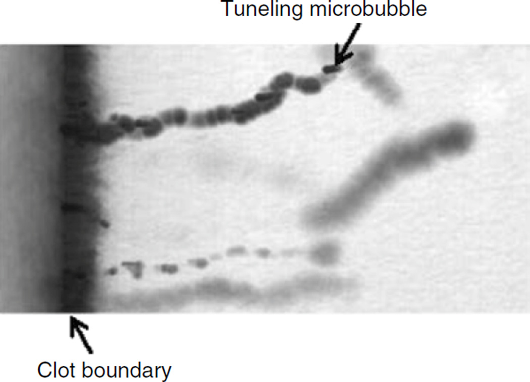
Microbubble tunneling though a fibrin clot (fluid-clot boundary at left) caused by acoustic radiation force (1 MHz, 0.4 MPa). The blurred, dark line is a motion artifact from deflection of the clot boundary due to radiation force (Acconcia et al. 2013)
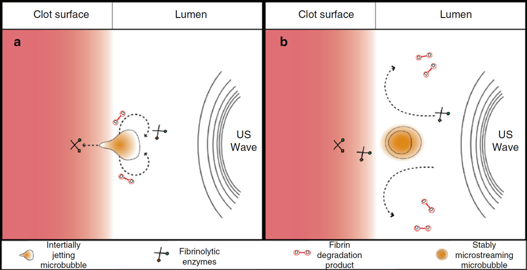
Interaction of cavitating microbubbles with thrombi, (a) Illustration of inertial cavitation. The asymmetric boundary condition causes a liquid jet to form during the final stages of collapse during inertial cavitation (Brujan et al. 2001). The jet will impinge on the thrombus, resulting in direct mechanical damage and erosion of clot surface, (b) Illustration of stable cavitation. Sustained bubble motion promotes strong fluid mixing through microstreaming (Elder 1959). Local pressure gradients around the microbubble cause enhanced mixing of the fibrinolytic enzymes, and removal of fibrin degradation products from the clot (Datta et al. 2008) (Figure Adapted from Sutton et al. (2013a))
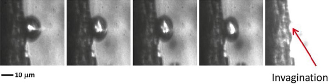
Interaction of inertially cavitating microbubble with thrombus (1 MHz, 1.5 MPa) recorded at 5 Mfps (200 ns interframe time). As the microbubble disappears at the completion of the inertial collapse, there is a residual “pit” at the thrombus site (Chen et al. 2014)
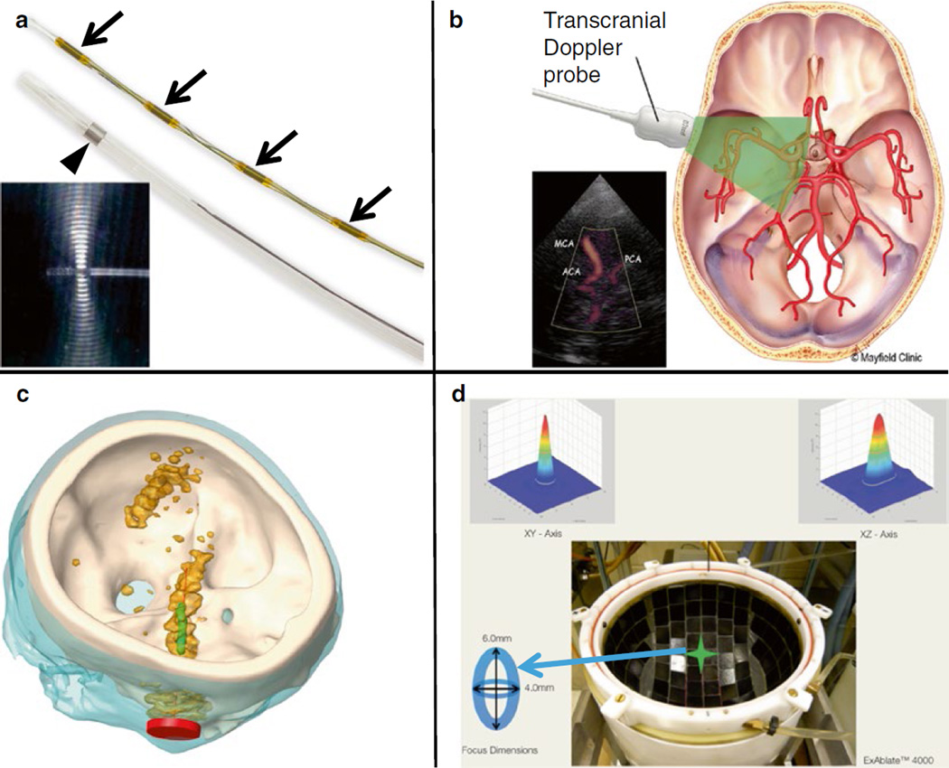
Ultrasound insonation schemes for sonothrombolysis. (a) Ultrasound-assisted thrombolysis catheter composed of a 5.2-Fr multi-sidehole drug infuser (arrowhead). Ultrasound elements along central core (small arrows) shown separately. During treatment, the central core is placed inside the infusion catheter (Engelberger and Kucher 2014). The omnidirectional ultrasound field promotes cavitation and acoustic streaming, driving thrombolytics into the clot. Images provided by EKOS Corporation (Bothell, WA, USA), (b) Transcranial Doppler. Schematic representation of the penetration of 2-MHz TCCS through the skull. Images reprinted with permission from Mayfield Clinic (Cincinnati, OH, USA). (c) Unfocused, Sub-megahertz Ultrasound. Simulated acoustic fields generated by an unfocused, transcranial sub-megahertz (120 kHz) ultrasound system in a human skull (white). The Ml segment of the MCA is shown in green and the transducer in red. The orange contour indicates the regions where the acoustic pressure is larger than half the maximum pressure in the Ml region of interest (Bouchoux et al. 2014). (d) MR-Guided Focused Ultrasound. Top view of ExAblate™ 4000 HIFU hemispherical 1000-element system for MR-guided transcranial focused ultrasound exposure. A sharp 4 mm×6 mm focus is created along the lateral and elevational orientations, respectively (Hölscher et al. 2013)
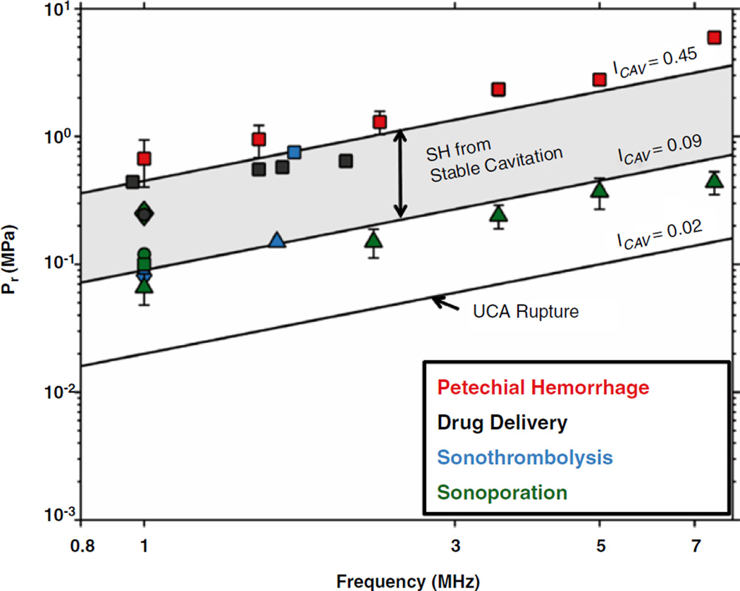
Comparison of cavitation index to selected bioeffects (Bader and Holland 2012). The line labeled ICAV=0.45 indicates the peak rarefractional pressure (Pr) required to initiate shell rupture of ultrasound contrast agents. The lines labeled ICAV=0.09 and ICAV=0.45 bordering the shaded region demarcate the parameter space over which subharmonic (SH) emissions from stable cavitation are likely. The cavitation index is well suited for predicting beneficial bioeffects associated with stable cavitation, such as drug delivery (Hitchcock et al. 2010; McDannold et al. 2008), sonoporation (Greenleaf et al. 1998; Miller and Dou 2004; Rahim et al. 2006; Juffermans et al. 2009) and sonothrombolysis (Porter et al. 2001; Prokop et al. 2007; Petit et al. 2012a). Beyond ICAV=0.45, subharmonic emissions originate from inertial cavitation. Bioeffects associated with inertial cavitation, such as petechial hemorrhage (Miller et al. 2008), occur at a cavitation index above 0.45
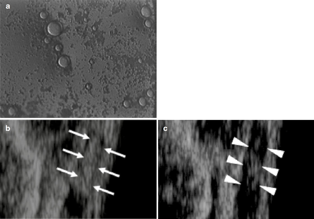
(a) Photograph of thrombus-targeted micro-bubble bound to human whole blood clot in-vitro. Non-targeted microbubbles did not bind to the clot (Unger et al. 1998). (b) Thrombus-targeted bubble liposomes (white arrows’) accumulate on thrombus within an occluded iliac artery in a rabbit model of thromboembolism. (c) After insonation of the thrombus-targeted bubble liposomes, flow was restored and the hyperechogenic area within the iliac artery was reduced (white arrowheads’) (Hagisawa et al. 2013)
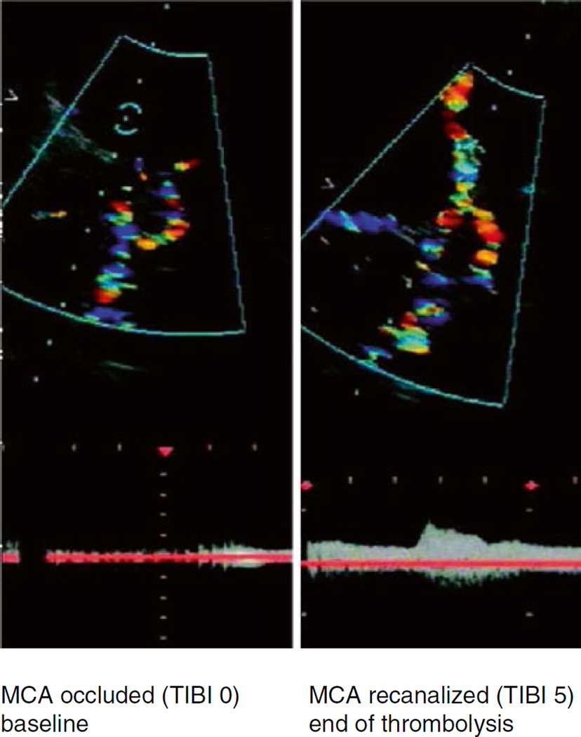
Transcranial color-coded sonography in an ischemic stroke patient with an MCA occlusion. Proximal middle cerebral artery stem occlusion (left) with complete recanalization 1 h after intravenous rt-PA plus TCCS exposure (right) (Eggers et al. 2003)
References
-
- Aaslid R, Markwalder TM, Nornes H. Noninvasive transcranial Doppler ultrasound recording of flow velocity in basal cerebral arteries. J Neurosurg. 1982;57:769–774. - PubMed
-
- Acconcia C, Leung BYC, Hynynen K, Goertz DE. Interactions between ultrasound stimulated microbubbles and fibrin clots. Appl Phys Lett. 2013;103:053701.
-
- Adeoye O, Clark JF, Khatri P, Wagner KR, Zuccarello M, Pyne-Geithman GJ. Do current animal models of intracerebral hemorrhage mirror the human pathology? Transl Stroke Res. 2010;2:17–25. - PubMed
-
- Alexandrov AV, Demchuk AM, Felberg RA, Christou I, Barber PA, Burgin WS, Malkoff M, Wojner AW, Grotta JC. High rate of complete recanalization and dramatic clinical recovery during tPA infusion when continuously monitored with 2-MHz transcranial Doppler monitoring. Stroke. 2000;31:610–614. - PubMed
-
- Alexandrov AV, Molina CA, Grotta JC, Garami Z, Ford SR, Alvarez-Sabin J, Montaner J, Saqqur M, Demchuk AM, Moyé LA, Hill MD, Wojner AW Clotbust Investigators. Ultrasound-enhanced systemic thrombolysis for acute ischemic stroke. N Engl J Med. 2004;351:2170–2178. - PubMed
Publication types
MeSH terms
Substances
Grants and funding
LinkOut - more resources
Full Text Sources
Other Literature Sources
Molecular Biology Databases
