. 2016 Mar 1;213(5):712-22.
doi: 10.1093/infdis/jiv499. Epub 2015 Oct 20.

Middle East Respiratory Syndrome Coronavirus Causes Multiple Organ Damage and Lethal Disease in Mice Transgenic for Human Dipeptidyl Peptidase 4

Affiliations

Middle East Respiratory Syndrome Coronavirus Causes Multiple Organ Damage and Lethal Disease in Mice Transgenic for Human Dipeptidyl Peptidase 4

Kun Li et al. J Infect Dis. .

Abstract

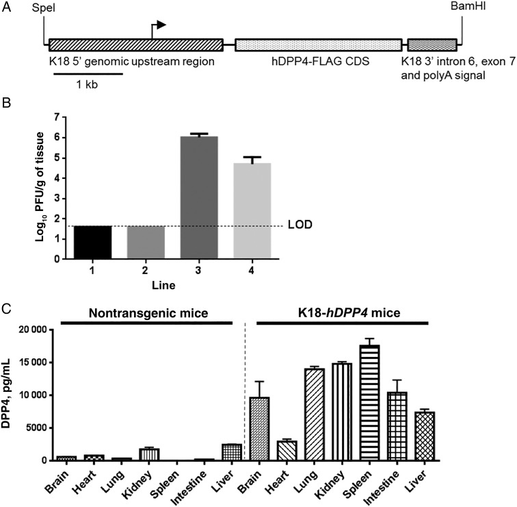
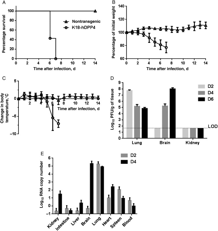
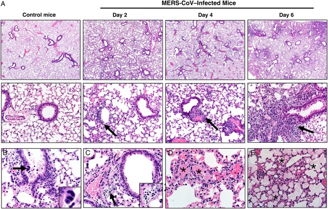
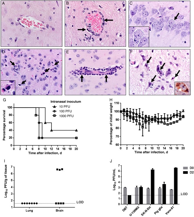
Middle East respiratory syndrome coronavirus (MERS-CoV) causes life-threatening disease. Dipeptidyl peptidase 4 (DPP4) is the receptor for cell binding and entry. There is a need for small-animal models of MERS, but mice are not susceptible to MERS because murine dpp4 does not serve as a receptor. We developed transgenic mice expressing human DPP4 (hDPP4) under the control of the surfactant protein C promoter or cytokeratin 18 promoter that are susceptible to infection with MERS-CoV. Notably, mice expressing hDPP4 with the cytokeratin 18 promoter developed progressive, uniformly fatal disease following intranasal inoculation. High virus titers were present in lung and brain tissues 2 and 6 days after infection, respectively. MERS-CoV-infected lungs revealed mononuclear cell infiltration, alveolar edema, and microvascular thrombosis, with airways generally unaffected. Brain disease was observed, with the greatest involvement noted in the thalamus and brain stem. Animals immunized with a vaccine candidate were uniformly protected from lethal infection. These new mouse models of MERS-CoV should be useful for investigation of early disease mechanisms and therapeutic interventions.

Keywords: DPP4/CD26; MERS; transgenic mice.


Figures

Figure 1.
Figure 1.
Generation and characterization of K18-hDPP4 mice. A, The hDPP4 coding sequence was cloned into a plasmid containing the 5′ and 3′ genomic regions of human cytokeratin 18 (K18). The K18 5′ genomic region consists of a 2.5-kb upstream genomic sequence, promoter, and first intron of the human K18 gene while the K18 3′ region consists of exon 6, intron 6, exon 7, and approximately 300 base pairs of 3′ untranslated region of the human K18 gene, including the K18 polyA signal. Immediately upstream of the hDPP4 start codon is a translational enhancer (TE) sequence from alfalfa mosaic virus. B, Four K18-hDPP4 transgenic founder lines were generated and intranasally inoculated with 1 × 10⁵ plaque-forming units (PFU) of Middle East respiratory syndrome coronavirus. Lung titers of founder mice were determined by plaque assay 3 days after infection. Data are mean ± standard deviation [SD] for 6–10 mice/line. C, Quantitative measurement of human DPP4 concentrations in tissues of nontransgenic or K18-hDPP4 founder line 3 by enzyme-linked immunosorbent assay. Data are mean ± SD for 3 mice. Abbreviation: LOD, limit of detection.
Figure 2.
Figure 2.
Middle East respiratory syndrome coronavirus (MERS-CoV) infection causes lethal disease in human cytokeratin 18–hDPP4 mice. A–C, Animals were inoculated intranasally with 1 × 10⁵ plaque-forming units (PFU) of MERS-CoV and survival (A), weight (B), and temperature (C) monitored daily. Data are for 4 nontransgenic mice and 14 K18-hDPP4 mice. D, MERS-CoV titers in indicated tissues at days 2, 4, and 6 after infection. Data are for 3–4 mice. E, The copy numbers of viral RNA in indicated tissues at days 2 and 4 after infection were analyzed by quantitative real-time polymerase chain reaction targeting regions within open reading frame 1a. Data are for 4 mice. All results are expressed as mean ± SD.
Figure 3.
Figure 3.
A, Lungs from control or Middle East respiratory syndrome coronavirus (MERS-CoV)–infected mice. MERS-CoV infection from days 2, 4, and 6 after infection consistently caused multifocal to patchy consolidation in lung with perivascular and peribronchiolar inflammation (arrows). Section were stained with hematoxylin-eosin (top panels, original magnification ×40; bottom panels, original magnification ×200). B, Airways were generally intact, with uncommon scattered sloughed cells (day 6). Note the very rare multinucleate cells (arrow and inset). C, Late in the course of infection (day 6), degenerating cells and cellular debris (arrow and inset) could be seen filling several lymphatics. D, Vascular thrombi (asterisks) were seen in most cases on days 4 and 6 after infection, with adjacent congestion and lesser amounts of necrosis and hemorrhage. E, Edema, characterized by eosinophilic fluid material in airspaces (asterisks), was progressively detected in some cases on days 4 and 6. Section were stained with hematoxylin-eosin (original magnification ×200 [A] and ×400 [B-E]).
Figure 4.
Figure 4.
Brain disease in Middle East respiratory syndrome coronavirus (MERS-CoV)–infected human cytokeratin 18 (K18)–hDPP4 and uninfected mice. A, Normal brain from an uninfected mouse. B, MERS-CoV caused lymphocytic perivascular cuffing in the infected brain. C, Infected neuron in hippocampus 6 days after infection. Note the granular degeneration and basophilic cytoplasmic inclusions (arrow and inset). D, Dying cells undergoing degeneration (arrows and inset; 6 days after infection) are detected in highly infected regions such as the thalamus or brain stem. E, Meningeal and perivascular cuffing included neutrophilic infiltrates (arrows; 6 days after infection). F, Several degenerating cells had small to granular basophilic cytoplasmic inclusions (arrows; 6 days after infection) that were stained with anti–MERS-CoV antibody (inset; brown). Note the neuropil rarefaction. Section were stained with hematoxylin-eosin (original magnification ×600). GI, Outcomes of K18-hDPP4 mice infected with different intranasal inocula of MERS-CoV. K18-hDPP4 mice received 1000, 100, or 10 plaque-forming units (PFU) of MERS-CoV and were monitored for survival (G) and weight (H). There were 5 mice/group. I, Lungs and brains of mice receiving 10 PFU were harvested 10 days after inoculation or when they lost 20% of body weight. A total of 3 of 7 MERS-CoV–infected mice showed high virus titers in the brains. J, MERS-CoV replicates in cells of the nervous system. Human central nervous system–derived cell lines (U-138 MG an SK-N-SH), primary porcine astrocytes, a murine astrocytoma cell line (DBT), and African green monkey kidney cells (Vero-81) were infected with MERS-CoV at a multiplicity of infection of 1. Titers from these cells immediately after infection (day 0) or 2 days after infection were determined by plaque assay. Data are mean ± SD for 3 replicates/condition. Abbreviation: LOD, limit of detection.
Figure 5.
Figure 5.
Immunization with Venezuelan equine encephalitis replicon particles (VRPs) expressing Middle East respiratory syndrome coronavirus (MERS-CoV) spike glycoprotein (VRP–MERS-S) or passive immunization protects human cytokeratin 18 (K18)–hDPP4 mice against MERS-CoV infection. A and B, K18-hDPP4 mice were immunized with 1 × 10⁵ infectious units (IU) of VRPs expressing green fluorescent protein (VRP-GFP) or VRP-MERS-S in the footpad and boosted with the same dose 4 weeks later. Mice were infected with 1 × 10⁵ plaque-forming units of MERS-CoV 2 weeks after the boost. C and D, For passive immunization, nontransgenic mice were immunized as described above. Sera were obtained 2 weeks after boosting and transferred into K18-hDPP4 mice intraperitoneally 1 day before infection with MERS-CoV. Survival and weights were recorded for active immunization (A and B) and passive immunization (C and D). Data are mean ± standard deviation (SD) for 5 mice/group. E and F, MERS-CoV titers in lung tissue (E) and brain tissue (F) 2, 4, and 6 days after infection in mice with or without passive immunization. Data are mean ± SD for 3 mice/group. Abbreviation: LOD, limit of detection.

References

    1. Zaki AM, van Boheemen S, Bestebroer TM, Osterhaus AD, Fouchier RA. Isolation of a novel coronavirus from a man with pneumonia in Saudi Arabia. N Engl J Med 2012; 367:1814–20. - PubMed
    1. Raj VS, Mou H, Smits SL et al. . Dipeptidyl peptidase 4 is a functional receptor for the emerging human coronavirus-EMC. Nature 2013; 495:251–4. - PMC - PubMed
    1. Zumla AI, Hui DS, Perlman S. Middle East respiratory syndrome. Lancet 2015; 386:995–1007. - PMC - PubMed
    1. Saad M, Omrani AS, Baig K et al. . Clinical aspects and outcomes of 70 patients with Middle East respiratory syndrome coronavirus infection: a single-center experience in Saudi Arabia. Int J Infect Dis 2014; 29:301–6. - PMC - PubMed
    1. Arabi YM, Arifi AA, Balkhy HH et al. . Clinical course and outcomes of critically ill patients with Middle East respiratory syndrome coronavirus infection. Ann Intern Med 2014; 160:389–97. - PubMed

Publication types

MeSH terms