

Mechanisms for greater insulin-stimulated glucose uptake in normal and insulin-resistant skeletal muscle after acute exercise
- PMID: 26487009
- PMCID: PMC4816200
- DOI: 10.1152/ajpendo.00416.2015
Mechanisms for greater insulin-stimulated glucose uptake in normal and insulin-resistant skeletal muscle after acute exercise
Abstract
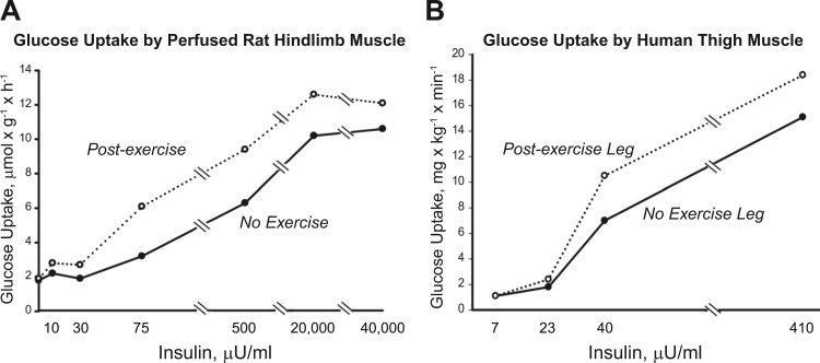
Enhanced skeletal muscle and whole body insulin sensitivity can persist for up to 24-48 h after one exercise session. This review focuses on potential mechanisms for greater postexercise and insulin-stimulated glucose uptake (ISGU) by muscle in individuals with normal or reduced insulin sensitivity. A model is proposed for the processes underlying this improvement; i.e., triggers initiate events that activate subsequent memory elements, which store information that is relayed to mediators, which translate memory into action by controlling an end effector that directly executes increased insulin-stimulated glucose transport. Several candidates are potential triggers or memory elements, but none have been conclusively verified. Regarding potential mediators in both normal and insulin-resistant individuals, elevated postexercise ISGU with a physiological insulin dose coincides with greater Akt substrate of 160 kDa (AS160) phosphorylation without improved proximal insulin signaling at steps from insulin receptor binding to Akt activity. Causality remains to be established between greater AS160 phosphorylation and improved ISGU. The end effector for normal individuals is increased GLUT4 translocation, but this remains untested for insulin-resistant individuals postexercise. Following exercise, insulin-resistant individuals can attain ISGU values similar to nonexercising healthy controls, but after a comparable exercise protocol performed by both groups, ISGU for the insulin-resistant group has been consistently reported to be below postexercise values for the healthy group. Further research is required to fully understand the mechanisms underlying the improved postexercise ISGU in individuals with normal or subnormal insulin sensitivity and to explain the disparity between these groups after similar exercise.
Keywords: AMP-activated protein kinase; Akt substrate of 160 kDa; glucose transporter 4; insulin sensitivity; physical activity.
Copyright © 2015 the American Physiological Society.
Figures


Similar articles
-
Exercise effects on γ3-AMPK activity, phosphorylation of Akt2 and AS160, and insulin-stimulated glucose uptake in insulin-resistant rat skeletal muscle.J Appl Physiol (1985). 2020 Feb 1;128(2):410-421. doi: 10.1152/japplphysiol.00428.2019. Epub 2020 Jan 16. J Appl Physiol (1985). 2020. PMID: 31944891 Free PMC article.
-
Exercise-Induced Improvement in Insulin-Stimulated Glucose Uptake by Rat Skeletal Muscle Is Absent in Male AS160-Knockout Rats, Partially Restored by Muscle Expression of Phosphomutated AS160, and Fully Restored by Muscle Expression of Wild-Type AS160.Diabetes. 2022 Feb 1;71(2):219-232. doi: 10.2337/db21-0601. Diabetes. 2022. PMID: 34753801 Free PMC article.
-
AS160 expression, but not AS160 Serine-588, Threonine-642, and Serine-704 phosphorylation, is essential for elevated insulin-stimulated glucose uptake by skeletal muscle from female rats after acute exercise.FASEB J. 2023 Jul;37(7):e23021. doi: 10.1096/fj.202300282RR. FASEB J. 2023. PMID: 37289137 Free PMC article.
-
Role of Akt substrate of 160 kDa in insulin-stimulated and contraction-stimulated glucose transport.Appl Physiol Nutr Metab. 2007 Jun;32(3):557-66. doi: 10.1139/H07-026. Appl Physiol Nutr Metab. 2007. PMID: 17510697 Review.
-
Roles of TBC1D1 and TBC1D4 in insulin- and exercise-stimulated glucose transport of skeletal muscle.Diabetologia. 2015 Jan;58(1):19-30. doi: 10.1007/s00125-014-3395-5. Epub 2014 Oct 4. Diabetologia. 2015. PMID: 25280670 Free PMC article. Review.
Cited by
-
Effectiveness of a multimodal intervention in functionally impaired older people with type 2 diabetes mellitus.J Cachexia Sarcopenia Muscle. 2019 Aug;10(4):721-733. doi: 10.1002/jcsm.12432. Epub 2019 Apr 23. J Cachexia Sarcopenia Muscle. 2019. PMID: 31016897 Free PMC article. Clinical Trial.
-
Exercise effects on γ3-AMPK activity, phosphorylation of Akt2 and AS160, and insulin-stimulated glucose uptake in insulin-resistant rat skeletal muscle.J Appl Physiol (1985). 2020 Feb 1;128(2):410-421. doi: 10.1152/japplphysiol.00428.2019. Epub 2020 Jan 16. J Appl Physiol (1985). 2020. PMID: 31944891 Free PMC article.
-
Tsg101 Is Involved in the Sorting and Re-Distribution of Glucose Transporter-4 to the Sarcolemma Membrane of Cardiac Myocytes.Cells. 2020 Aug 21;9(9):1936. doi: 10.3390/cells9091936. Cells. 2020. PMID: 32839388 Free PMC article.
-
The Relationship of Lean Body Mass With Aging to the Development of Diabetes.J Endocr Soc. 2020 Apr 30;4(7):bvaa043. doi: 10.1210/jendso/bvaa043. eCollection 2020 Jul 1. J Endocr Soc. 2020. PMID: 32666006 Free PMC article.
-
Postexercise improvement in glucose uptake occurs concomitant with greater γ3-AMPK activation and AS160 phosphorylation in rat skeletal muscle.Am J Physiol Endocrinol Metab. 2018 Nov 1;315(5):E859-E871. doi: 10.1152/ajpendo.00020.2018. Epub 2018 Aug 21. Am J Physiol Endocrinol Metab. 2018. PMID: 30130149 Free PMC article.
References
-
- Arias EB, Kim J, Funai K, Cartee GD. Prior exercise increases phosphorylation of Akt substrate of 160 kDa (AS160) in rat skeletal muscle. Am J Physiol Endocrinol Metab 292: E1191–E1200, 2007. - PubMed
-
- Betts JJ, Sherman WM, Reed MJ, Gao JP. Duration of improved muscle glucose uptake after acute exercise in obese Zucker rats. Obes Res 1: 295–302, 1993. - PubMed
-
- Bonen A, Tan MH, Watson-Wright WM. Effects of exercise on insulin binding and glucose metabolism in muscle. Can J Physiol Pharmacol 62: 1500–1504, 1984. - PubMed
Publication types
MeSH terms
Substances
Grants and funding
LinkOut - more resources
Full Text Sources
Other Literature Sources
Medical