. 2016 May;9(3):787-97.
doi: 10.1038/mi.2015.102. Epub 2015 Oct 21.

Host and microbiota interactions are critical for development of murine Crohn's-like ileitis

Affiliations

Host and microbiota interactions are critical for development of murine Crohn's-like ileitis

M Roulis et al. Mucosal Immunol. 2016 May.

Abstract

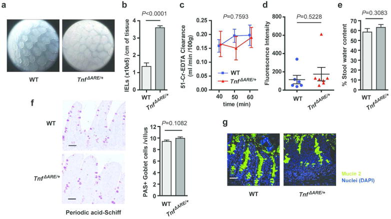
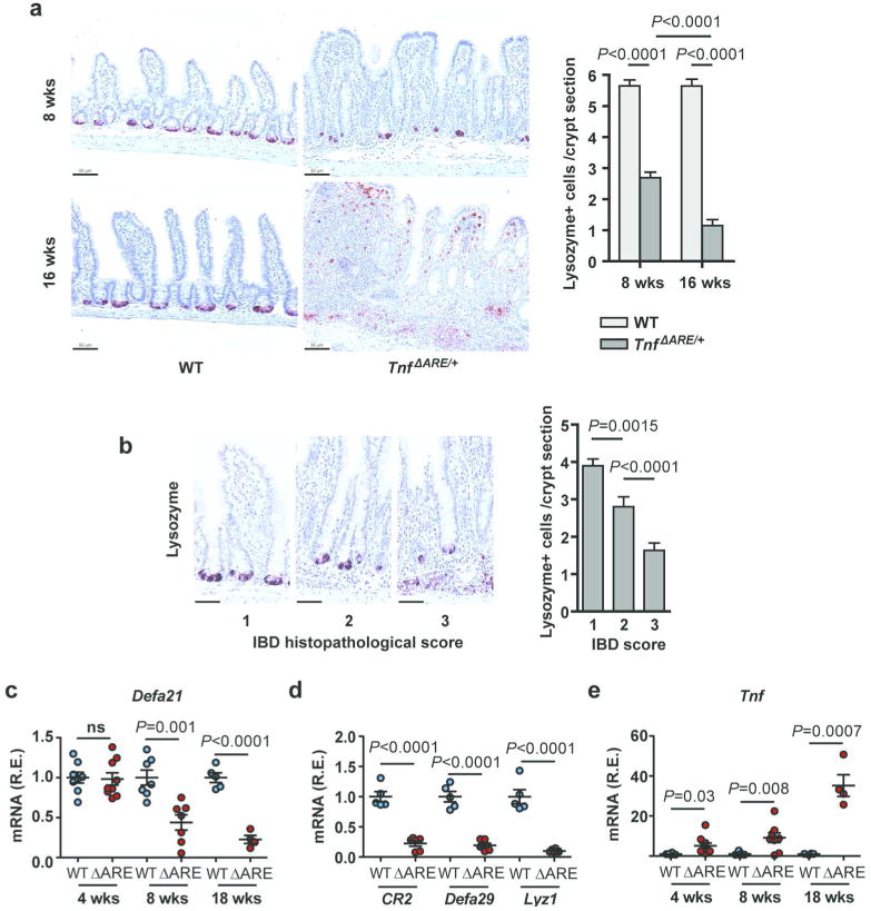
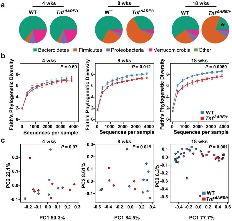
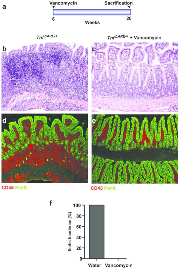
Deregulation of host-microbiota interactions in the gut is a pivotal characteristic of Crohn's disease. It remains unclear, however, whether commensals and/or the dysbiotic microbiota associated with pathology in humans are causally involved in Crohn's pathogenesis. Here, we show that Crohn's-like ileitis in Tnf(ΔARE/+) mice is microbiota-dependent. Germ-free Tnf(ΔARE/+) mice are disease-free and the microbiota and its innate recognition through Myd88 are indispensable for tumor necrosis factor (TNF) overexpression and disease initiation in this model. The epithelium of diseased mice shows no major defects in mucus barrier and paracellular permeability. However, Tnf(ΔARE/+) ileitis associates with the reduction of lysozyme-expressing Paneth cells, mediated by adaptive immune effectors. Furthermore, we show that established but not early ileitis in Tnf(ΔARE/+) mice involves defective expression of antimicrobials and dysbiosis, characterized by Firmicutes expansion, including epithelial-attaching segmented filamentous bacteria, and decreased abundance of Bacteroidetes. Microbiota modulation by antibiotic treatment at an early disease stage rescues ileitis. Our results suggest that the indigenous microbiota is sufficient to drive TNF overexpression and Crohn's ileitis in the genetically susceptible Tnf(ΔARE/+) hosts, whereas dysbiosis in this model results from disease-associated alterations including loss of lysozyme-expressing Paneth cells.


Conflict of interest statement

The authors declare that they have no conflict of interest.

Figures

Figure 1
Figure 1. Germ-free TnfΔARE/+ mice are completely rescued from TNF overexpression in the intestine and Crohn's-like IBD pathology
(a) Histological analysis was performed in the terminal ileum of n = 6 TnfΔARE/+ mice raised under germ-free conditions at the age of 15 weeks. N = 7 TnfΔARE/+ mice raised under Specific Pathogen Free (SPF) conditions (11 weeks) were used as controls. Representative H&E-stained sections are shown. (Scale bars: 100 μm). (b) Histopathological analysis performed in the terminal ileum of germ-free and SPF control TnfΔARE/+ mice examined at the age of 15 and 11 weeks respectively. Statistical significance was calculated by Kruskal-Wallis one-way analysis of variance. (c) Quantification of TNF gene expression in whole tissue biopsies from the terminal ileum of TnfΔARE/+ mice reared under germ-free and SPF conditions, examined at the age of 15 and 11 weeks respectively. Statistical significance was calculated with two-tailed Mann-Whitney test. All data shown represent mean ± SEM.
Figure 2
Figure 2. Myd88 mediates TNF overexpression and IBD pathogenesis in TnfΔARE/+ mice
(a) Crohn's-like pathology is attenuated in the terminal ileum of 16-week-old TnfΔARE/+Myd88-/- mice as compared to TnfΔARE/+ littermate controls. Representative H&E-stained sections are shown. (Scale bars: 50 μm). (b) Histopathological analysis performed in the terminal ileum of TnfΔARE/+Myd88-/- and TnfΔARE/+ control mice at the age of 16 weeks. Statistical significance was calculated with Mann-Whitney test. (** P < 0.01) (c) Thioglycollate-elicited peritoneal macrophages (see SI Methods) were isolated from 16-week-old TnfΔARE/+, TnfARE/+Myd88-/-, Myd88-/- and WT control mice (n = 3 mice per genotype) and stimulated with LPS for 6 hours. TNF secretion was measured by ELISA. Macrophages isolated from TnfΔARE/+Myd88-/- mice show defective LPS-induced TNF secretion. Statistical significance was calculated with unpaired Welch t test. (** P < 0.01) All data shown represent mean ± SEM.
Figure 3
Figure 3. Epithelial barrier function in the ileum of diseased TnfΔARE/+ mice
(a) Endoscopic pictures from the terminal ileum of 12 week-old TnfΔARE/+ mice and WT controls. Representative of n = 2 independent experiments. (b) Quantification of intraepithelial lymphocytes (IELs) isolated from the ileum of 12 week-old TnfΔARE/+ mice and WT controls (n=5 each). Unpaired two-tailed Student t-test (*** P < 0.001) (c) Epithelial paracellular permeability, as measured by blood to lumen 51Cr-EDTA clearance in the terminal ileum of 12 week-old TnfΔARE/+ mice (n=4) and WT controls (n=4). No significant difference (ns) was found between the two groups by Two-way ANOVA. (d) Epithelial barrier permeability of 12 week-old TnfΔARE/+ mice (n=7) and WT controls (n=6), as measured by lumen to blood passage of orally-delivered 20kDa FITC-dextran. (e) Stool water content of TnfΔARE/+ mice (n=24) and WT littermates (n=18) at the age of 16 weeks. No significant (ns) differences were found by unpaired two-tailed Student t-test. (f-g) Goblet cell numbers and mucus secretion show no major defects in the inflamed ileum of 12-week-old TnfΔARE/+ mice compared to littermate WT controls (n=3 per group) as shown by Periodic acid-Schiff (PAS)-staining (e) and mucin 2 immunostaining (f). Unpaired two-tailed Student t-test. (Scale bars: 50 μm).
Figure 4
Figure 4. Loss of Paneth cells and altered expression of antimicrobials in the ileum of diseased TnfΔARE/+ mice
(a) Immunohistochemistry for Lysozyme for the quantification of Paneth cells per crypt in the terminal ileum of TnfΔARE/+ mice at the age of 8 and 16 weeks and littermate WT controls (n=3/group). Two-tailed Mann-Whitney test (*** P < 0.001). (b) Paneth cell numbers are significantly reduced as the local IBD histopathological score increases in the crypts of 8 week-old TnfΔARE/+ mice (n=3). A total of n=108 well-oriented crypts were examined. Indicative lysozyme immunostaining pictures are shown for each disease stage. (Scale bars: 33 μm). Statistical significance was calculated by two-tailed Mann-Whitney test (** P < 0.01). (c) Relative mRNA expression of Defa21 in the last 2 cm of the terminal ileum of WT and TnfΔARE/+ mice at different ages (n= 4-8/group). Two-tailed Student t-test. (d) Relative expression of antimicrobials Cr2, Defa29 and Lyzc1 in whole tissue extracts from the last 2 cm of the ileum of WT and TnfΔARE/+ mice at 18 wks (n= 6/group). Two-tailed Student t-test. (e) Relative expression of Tnf in whole tissue extracts from the last 2 cm of the ileum of WT and TnfΔARE/+ mice at different ages (n= 4-8/group). Two-tailed Student t-test. R.E., Relative Expression. Each dot represents an animal. All data shown represent mean ± SEM.
Figure 5
Figure 5. Biome analysis of TnfΔARE/+ mice
(a) Relative abundance of phyla present in the terminal ileum of WT and TnfΔARE/+ mice at 4 (W, n = 6; T, n = 9), 8 (W, n = 9; T, n = 7), and 18 (W, n = 19; T, n = 20) weeks of age. Data shown represent the most abundant phyla, while low abundant and unclassified OTUs were grouped in ‘Other’, * represents a significant (q < 0.05) change in abundance between age-matched WT and TnfΔARE/+ mice. (b) Alpha diversity in TnfARE/+ mice at 4, 8 and 18 weeks of age. Statistical significance was calculated with Mann-Whitney test. (c) Weighted Unifrac distances between TnfΔARE/+ and co-housed WT litter-mate controls at 4, 8 and 18 wks of age. Statistical significance was calculated with Adonis test.
Figure 6
Figure 6. Increased abundance of Candidatus Arthromitus in TnfΔARE/+ mice
(a) In total 139 OTUs were detected across all samples. Pearson hierarchical clustering of the abundance profiles of the 23 OTUs that were significantly altered in the terminal ileum of TnfΔARE/+ mice compared to co-housed WT littermate controls at 4, 8 and 18 weeks. 4 weeks: WT (n = 6), TnfΔARE/+ (n = 9); 8 weeks: WT (n = 9), TnfΔARE/+ (n = 7); 18 weeks: WT (n = 19), TnfΔARE/+ (n = 20). Two OTUs, 376862 and 425767, were significantly enriched at 18 weeks; both represent the genus Candidatus Arthromitus. Each column represents a different mouse. Left colored boxes indicate the family/genus taxonomy classification (T). Right colored boxes indicate if the comparison (p) between WT and TnfΔARE/+ shows significant difference (blue, P < 0.05). (b) LOESS regression of the median read counts of the S24-7/- and Clostridiaceae/Candidatus Arthromitus OTUs shown in (a). Shaded region represents standard error. (c) Boxplots showing the relative abundance of the two OTUs (376862, 425767) enriched in TnfΔARE/+ mice at 18 weeks of age.
Figure 7
Figure 7. Increased number of mucosa-associated bacteria in the terminal ileum of TnfΔARE/+ mice
(a-b) Visualization of bacterial localization in the distal ileum of WT and TnfΔARE/+ mice at 18 wks. Transversal sections of the distal ileum were hybridized with a eubacterial 16S probe (red) and counterstained with Pan-keratin antibody to visualize the intestinal epithelial cells (green) and DAPI for nuclear staining (blue). Notice the increased attachment of bacteria to the epithelium of TnfΔARE/+ mice when compared to WT animals. Shown are representative images (a) and quantification of 5 mice per group (b). (c-d) Bacterial content in the stool (c) and in the distal ileum mucosa (d) of WT (n = 4) and TnfΔARE/+ mice (n = 5) at 18 wks of age determined by pan-bacterial 16S qPCR amplification relative to host ubiquitin. (e) Relative expression of mucosa-associated SFB in the terminal ileum of WT and TnfΔARE/+ mice at 18 weeks of age. R.E., Relative Expression. Each dot presents an animal. Data are expressed as mean ± SEM. Statistical significance was calculated with two-tailed Student t-test. Scale bars = 50 μm.
Figure 8
Figure 8. Antibiotics treatment rescues ileitis in TnfΔARE/+ mice
(a) Diagram of antibiotic treatment. TnfΔARE/+ mice at 6 weeks of age were treated with water (n=5) or vancomycin (n=5). Animals were sacrificed at 20 weeks of age. Inflammatory infiltrates in the ileum of TnfΔARE/+ mice were completely abrogated in animals treated with vancomycin (c) when compared to water treated controls (b). All mice (n = 5 per group) were analyzed histologically and representative H&E-stained sections are shown. (d-e) Immunostaining of CD45 and Pan-keratin in the terminal ileum of TnfΔARE/+ mice treated with water (n = 5) (d) or vancomycin (n =5) (e), sacrificed at 20 weeks of age. (f) Ileitis incidence in 20-week-old TnfΔARE/+ mice treated with water (n = 5) or vancomycin (n = 5).

References

    1. Xavier RJ, Podolsky DK. Unravelling the pathogenesis of inflammatory bowel disease. Nature. 2007;448(7152):427–434. - PubMed
    1. Johansson ME, Larsson JM, Hansson GC. The two mucus layers of colon are organized by the MUC2 mucin, whereas the outer layer is a legislator of host-microbial interactions. Proc Natl Acad Sci U S A. 2011;108 Suppl 1:4659–4665. - PMC - PubMed
    1. Bevins CL, Salzman NH. Paneth cells, antimicrobial peptides and maintenance of intestinal homeostasis. Nat Rev Microbiol. 2011;9(5):356–368. - PubMed
    1. Pearson C, Uhlig HH, Powrie F. Lymphoid microenvironments and innate lymphoid cells in the gut. Trends in immunology. 2012;33(6):289–296. - PubMed
    1. Frank DN, St Amand AL, Feldman RA, Boedeker EC, Harpaz N, Pace NR. Molecular-phylogenetic characterization of microbial community imbalances in human inflammatory bowel diseases. Proc Natl Acad Sci U S A. 2007;104(34):13780–13785. - PMC - PubMed

Publication types

MeSH terms

Substances