

Proteomic analysis of neurons microdissected from formalin-fixed, paraffin-embedded Alzheimer's disease brain tissue
- PMID: 26487484
- PMCID: PMC4614382
- DOI: 10.1038/srep15456
Proteomic analysis of neurons microdissected from formalin-fixed, paraffin-embedded Alzheimer's disease brain tissue
Abstract
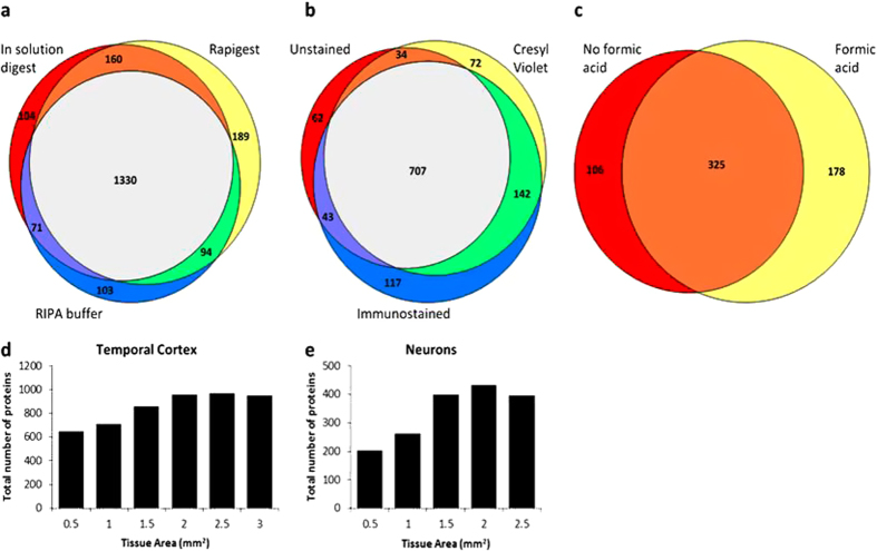
The vast majority of human tissue specimens are formalin-fixed, paraffin embedded (FFPE) archival samples, making this type of tissue a potential gold mine for medical research. It is now accepted that proteomics can be done using FFPE tissue and can generate similar results as snap-frozen tissue. However, the current methodology requires a large amount of starting protein, limiting the questions that can be answered in these types of proteomics studies and making cell-type specific proteomics studies difficult. Cell-type specific proteomics has the potential to greatly enhance understanding of cell functioning in both normal and disease states. Therefore, here we describe a new method that allows localized proteomics on individual cell populations isolated from FFPE tissue sections using laser capture microdissection. To demonstrate this technique we microdissected neurons from archived tissue blocks of the temporal cortex from patients with Alzheimer's disease. Using this method we identified over 400 proteins in microdissected neurons; on average 78% that were neuronal and 50% that were associated with Alzheimer's disease. Therefore, this technique is able to provide accurate and meaningful data and has great potential for any future study that wishes to perform localized proteomics using very small amounts of archived FFPE tissue.
Figures
References
-
- Maes E. et al. Analysis of the formalin-fixed paraffin-embedded tissue proteome: pitfalls, challenges, and future prospectives. Amino. Acids 45, 205–218 (2013). - PubMed
-
- Fowler C. B., O’Leary T. J. & Mason J. T. Toward improving the proteomic analysis of formalin-fixed, paraffin-embedded tissue. Expert. Rev. Proteomics. 10, 389–400 (2013). - PubMed
-
- Tanca A., Pagnozzi D. & Addis M. F. Setting proteins free: progresses and achievements in proteomics of formalin-fixed, paraffin-embedded tissues. Proteomics. Clin. Appl. 6, 7–21 (2012). - PubMed
-
- Rekhter M. D. & Chen J. Molecular analysis of complex tissues is facilitated by laser capture microdissection: critical role of upstream tissue processing. Cell Biochem. Biophys. 35, 103–113 (2001). - PubMed
-
- Molina M. et al. Enrichment of single neurons and defined brain regions from human brain tissue samples for subsequent proteome analysis. J Neural. Transm. 122, 993–1005 (2015). - PubMed
Publication types
MeSH terms
Grants and funding
LinkOut - more resources
Full Text Sources
Other Literature Sources
Medical
