Comparative Study
. 2016 Jun;27(6):1609-16.
doi: 10.1681/ASN.2014111059. Epub 2015 Oct 20.

Autophagy Induces Prosenescent Changes in Proximal Tubular S3 Segments

Affiliations
Comparative Study

Autophagy Induces Prosenescent Changes in Proximal Tubular S3 Segments

Arpita Baisantry et al. J Am Soc Nephrol. 2016 Jun.

Abstract

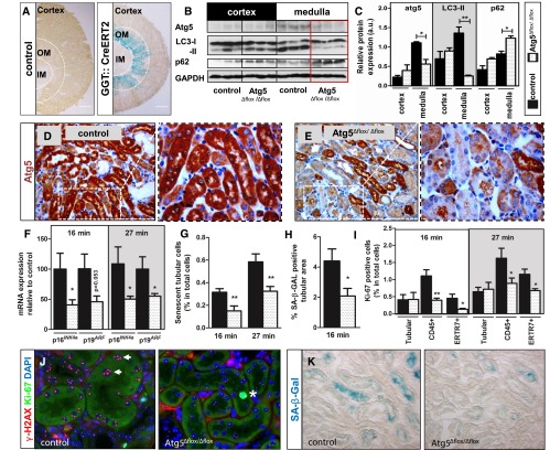
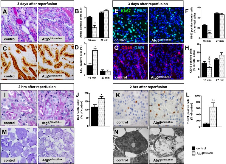
Evidence suggests that autophagy promotes the development of cellular senescence. Because cellular senescence contributes to renal aging and promotes the progression from AKI to CKD, we investigated the potential effect of tubular autophagy on senescence induction. Compared with kidneys from control mice, kidneys from mice with conditional deletion of autophagy-related 5 (Atg5) for selective ablation of autophagy in proximal tubular S3 segments (Atg5(Δ) (flox/) (Δ) (flox)) presented with significantly less tubular senescence, reduced interstitial fibrosis, and superior renal function 30 days after ischemia/reperfusion injury. To correlate this long-term outcome with differences in the early injury process, kidneys were analyzed 2 hours and 3 days after reperfusion. Notably, compared with kidneys of control mice, Atg5(Δ) (flox/) (Δ) (flox) kidneys showed more cell death in outer medullary S3 segments at 2 hours but less tubular damage and inflammation at day 3. These data suggest that the lack of autophagy prevents early survival mechanisms in severely damaged tubular cells. However, if such compromised cells persist, then they may lead to maladaptive repair and proinflammatory changes, thereby facilitating the development of a senescent phenotype and CKD.

Keywords: acute renal failure; fibrosis; ischemia/reperfusion; pathophysiology of renal disease and progression; progression of renal failure; proximal tubule.


Figures

Figure 1.
Figure 1.
Targeted Atg5 deletion impairs activation of autophagy and diminishes development of cellular senescence after I/R in S3 segments of kidney proximal tubules. (A) GGT::Cre-ERT2 mice expressing tamoxifen–inducible Cre recombinase were crossed with ROSA26 (R26R) reporter mice and analyzed for LacZ expression. GGT::Cre-ERT2 mice were crossed with Atg5flox/flox mice to generate animals with deletion of the Atg5 gene in the S3 segment. Three weeks after tamoxifen induction, Atg5Δflox/Δflox mice underwent 16- or 27-minute renal I/R injury. Scale bar, 500 µm. (B and C) Representative immunoblots and corresponding densitometry show a reduction of Atg5, a loss of LC3 lipidation, and an accumulation of p62 in the outer medulla of Atg5Δflox/Δflox kidneys at 16 hours after 16 minutes of renal clamping. (D and E) Representative immunohistochemistry of Atg5 in the outer medulla revealing reduced Atg5 expression in Atg5Δflox/Δflox kidneys versus control kidneys. (F) Quantitative real-time PCR showing relative expression of established senescence markers 30 days after I/R. (G) Quantification of γ-H2AX+/Ki67 cells. (H) Quantification of SA-β-GAL–positive area. (I) Quantification of Ki67–positive tubular and interstitial cells (fibroblast marker ERTR7 and leukocyte marker CD45) showing significantly fewer proliferating interstitial cells in Atg5Δflox/Δflox kidneys. (J) Representative coimmunostaining from outer medulla for γ-H2AX (red) and Ki67 (green) reveals fewer senescent tubular cells without Ki67 and five or more γ-H2A.X foci (arrows) in Atg5Δflox/Δflox kidneys. A Ki67–positive proliferating tubular cell is marked with an asterisk. (K) Representative pictures from the outer medulla showing diminished SA-β-GAL staining in Atg5Δflox/Δflox kidneys after 16 minutes of clamping. Data are presented as mean values±SEMs (n=4 in C, n=10 in F–I). Original magnifications, ×200 in D, E, and K; ×630 in J. *P<0.05; **P<0.01. DAPI, 4′,6-diamidino-2-phenylindole; IM, inner medulla; OM, outer medulla.
Figure 2.
Figure 2.
Atg5 deletion in S3 segments leads to better recovery of GFR, diminished tubular atrophy, less interstitial fibrosis, and reduced inflammation at day 30 after I/R. Control and Atg5Δflox/Δflox mice underwent 16 or 27 minutes of renal I/R and were analyzed at day 30. (A) Representative pictures from the outer medulla and (B) corresponding analyses showing reduced tubular atrophy in periodic acid–Schiff staining, (C) a higher percentage of intact LTL–positive proximal tubules, and (D and E) less interstitial matrix expansion by Masson Trichrome staining in Atg5Δflox/Δflox kidneys. (F) Quantitative real-time PCR showing reduced expression of established renal damage markers Kim-1 and NGAL in Atg5Δflox/Δflox kidneys. (G and H) Representative immunostaining and quantification from the outer medulla showing reduced numbers of CD45-positive cells in Atg5Δflox/Δflox kidneys. (I) Transcutaneous GFR measurements of control and Atg5Δflox/Δflox animals at indicated time points after 16 minutes of bilateral renal clamping. Data are presented as mean values±SEMs (n=10). Original magnifications, ×400 in A, D, and G. *P<0.05; **P<0.01; ***P<0.001. DAPI, 4′,6-diamidino-2-phenylindole.
Figure 3.
Figure 3.
The effect of S3 segment–specific Atg5 deletion on early I/R injury is dependent on the duration of I/R. Control and Atg5Δflox/Δflox mice underwent 16 or 27 minutes of renal I/R and were analyzed at day 3. (A) Representative periodic acid–Schiff staining images from the outer medulla of 16-minute clamped kidneys and (B) morphologic damage scoring showing reduced acute injury in Atg5Δflox/Δflox kidneys. (C) Representative LTL staining images from the outer medulla of 16-minute clamped kidneys and (D) quantification showing a higher percentage of LTL-positive area in Atg5Δflox/Δflox kidneys. (E) Representative Ki67 immunostaining images from the outer medulla of 16-minute clamped kidneys and (F) quantification showing reduced tubular proliferation in Atg5Δflox/Δflox kidneys. (G) Representative CD45 immunostaining images from the outer medulla of 16-minute clamped kidneys and (H) quantification showing reduced immune infiltration in Atg5Δflox/Δflox kidneys. Control and Atg5Δflox/Δflox mice underwent 16 minutes of renal I/R and were analyzed at 2 hours after reperfusion. (I) Representative periodic acid–Schiff staining images from the outer medulla and (J) stereologic quantification of dead tubular cells showing enhanced cell death in Atg5Δflox/Δflox kidneys after 16 minutes of clamping. (K) Representative TUNEL assay images from the outer medulla and (L) quantification showing a significant increase in the number of apoptotic tubular cells in Atg5Δflox/Δflox kidneys after 16 minutes of clamping. (M) Representative Toluidine blue–stained Epon sections and (N) electron microscopy images from control and Atg5Δflox/Δflox kidneys after 27 minutes of clamping indicating more severe damage in Atg5Δflox/Δflox kidneys as reflected by dead lysing tubular epithelial cells (asterisks). Data are presented as mean values±SEMs (n=7–8 in day 3 I/R samples, and n=5 in 2-hour I/R samples). Original magnifications, ×400 in A, C, E, G, and M; ×630 in I and K. Scale bar, 10 µm in N. *P<0.05; **P<0.01; ***P<0.001. DAPI, 4′,6-diamidino-2-phenylindole.

References

    1. Belayev LY, Palevsky PM: The link between acute kidney injury and chronic kidney disease. Curr Opin Nephrol Hypertens 23: 149–154, 2014 - PMC - PubMed
    1. Schmitt R, Melk A: New insights on molecular mechanisms of renal aging. Am J Transplant 12: 2892–2900, 2012 - PubMed
    1. Braun H, Schmidt BM, Raiss M, Baisantry A, Mircea-Constantin D, Wang S, Gross ML, Serrano M, Schmitt R, Melk A: Cellular senescence limits regenerative capacity and allograft survival. J Am Soc Nephrol 23: 1467–1473, 2012 - PMC - PubMed
    1. Clements ME, Chaber CJ, Ledbetter SR, Zuk A: Increased cellular senescence and vascular rarefaction exacerbate the progression of kidney fibrosis in aged mice following transient ischemic injury. PLoS One 8: e70464, 2013 - PMC - PubMed
    1. Westhoff JH, Schildhorn C, Jacobi C, Hömme M, Hartner A, Braun H, Kryzer C, Wang C, von Zglinicki T, Kränzlin B, Gretz N, Melk A: Telomere shortening reduces regenerative capacity after acute kidney injury. J Am Soc Nephrol 21: 327–336, 2010 - PMC - PubMed

Publication types

LinkOut - more resources