. 2015 Nov 3;112(44):13573-8.
doi: 10.1073/pnas.1508273112. Epub 2015 Oct 20.

Knockout of Lmod2 results in shorter thin filaments followed by dilated cardiomyopathy and juvenile lethality

Affiliations

Knockout of Lmod2 results in shorter thin filaments followed by dilated cardiomyopathy and juvenile lethality

Christopher T Pappas et al. Proc Natl Acad Sci U S A. .

Abstract

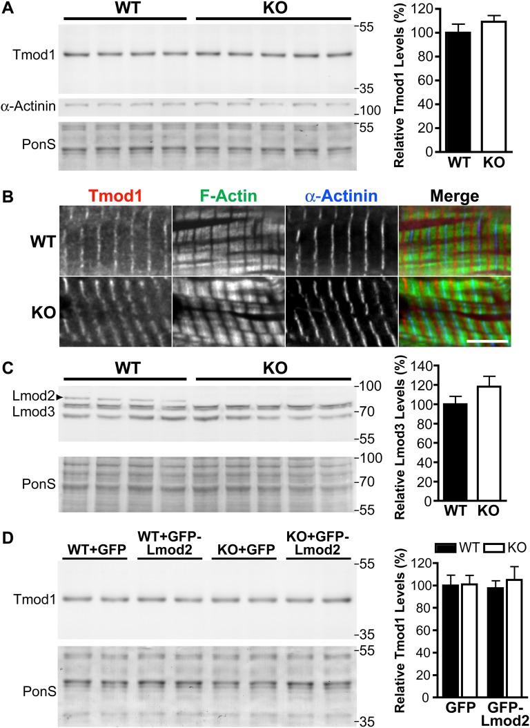
Leiomodin 2 (Lmod2) is an actin-binding protein that has been implicated in the regulation of striated muscle thin filament assembly; its physiological function has yet to be studied. We found that knockout of Lmod2 in mice results in abnormally short thin filaments in the heart. We also discovered that Lmod2 functions to elongate thin filaments by promoting actin assembly and dynamics at thin filament pointed ends. Lmod2-KO mice die as juveniles with hearts displaying contractile dysfunction and ventricular chamber enlargement consistent with dilated cardiomyopathy. Lmod2-null cardiomyocytes produce less contractile force than wild type when plated on micropillar arrays. Introduction of GFP-Lmod2 via adeno-associated viral transduction elongates thin filaments and rescues structural and functional defects observed in Lmod2-KO mice, extending their lifespan to adulthood. Thus, to our knowledge, Lmod2 is the first identified mammalian protein that functions to elongate actin filaments in the heart; it is essential for cardiac thin filaments to reach a mature length and is required for efficient contractile force and proper heart function during development.

Keywords: actin-thin filaments; cardiomyopathy; cytoskeletal dynamics.


Conflict of interest statement

The authors declare no conflict of interest.

Figures

Fig. 1.
Fig. 1.
Lmod2-KO mice die before weaning with no detectable Lmod2 protein in the heart. Lmod2 expression is restricted to striated muscle in the mouse embryo. (A) Survival curve of Lmod2+/+ (WT, black line) and Lmod2−/− (KO, gray line) mice. The KO curve is significantly different from WT, P < 0.0001, log-rank test. (B) Genotyping with Lmod2 and LacZ cassette-specific primers. WT mice produce a 231-bp Lmod2 band, KO mice produce a 684-bp LacZ cassette band, and heterogeneous (HET) mice produce both bands. (C) Immunoblots of LV lysate from P1 Lmod2 WT and KO mice. Lysate was probed with anti-Lmod2 and anti-GAPDH antibodies. (D) β-Gal staining of Lmod2+/− embryos at E8.5–E10.5. Arrows denote the heart, arrowheads the pharyngeal arches, and asterisks the somites. (Scale bars: 0.5 mm.)
Fig. S1.
Fig. S1.
Strategy used by Regeneron Pharmaceuticals to knock out the Lmod2 gene in mice. Lines represent introns, and boxes indicate exons; filled boxes designate the coding sequence. Homologous recombination results in the replacement of the complete coding sequence of Lmod2 with a cassette containing the LacZ gene from Escherichia coli. The locations of primers used in genotyping are specified above their corresponding exons (Lmod2 GP and LacZ GP).
Fig. 2.
Fig. 2.
Lmod2-KO mice have shorter cardiac thin filaments. (A, Upper) Representative image of F-actin stain from WT and Lmod2-KO stretched LV tissue at P6; pink lines denote a gap in F-actin staining across the M line (center of sarcomere). B, barbed end; P, pointed end. (Scale bar: 1 μm.) (Lower) An example of the intensity profiles used by the DDecon analysis program to determine thin filament length accurately. (B) Thin filament (TF) lengths in the LV of WT (black bars) and Lmod2-KO (white bars) mice at various developmental time points; n = 3 or 4. (C) Thin filament lengths in the EDL and soleus muscles of WT (black bars) and Lmod2-KO (white bars) mice at P15; n = 2. (D) Thin filament lengths from neonatal mouse cardiomyocytes (NMCM) in culture isolated from WT (black bars) and Lmod2-KO (white bars) hearts followed by transduction with GFP or GFP-Lmod2 Adv at a multiplicity of infection (MOI) of 2; n = ∼110 total measurements from 8–12 cells per culture, three cultures. All values are mean ± SEM.
Fig. S2.
Fig. S2.
Determination of the relative protein levels of GFP-Lmod2 that rescue the thin filament length deficit in Lmod2-KO cardiomyocytes in culture and localization of GFP-Lmod2. (A, Left) Immunoblot analysis of Lmod2 protein levels in WT and Lmod2-KO neonatal cardiomyocytes transduced with 2 MOI of GFP or GFP-Lmod2 Adv. Note: Endogenous Lmod2 runs between 70–100 kDa and GFP-Lmod2 between 100–130 kDa. (Right) Mean relative Lmod2 protein expression in two cultures ± SEM. (B) Rat cardiomyocytes plated on flexible culture plates (Flexcell Inc.), transduced with GFP-Lmod2 and subjected to ∼20% equibiaxial stretch before fixation. Costaining for α-actinin marks the Z-disk. (Scale bar: 2 μm.)
Fig. 3.
Fig. 3.
Lmod2-KO hearts display large ventricular lumens, thin ventricular walls, and reduced systolic performance. (A) Longitudinal (Upper) and transverse (Lower) sections of P15 paraffin-embedded hearts stained with Masson's Trichrome. RV, right ventricle. (Scale bar: 0.5 mm.) (BD) Echocardiography analysis of WT (black bars) and Lmod2-KO (white bars) hearts at P2, P6, and P15. (B) LV posterior wall in diastole. (C) LV end diastolic diameter. (D) Ejection fraction (EF). Data are shown as mean ± SEM; n = 6 or 7.
Fig. S3.
Fig. S3.
Cardiomyocytes are significantly longer in Lmod2-KO mice. Length and width measurements of cardiomyocytes isolated from P19 WT (black bars) and KO (white bars) mice. Mean ± SEM; n = 3; *P < 0.05, Student's t test.
Fig. S4.
Fig. S4.
Expression of four of six molecular markers of heart failure is not altered significantly at P15, but all markers are significantly changed at P19. RT-qPCR analysis of gene expression in the LV of WT and Lmod2-KO mice at P15 (A) and P19 (B). Data are shown as mean ± SEM; n = 5–11.
Fig. S5.
Fig. S5.
The hearts of Lmod2-KO mice present with few detectable defects early in neonatal development but exhibit myofibril disarray and common features of DCM ∼2 wk later by EM. (A) Electron micrographs of cardiac LV tissue from P6 WT and Lmod2-KO mice. (Scale bar: 1 μm.) (B) LV micrographs of P20 WT (a, c, e, and g) and KO (b, d, f, and h) mice. (a and c) WT myocardium has compact filaments, with sarcomeres in register and mitochondria with intact cristae. (b and d) Lmod2-KO mouse hearts present with decreased mitochondrial (M) number, mitochondrial swelling with disorganized cristae, and dilated sarcoplasmic reticulum (SR). (e and f) Lmod2-KO intercalated discs have a significantly higher degree of convolutions compared with WT (arrows). (g and h) The sarcoplasmic reticulum (SR) is dilated in Lmod2-KO mice, and the mitochondria show degradation. (Scale bars: 2 μm in a and b; 1 μm in ch.) (C) Skeletal muscle of the KO mice does not display any detectable defects. Electron micrographs of EDL muscle at P16. Z, Z-disk. (Scale bar: 1 μm.)
Fig. S6.
Fig. S6.
Tmod1 and Lmod3 protein levels and Tmod1 localization are not significantly altered in Lmod2-KO mice, and the introduction of GFP-Lmod2 AAV does not alter Tmod1 protein levels. (A, Left) Immunoblot analysis of Tmod1 of protein levels in WT and Lmod2-KO mice at P9. (Right) Mean relative Tmod1 protein expression in four or five mice ± SEM. (B) Immunofluorescence staining of Tmod1 in the LV of WT and Lmod2-KO mice at P6. Staining for α-actinin marks the Z-disk. (Scale bar: 5 μm.) (C, Left) Immunoblot analysis of Lmod3 protein levels in WT and Lmod2-KO mice at P9. (Right) Mean relative Lmod3 protein expression in four or five mice ± SEM. Note: The Lmod3 antibody appears to cross-react with Lmod2 (top bands that disappear in the KO). (D) Immunoblot analysis of Tmod1 in the LV of WT and Lmod2-KO mice injected with GFP or GFP-Lmod2 AAV. (Left) Representative blot of two animals in each group. (Right) Mean relative Tmod1 protein expression in four to six animals ± SEM.
Fig. 4.
Fig. 4.
Lmod2-KO cardiomyocytes have reduced contractile force. (A) Contractile force of WT (black bar) and Lmod2-KO (white bar) neonatal cardiomyocytes plated on micropillar arrays. Data are shown as mean ± SEM; n = ∼70 cells from four cultures. (B) Contractile force of WT (black bar) and KO (white bar) neonatal cardiomyocytes transduced with GFP, and KO neonatal cardiomyocytes transduced with GFP-Lmod2 (gray vertically striped bar). n = 30–70 cells from two or three cultures.
Fig. S7.
Fig. S7.
Micropillar array fabrication. (A) Schematic of micropillar fabrication process. (B) Lateral deflection of each pillar (δ) is measured and then multiplied by the pillar’s spring constant (k = 3πED4/64L3) to obtain a force value.
Fig. 5.
Fig. 5.
Introduction of GFP-Lmod2 AAV rescues Lmod2-KO mice structurally and functionally. (A) Immunoblot analysis of Lmod2 protein levels in the LV of WT and KO mice injected with GFP or GFP-Lmod2 AAV. (Left) Representative blots of two animals in each group. Note: Endogenous Lmod2 runs between 70–100 kDa and GFP-Lmod2 between 100–130 kDa. (Right) Mean relative Lmod2 protein expression ± SEM; n = 4–6 animals. (B) LV thin filament lengths. (C) RT-qPCR of molecular markers of heart failure. Note: Because of a high degree of variation, ANF was not statistically significantly up-regulated in the KO mice injected with GFP-AAV. (D) Echocardiographic analysis of injected mice. Wall thickness, LV posterior wall in diastole; LV diameter, LV end diastolic diameter; EF, ejection fraction. All analyses are of P17–P19 mice. Data are shown as mean ± SEM; n = 5–6.
Fig. 6.
Fig. 6.
Lmod2 enhances actin incorporation and dynamics at thin filament pointed ends. (A) Microinjection of Rho-actin in GFP (Upper) and GFP-Lmod2 overexpressing (OE) (Lower) neonatal rat cardiomyocytes. Staining for α-actinin marks the Z-disk where the barbed ends of the actin filament are located (pink arrows); pointed ends are denoted by blue arrowheads. Note: GFP-Lmod2 often localizes to the pointed end and along the length of the thin filament but is excluded from the Z-disk; the non–pointed-end localization is likely of low affinity and/or nonspecific (see ref. 13). (Scale bar: 1 μm.) (A′) Plot profile of Rho-actin in cells transduced with GFP (pink) and GFP-Lmod2 (orange). (BD) FRAP of GFP-cardiac actin in rat cardiomyocytes transduced with mCherry or mCherry-Lmod2. (B) Representative images of rat cardiomyocytes before and after photo bleaching. Barbed (pink arrow) and pointed (blue arrowheads) ends of the actin filaments are marked. (Scale bar: 1 μm.) (C) Mean relative recovery following photobleaching over time ± SEM. (D) Mean slow and fast mobile fractions ± SEM; n = 9–15. *P < 0.05, ****P < 0.0001.

Similar articles

Cited by

References

    1. Gokhin DS, Fowler VM. Tropomodulin capping of actin filaments in striated muscle development and physiology. J Biomed Biotechnol. 2011;2011:103069. - PMC - PubMed
    1. Fowler VM, Sussmann MA, Miller PG, Flucher BE, Daniels MP. Tropomodulin is associated with the free (pointed) ends of the thin filaments in rat skeletal muscle. J Cell Biol. 1993;120(2):411–420. - PMC - PubMed
    1. Weber A, Pennise CR, Babcock GG, Fowler VM. Tropomodulin caps the pointed ends of actin filaments. J Cell Biol. 1994;127(6 Pt 1):1627–1635. - PMC - PubMed
    1. Gregorio CC, Weber A, Bondad M, Pennise CR, Fowler VM. Requirement of pointed-end capping by tropomodulin to maintain actin filament length in embryonic chick cardiac myocytes. Nature. 1995;377(6544):83–86. - PubMed
    1. Gregorio CC, Fowler VM. Mechanisms of thin filament assembly in embryonic chick cardiac myocytes: Tropomodulin requires tropomyosin for assembly. J Cell Biol. 1995;129(3):683–695. - PMC - PubMed

Publication types

MeSH terms