. 2015 Dec 18;290(51):30240-52.
doi: 10.1074/jbc.M115.693978. Epub 2015 Oct 20.

N-helix and Cysteines Inter-regulate Human Mitochondrial VDAC-2 Function and Biochemistry

Affiliations

N-helix and Cysteines Inter-regulate Human Mitochondrial VDAC-2 Function and Biochemistry

Svetlana Rajkumar Maurya et al. J Biol Chem. .

Abstract

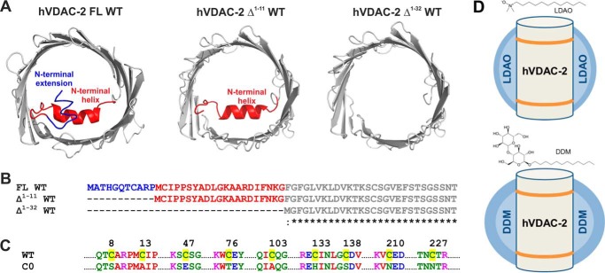
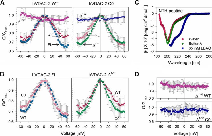
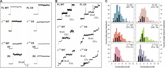
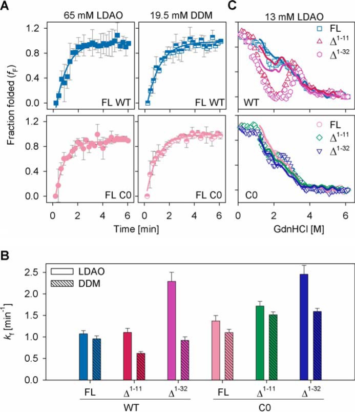
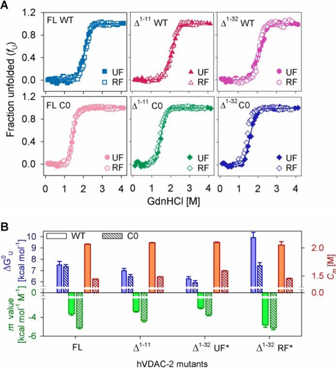
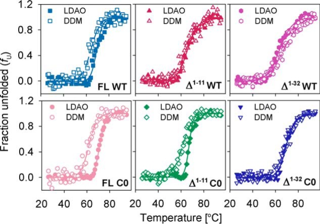
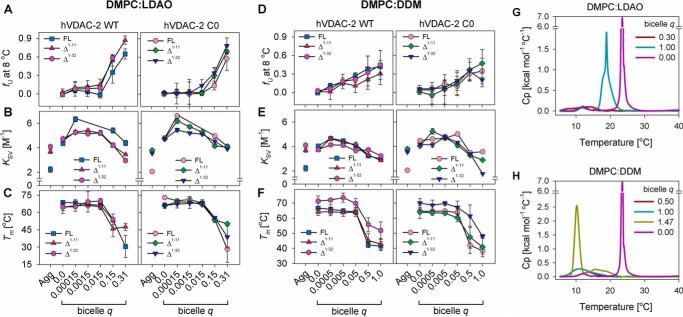
Human voltage-dependent anion channel-2 (hVDAC-2) functions primarily as the crucial anti-apoptotic protein in the outer mitochondrial membrane, and additionally as a gated bidirectional metabolite transporter. The N-terminal helix (NTH), involved in voltage sensing, bears an additional 11-residue extension (NTE) only in hVDAC-2. In this study, we assign a unique role for the NTE as influencing the chaperone-independent refolding kinetics and overall thermodynamic stability of hVDAC-2. Our electrophysiology data shows that the N-helix is crucial for channel activity, whereas NTE sensitizes this isoform to voltage gating. Additionally, hVDAC-2 possesses the highest cysteine content, possibly for regulating reactive oxygen species content. We identify interdependent contributions of the N-helix and cysteines to channel function, and the measured stability in micellar environments with differing physicochemical properties. The evolutionary demand for the NTE in the presence of cysteines clearly emerges from our biochemical and functional studies, providing insight into factors that functionally demarcate hVDAC-2 from the other VDACs.

Keywords: N-terminal domain; gating; ion channel; membrane protein; protein folding; thermodynamics; voltage-dependent anion channel (VDAC).


Figures

FIGURE 1.
FIGURE 1.
hVDAC-2 N-terminal deletion mutants and detergent systems used in this study. A, the I-TASSER (57) modeled structure of the hVDAC-2 barrel (gray) is shown on the left, with NTE in blue and NTH in red. Middle and right panels show the I-TASSER modeled structure of hVDAC-2 Δ1–11 and Δ1–32 WT, respectively. B, multiple sequence alignment of the N-terminal mutants of hVDAC-2, and color coded according to A. C, comparison of hVDAC-2 WT and C0 sequences, highlighting the nine cysteines of WT in yellow, and showing the corresponding replacements in C0. All residues have been color coded according to their physicochemical properties. D, schematic representation of the hVDAC-2 barrel in LDAO (upper panel) and DDM (lower panel) micelles. The hydrophobic core of the micelles is shown in light blue and the polar headgroups in dark blue. The hydrophobic core of the protein is demarcated by orange lines. Note the spherical and oblate shapes of LDAO and DDM micelles, respectively. Chemical structures of LDAO and DDM are also provided to highlight the size of the headgroups.
FIGURE 2.
FIGURE 2.
Channel conductance measurements of hVDAC-2 and its mutants in lipid bilayers. A, G/Gmax plots for WT proteins (left panels) and C0 constructs (right panels), obtained in response to a voltage gradient ranging from +60 to −60 mV in DiPhPC + 0.1% cholesterol membrane. The data shows the importance of the NTH in voltage gating of hVDAC-2. In both the WT and C0 barrels loss of voltage dependence is seen upon deletion of the N-helix. WT constructs: FL (square), Δ1–11 (triangle), and Δ1–32 (hexagon); C0 constructs: FL (circle), Δ1–11 (diamond), and Δ1–32 (inverted triangle). B, comparison of the G/Gmax plots of WT and C0 mutants of FL hVDAC-2 (left panel) and Δ1–11 hVDAC-2 (right panel). C, the NTH peptide is unstructured (random coil conformation) in both water and Buffer A, and shows a helical conformation only in 65 mm LDAO (in Buffer A), when measured using far-UV CD. The data suggests that NTH can interact with the lipid environment in hVDAC-2, which can promote NTH structuring. D, exogenously supplemented NTH peptide is unable to restore voltage-dependent gating in Δ1–32 mutants. Experiments were carried out with protein:peptide ratios ranging from 1:10 to 1:1000 and results for 1:1000 ratio is shown. Error bars in panels A, B, and D are derived from at least four independent experiments.
FIGURE 3.
FIGURE 3.
Channel insertion events of hVDAC-2 and its mutants. A, representative single channel insertion events of hVDAC-2 mutants in DiPhPC + 0.1% cholesterol membrane at 10 mV. Two events are shown for Δ1–32 mutants, representing insertion at ∼2 nS ((a), subconductance state) and ∼4 nS ((b), open state). B, representative stepwise insertion events of hVDAC-2 mutants. C, histograms obtained from channel insertion events; those depicted in color are obtained from stepwise insertion events shown in B with the counts given on the left. Histograms in black are derived from single channel insertion events shown in A, with the counts shown on the right. Insertion of a few VDAC channels in the DiPhPC membrane promotes further channel insertion events. Hence, simultaneous insertion of 2–4 channels gives us a broad range in the conductance histogram derived from stepwise insertions. Both methods show that the removal of NTE + NTH gives rise to higher instances of channel insertions in the subconductance state. Numbers in brackets indicate total number of insertion events considered for deriving the histograms from stepwise and single channel insertion events, respectively.
FIGURE 4.
FIGURE 4.
Re/unfolding experiments in LDAO and DDM micelles. A, refolding kinetics of hVDAC-2 WT (upper panel) and C0 (lower panel) in 65 mm LDAO (left) and 19.5 mm DDM (right) measured using changes in Trp fluorescence anisotropy. Solid lines represent fits to a single exponential function, used to derive the rate of refolding (kf) shown in B. The differences obtained upon comparison of FL and Δ1–11 with Δ1–32, and WT with C0 constructs in LDAO or DDM are significant (p value: <0.05, derived using one-way analysis of variance method). However, taking into consideration the rapid refolding kinetics of all hVDAC-2 constructs and the experimental dead time (see “Experimental Procedures”), these data must be interpreted with extreme caution. C, chemical denaturation studies in 13 mm LDAO using GdnHCl, at 25 °C. The unfolding and refolding curves are represented using symbols and lines, respectively, and were recorded after 16 h of incubation at 25 °C. Open symbols are used and error bars are hidden for clarity. The symbol/color schemes are retained from Fig. 2A. The Δ1–11 and Δ1–32 WT mutants are most affected, and considerable protein aggregation is observed. Although C0 and its mutants also aggregate, the fluorescence intensity is not drastically affected, possibly due to the different nature of aggregates in this case. Error bars in A denote S.D. values derived from independent experiments and in B show the goodness of fit.
FIGURE 5.
FIGURE 5.
Chemical denaturation studies of hVDAC-2 in DDM and LDAO. A, the unfolding and refolding curves of hVDAC-2 mutants after a 24-h incubation at 25 °C in DDM shows no hysteresis in FL and Δ1–11 constructs. Mild hysteresis is observed in Δ1–32 constructs, and apparent unfolding (UF*) and refolding (RF*) thermodynamic parameters were derived. Solid lines represent fits to the two-state equation (used to derive the thermodynamic parameters in B) and have been only shown for the unfolding curves. B, summary of thermodynamic parameters in 3.9 mm DDM. The free energy of unfolding is shown in blue bars, m value in green, and Cm in red. Asterisk (*) in Δ1–32 mutants represents the apparent thermodynamic parameters. For convenient comparison, the sign of ΔGapp0 and m values for RF* for Δ1–32 are reversed. In panel A errors have not been shown for purpose of clarity and those in panel B represent goodness of fit.
FIGURE 6.
FIGURE 6.
Thermal denaturation studies of hVDAC-2. Data were obtained from 13 mm LDAO (filled symbols) and 3.9 mm DDM (open symbols) and the parameters derived from the data and fits are summarized in Table 4. Dashed and solid lines denote fits to the two-state equation for LDAO and DDM, respectively. Overall, removal of the NTH drastically affects the cooperativity in both LDAO and DDM. Error bars have been omitted for clarity.
FIGURE 7.
FIGURE 7.
Stability of hVDAC-2 in DMPC:LDAO and DMPC:DDM bicelles. A and D, comparison of the unfolded fraction (fU) determined in bicellar systems from MRE215, measured at 8 °C using CD, with increasing DMPC concentrations. B and E, Stern-Volmer constants (KSV) obtained from acrylamide quenching studies of the refolded proteins in DMPC:LDAO and DMPC:DDM at 25 °C are depicted, and compared with the aggregated protein (Agg). C and F, dependence of the Tm of the refolded proteins to bicelle q, are shown for DMPC:LDAO and DMPC:DDM, and are derived from changes in MRE215. A–F, symbol/color schemes retained from Fig. 2A. Error bars depict the S.D. obtained from independent experiments. The data shown in panels A–C for refolded FL WT and FL C0 in DMPC:LDAO was originally published in Ref. , and has been reused here with permission, for comparison with the other constructs. G and H, differential scanning calorimetry profiles of empty DMPC:LDAO and DMPC:DDM bicelles, respectively, compared with pure DMPC vesicles (purple line).
FIGURE 8.
FIGURE 8.
Cladogram comparing sequence similarities in VDAC-2 across various organisms, created using ClustalW2 Phylogeny (58, 59). The 10 organisms with a cysteine-rich VDAC-2 sequence that evolved from a common root (*) and show sequence conservation (see supplemental Fig. S1) are boxed. We believe that during sequence divergence, a Cys-rich barrel was selected earlier in evolution, after which the NTE was added to the primary sequence of VDAC-2. As a result, the conserved cysteines (Cys-133 and Cys-138, numbered according to hVDAC-2) are retained across all organisms that evolved from the root, but sequence similarity across the N-terminal residues is restricted to the higher mammals (see supplemental Fig. S1 for the alignment). Across different VDAC isoforms Cys-138 (numbered according to hVDAC-2) shows conservation in the first 10 organisms in both VDAC-1 and -2 (see supplemental Fig. S1 and S2), whereas Cys-133 (numbered according to hVDAC-2) is conserved in VDAC-2 and -3 (see supplemental Fig. S2 and S3).

Similar articles

Cited by

References

    1. Messina A., Reina S., Guarino F., and De Pinto V. (2012) VDAC isoforms in mammals. Biochim. Biophys. Acta 1818, 1466–1476 - PubMed
    1. Shoshan-Barmatz V., Ben-Hail D., Admoni L., Krelin Y., and Tripathi S. S. (2015) The mitochondrial voltage-dependent anion channel 1 in tumor cells. Biochim. Biophys. Acta 1848, 2547–2575 - PubMed
    1. Shoshan-Barmatz V., De Pinto V., Zweckstetter M., Raviv Z., Keinan N., and Arbel N. (2010) VDAC, a multi-functional mitochondrial protein regulating cell life and death. Mol. Aspects Med. 31, 227–285 - PubMed
    1. Bayrhuber M., Meins T., Habeck M., Becker S., Giller K., Villinger S., Vonrhein C., Griesinger C., Zweckstetter M., and Zeth K. (2008) Structure of the human voltage-dependent anion channel. Proc. Natl. Acad. Sci. U.S.A. 105, 15370–15375 - PMC - PubMed
    1. Hiller S., Garces R. G., Malia T. J., Orekhov V. Y., Colombini M., and Wagner G. (2008) Solution structure of the integral human membrane protein VDAC-1 in detergent micelles. Science 321, 1206–1210 - PMC - PubMed

Publication types

MeSH terms