. 2015 Dec 4;290(49):29259-69.
doi: 10.1074/jbc.M115.691147. Epub 2015 Oct 20.

The Cysteine-rich Domain of the DHHC3 Palmitoyltransferase Is Palmitoylated and Contains Tightly Bound Zinc

Affiliations

The Cysteine-rich Domain of the DHHC3 Palmitoyltransferase Is Palmitoylated and Contains Tightly Bound Zinc

Colin D Gottlieb et al. J Biol Chem. .

Abstract

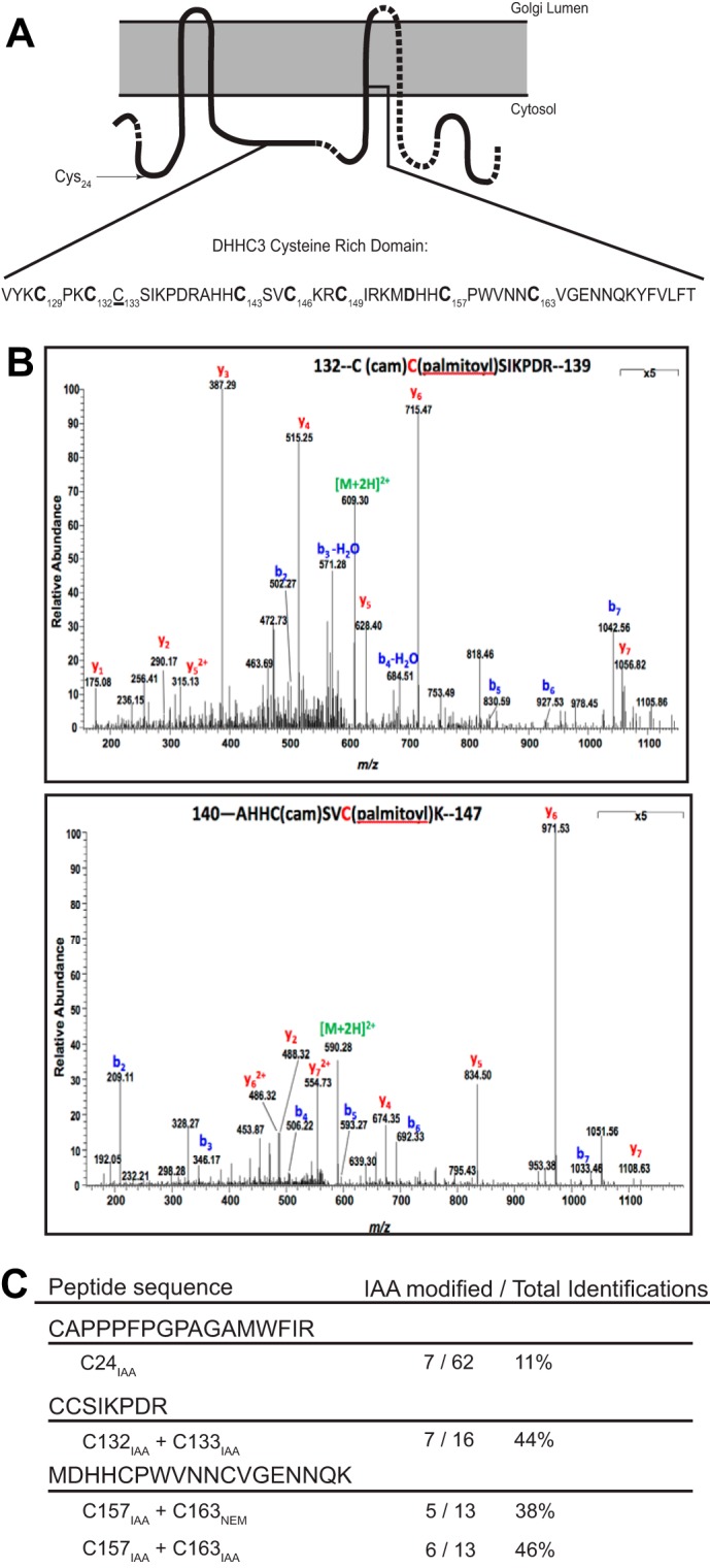
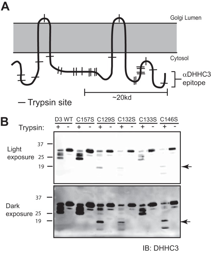
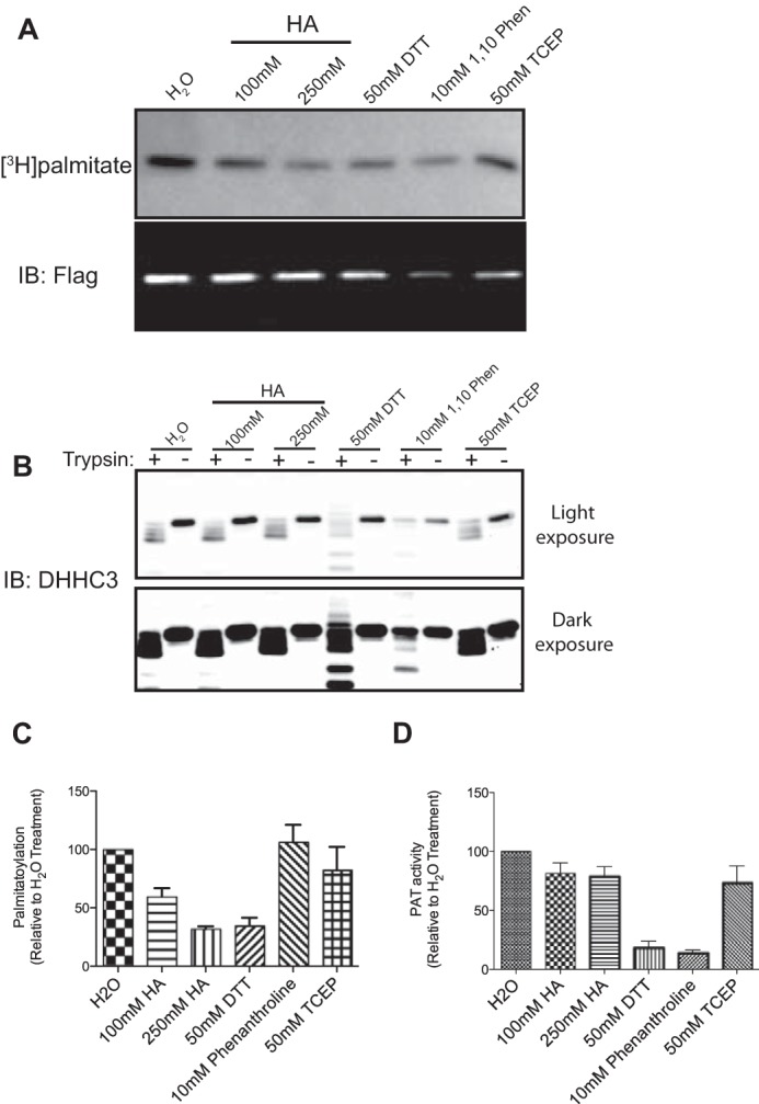
DHHC palmitoyltransferases catalyze the addition of the fatty acid palmitate to proteins on the cytoplasmic leaflet of cell membranes. There are 23 members of the highly diverse mammalian DHHC protein family, all of which contain a conserved catalytic domain called the cysteine-rich domain (CRD). DHHC proteins transfer palmitate via a two-step catalytic mechanism in which the enzyme first modifies itself with palmitate in a process termed autoacylation. The enzyme then transfers palmitate from itself onto substrate proteins. The number and location of palmitoylated cysteines in the autoacylated intermediate is unknown. In this study, we present evidence using mass spectrometry that DHHC3 is palmitoylated at the cysteine in the DHHC motif. Mutation of highly conserved CRD cysteines outside the DHHC motif resulted in activity deficits and a structural perturbation revealed by limited proteolysis. Treatment of DHHC3 with chelating agents in vitro replicated both the specific structural perturbations and activity deficits observed in conserved cysteine mutants, suggesting metal ion-binding in the CRD. Using the fluorescent indicator mag-fura-2, the metal released from DHHC3 was identified as zinc. The stoichiometry of zinc binding was measured as 2 mol of zinc/mol of DHHC3 protein. Taken together, our data demonstrate that coordination of zinc ions by cysteine residues within the CRD is required for the structural integrity of DHHC proteins.

Keywords: S-acylation; acyltransferase; fatty acylation; mass spectrometry (MS); post-translational modification (PTM); protein palmitoylation; zinc.


Figures

FIGURE 1.
FIGURE 1.
Multiple palmitoylated cysteines in DHHC3 can be identified by mass spectrometry. A, diagram of the predicted topology of DHHC3. The portions drawn with a solid line indicate sequence coverage observed with the tandem C4-C18 LC/MS/MS method described under “Experimental Procedures.” The CRD sequence is displayed below, with highly conserved cysteines in boldface type. The location of unconserved cysteines 24 (arrow) and 133 (underlined) is indicated. B, direct MS/MS identification of DHHC3 palmitoyl-peptides from a tryptic digest of DHHC3 purified from insect cells. A mass shift (238.2300 atomic mass units) was identified on Cys-133 and Cys-146 corresponding to the molecular weight of palmitate. C, peptides identified in the MS/MS analysis of an acyl switch assay performed on DHHC3 purified from insect cells. Unmodified cysteines were blocked with N-ethylmaleimide, and palmitate was replaced with iodoacetamide (IAA) in a hydroxylamine-dependent manner. The frequency of carbamidomethyl (iodoacetamide-modified) cysteine identifications, relative to total peptide identifications, is listed. Carbamidomethyl-Cys-157 was identified in 11 out of 13 identified peptides observed in this analysis.
FIGURE 2.
FIGURE 2.
Mutation of cysteines 146 and 157, but not other CRD cysteines, reduces the palmitoylation level of DHHC3 in cells. A, total palmitoylation levels of purified DHHC3 WT and DHHC3 mutants were detected using acyl-biotin exchange. Palmitate on DHHC3 was replaced with biotin-HPDP in a hydroxylamine-dependent manner. Increasing amounts of each construct were resolved by SDS-PAGE in order to compare the signal from different constructs. Biotinylated DHHC3 was then blotted for both total protein (IB: Flag) and for biotinylation levels (IB: streptavidin). B, the average streptavidin signals from the best-normalized amounts of each construct are displayed. Mutation of Cys-146 and Cys-157 reduced palmitoylation of DHHC3, as indicated by streptavidin-FITC signals. Mean and S.E. (error bars) are shown for three or four experiments for each protein, and p values were calculated using two-tailed Student's t tests.
FIGURE 3.
FIGURE 3.
Mutation of conserved cysteines in the CRD results in PAT activity deficits. PAT activity of purified DHHC3 WT and cysteine mutant proteins was assayed by combining 20–30 nm purified enzyme with 1 μm [3H]palmitoyl-CoA and 1 μm myristoylated Gαi as a protein substrate. A, [3H]palmitate transferred to Gαi was measured by liquid scintillation counting as described under “Experimental Procedures.” Non-enzymatic acylation of Gαi was measured in the absence of DHHC3 and subtracted from reactions containing enzyme. Enzyme activity was normalized to WT DHHC3. Mean and S.E. (error bars) are shown for 3–5 experiments for each mutant protein assayed. B, fluorography analysis demonstrates that both autoacylation (DHHC3 band) and transfer activity (Gαi band) are reduced in conserved cysteine mutants (C157S, C129S, C132S, and C146S) but not in the unconserved C133S mutant. Results are representative of at least three experiments.
FIGURE 4.
FIGURE 4.
Transpalmitoylation assays suggest that the mutation of conserved cysteines disrupts the tertiary structure of DHHC3. A, membranes were isolated from Sf9 insect cells expressing a mutant DHHC3-FLAG/His6 protein (bottom band) alone or in combination with a Myc3/His10-tagged DHHC3 WT protein (higher molecular weight band). Isolated membranes were normalized for total protein and combined with 1 μm [3H]palmitoyl-CoA (final). The top panel shows a representative fluorographic analysis of the amount of [3H]palmitate accumulated on WT and mutant DHHC3. Protein levels of DHHC3 are displayed in the immunoblot (bottom). Results are representative of at least three experiments. B, densitometry analysis of the average fluorography band produced by each mutant DHHC3 protein when palmitoylated in trans by DHHC3 WT. Error bars, S.E. for at least three experiments; *, p < 0.05, resulting from a two-tailed Student's t test comparing each double mutant with the C157S single mutant.
FIGURE 5.
FIGURE 5.
Limited proteolysis assays identify structural perturbations in conserved CRD cysteine mutants of DHHC3. A, diagram of the predicted topology of DHHC3. The approximate locations of potential trypsin cleavage sites are marked with perpendicular lines. The location of the DHHC3 antibody epitope is indicated. B, purified WT DHHC3 or single cysteine mutants of DHHC3 were combined with trypsin and incubated on ice for 45 min. The reactions were stopped with sample buffer containing soybean trypsin inhibitor and resolved by blotting with an antibody raised against the C-terminal 15 amino acids of DHHC3 (29). Partial proteolysis of DHHC3 mutants C129S, C132S, and C146S produces a ∼20-kDa fragment (arrowhead) that is absent in the proteolytic patterns of DHHC3 WT, DHHS3(C157S), and DHHC3(C133S). Results are representative of at least three experiments. IB, immunoblot.
FIGURE 6.
FIGURE 6.
Treatment with chelating reagents replicates characteristics of conserved CRD cysteine mutants. DHHC3 WT was metabolically labeled with [3H]palmitate in Sf9 insect cells to enable monitoring of palmitate removal. Radiolabeled protein was purified using Ni-NTA resin. Purified protein was buffer-exchanged to remove imidazole and then treated with DTT or two different concentrations of HA to remove palmitate from the enzyme. 1,10-Phenanthroline and TCEP were included to control for the chelating activity and the reducing activity of DTT, respectively. A, residual [3H]palmitate labeling following treatment is shown in the top panel; an immunoblot (IB) of protein levels is displayed below. B, chemically treated DHHC3 was subjected to limited proteolysis and analyzed by an immunoblot with a DHHC3 antibody. Both a light and dark exposure of the results are presented. Treatment with DTT and 1,10-phenanthroline, but not HA or TCEP, replicates the limited proteolysis pattern of conserved CRD cysteine mutants (C129S, C132S, and C146S) shown in Fig. 5B. C, quantification of palmitate remaining on DHHC3 post-treatment (normalized to protein), relative to an H2O-treated control. Mean and S.E. (error bars) are shown for three experiments. D, the relative activity of DHHC3 following chemical treatment in these experiments is shown. Chemically treated DHHC3 was buffer-exchanged into a non-denaturing buffer suitable for PAT assays and divided into two aliquots. One aliquot was heat-inactivated to control for spontaneous acylation, and the second was held on ice. PAT activity was assayed as described under “Experimental Procedures.” Spontaneous acylation was subtracted from the value for each experimental sample. PAT activity of DHHC3 treated with water was set at 100%, and the activities of chemically treated samples were normalized to that value. Mean and S.E. are shown for three experiments.
FIGURE 7.
FIGURE 7.
Direct detection of zinc bound to purified DHHC3. A, the fluorescent zinc indicator mag-fura-2 responds to increasing zinc concentrations by shifting peak fluorescence from 375 to 325 nm on an excitation wavelength spectrum. B, at low micromolar ion concentrations, the ratio of fluorescence emitted by mag-fura-2 at excitation wavelengths 325 and 350 nm responds in a positive linear correlation to free Zn2+, very weakly to free Ca2+, and not at all to Cu2+, Fe2+, Mg2+, Ni2+, or Mn2+. C, FLAG-tagged DHHC3 was purified to near homogeneity using ANTI-FLAG® M2-agarose affinity gel (Sigma). Elutions were pooled and applied to a preparative SEC. Fractions containing a monodisperse population of DHHC3 (shaded area under the curve) were selected and pooled. D, the pool was confirmed to be monodisperse by applying a sample to an analytical SEC monitored by intrinsic tryptophan fluorescence. To quantify zinc bound to DHHC3-FLAG, aliquots from the monodisperse pool were digested overnight with proteinase K to release zinc from the protein. Free zinc in these samples was detected with mag-fura-2 and compared with a buffer-matched standard curve of known zinc concentrations. E, total DHHC-FLAG protein was quantified in parallel samples (minus proteinase K) as described under “Experimental Procedures.” The zinc and protein concentrations are reported as concentrations ± S.E. and suggest that each molecule of DHHC3 binds two zinc ions.

Similar articles

Cited by

References

    1. Roth A. F., Wan J., Bailey A. O., Sun B., Kuchar J. A., Green W. N., Phinney B. S., Yates J. R. 3rd, and Davis N. G. (2006) Global analysis of protein palmitoylation in yeast. Cell 125, 1003–1013 - PMC - PubMed
    1. Kang R., Wan J., Arstikaitis P., Takahashi H., Huang K., Bailey A. O., Thompson J. X., Roth A. F., Drisdel R. C., Mastro R., Green W. N., Yates J. R. 3rd, Davis N. G., and El-Husseini A. (2008) Neural palmitoyl-proteomics reveals dynamic synaptic palmitoylation. Nature 456, 904–909 - PMC - PubMed
    1. Martin B. R., and Cravatt B. F. (2009) Large-scale profiling of protein palmitoylation in mammalian cells. Nat. Methods 6, 135–138 - PMC - PubMed
    1. Sanders S. S., Martin D. D., Butland S. L., Lavallée-Adam M., Calzolari D., Kay C., Yates J. R. 3rd, and Hayden M. R. (2015) Curation of the mammalian palmitoylome indicates a pivotal role for palmitoylation in diseases and disorders of the nervous system and cancers. PLoS Comput. Biol. 11, e1004405. - PMC - PubMed
    1. Salaun C., Greaves J., and Chamberlain L. H. (2010) The intracellular dynamic of protein palmitoylation. J. Cell Biol. 191, 1229–1238 - PMC - PubMed