

The key target of neuroprotection after the onset of ischemic stroke: secretory pathway Ca(2+)-ATPase 1
- PMID: 26487855
- PMCID: PMC4590240
- DOI: 10.4103/1673-5374.162760
The key target of neuroprotection after the onset of ischemic stroke: secretory pathway Ca(2+)-ATPase 1
Abstract
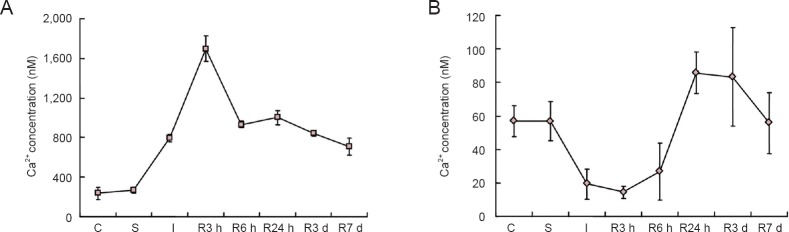
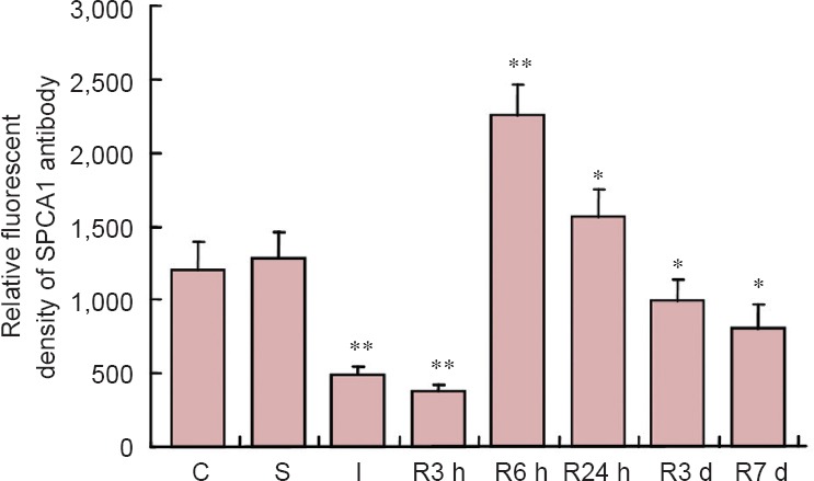
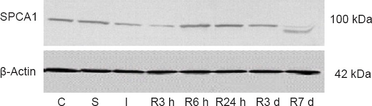
The regulatory mechanisms of cytoplasmic Ca(2+) after myocardial infarction-induced Ca(2+) overload involve secretory pathway Ca(2+)-ATPase 1 and the Golgi apparatus and are well understood. However, the effect of Golgi apparatus on Ca(2+) overload after cerebral ischemia and reperfusion remains unclear. Four-vessel occlusion rats were used as animal models of cerebral ischemia. The expression of secretory pathway Ca(2+)-ATPase 1 in the cortex and hippocampus was detected by immunoblotting, and Ca(2+) concentrations in the cytoplasm and Golgi vesicles were determined. Results showed an overload of cytoplasmic Ca(2+) during ischemia and reperfusion that reached a peak after reperfusion. Levels of Golgi Ca(2+) showed an opposite effect. The expression of Golgi-specific secretory pathway Ca(2+)-ATPase 1 in the cortex and hippocampus decreased before ischemia and reperfusion, and increased after reperfusion for 6 hours. This variation was similar to the alteration of calcium in separated Golgi vesicles. These results indicate that the Golgi apparatus participates in the formation and alleviation of calcium overload, and that secretory pathway Ca(2+)-ATPase 1 tightly responds to ischemia and reperfusion in nerve cells. Thus, we concluded that secretory pathway Ca(2+)-ATPase 1 plays an essential role in cytosolic calcium regulation and its expression can be used as a marker of Golgi stress, responding to cerebral ischemia and reperfusion. The secretory pathway Ca(2+)-ATPase 1 can be an important neuroprotective target of ischemic stroke.
Keywords: Ca2+ pump; Golgi Ca2+; Golgi apparatus; Golgi stress; NSFC grant; brain injury; cytoplasmic Ca2+; global cerebral ischemia; homeostasis; nerve regeneration; neural protection; neural regeneration; secretory pathway Ca2+-ATPase 1.
Conflict of interest statement
Figures
Similar articles
-
Changes in secretory pathway Ca(2+)-ATPase 2 following focal cerebral ischemia/reperfusion injury.Neural Regen Res. 2013 Jan 5;8(1):76-82. doi: 10.3969/j.issn.1673-5374.2013.01.010. Neural Regen Res. 2013. PMID: 25206375 Free PMC article.
-
A New Approach of Short Wave Protection against Middle Cerebral Artery Occlusion/Reperfusion Injury via Attenuation of Golgi Apparatus Stress by Inhibition of Downregulation of Secretory Pathway Ca(2+)-ATPase Isoform 1 in Rats.J Stroke Cerebrovasc Dis. 2016 Jul;25(7):1813-1822. doi: 10.1016/j.jstrokecerebrovasdis.2016.03.033. Epub 2016 Apr 25. J Stroke Cerebrovasc Dis. 2016. PMID: 27133772
-
Olfactory Mucosa Mesenchymal Stem Cells Alleviate Cerebral Ischemia/Reperfusion Injury Via Golgi Apparatus Secretory Pathway Ca2+ -ATPase Isoform1.Front Cell Dev Biol. 2020 Oct 30;8:586541. doi: 10.3389/fcell.2020.586541. eCollection 2020. Front Cell Dev Biol. 2020. PMID: 33195239 Free PMC article.
-
Cross-talk of intracellular calcium stores in the response to neuronal ischemia and ischemic tolerance.Gen Physiol Biophys. 2009;28 Spec No Focus:F104-14. Gen Physiol Biophys. 2009. PMID: 20093720 Review.
-
PMR1/SPCA Ca2+ pumps and the role of the Golgi apparatus as a Ca2+ store.Pflugers Arch. 2003 May;446(2):148-53. doi: 10.1007/s00424-003-1011-5. Epub 2003 Feb 15. Pflugers Arch. 2003. PMID: 12739151 Review.
Cited by
-
Aerobic exercise combined with huwentoxin-I mitigates chronic cerebral ischemia injury.Neural Regen Res. 2017 Apr;12(4):596-602. doi: 10.4103/1673-5374.205099. Neural Regen Res. 2017. PMID: 28553340 Free PMC article.
-
OM-MSCs Alleviate the Golgi Apparatus Stress Response following Cerebral Ischemia/Reperfusion Injury via the PEDF-PI3K/Akt/mTOR Signaling Pathway.Oxid Med Cell Longev. 2021 Nov 13;2021:4805040. doi: 10.1155/2021/4805040. eCollection 2021. Oxid Med Cell Longev. 2021. PMID: 34815829 Free PMC article.
-
BCL2L13: physiological and pathological meanings.Cell Mol Life Sci. 2021 Mar;78(6):2419-2428. doi: 10.1007/s00018-020-03702-9. Epub 2020 Nov 17. Cell Mol Life Sci. 2021. PMID: 33201252 Free PMC article. Review.
-
Pathways Involved in Oxygen Glucose Deprivation Damage of Astrocytes.J Mol Neurosci. 2017 Jan;61(1):115-122. doi: 10.1007/s12031-016-0832-6. Epub 2016 Sep 6. J Mol Neurosci. 2017. PMID: 27601172
-
Crosstalk among Calcium ATPases: PMCA, SERCA and SPCA in Mental Diseases.Int J Mol Sci. 2021 Mar 10;22(6):2785. doi: 10.3390/ijms22062785. Int J Mol Sci. 2021. PMID: 33801794 Free PMC article. Review.
References
-
- Ausubel FM, Brent R, Kingston RE. Short Protocol of Molecular Biology. 5th Edition. Singapore: Johns-Wiley Press, Singapore; 2008.
-
- Behne MJ, Tu CL, Aronchik I, Epstein E, Bench G, Bikle DD, Pozzan T, Mauro TM. Human keratinocyte ATP2C1 localizes to the Golgi and controls Golgi Ca 2+ stores. J Invest Dermatol. 2003;121:688–694. - PubMed
-
- Berridge MJ, Lipp P, Bootman MD. The versatility and universality of calcium signalling. Nature Rev Mol Cell Biol. 2000;1:11–21. - PubMed
-
- Berridge MJ, Bootman MD, Roderick HL. Calcium signaling: dynamics, homeostasis and remodeling. Nat Rev. 2003;4:517–529. - PubMed
LinkOut - more resources
Full Text Sources
Other Literature Sources
Miscellaneous