

Improvement of Distribution and Osteogenic Differentiation of Human Mesenchymal Stem Cells by Hyaluronic Acid and β-Tricalcium Phosphate-Coated Polymeric Scaffold In Vitro
- PMID: 26487981
- PMCID: PMC4599126
- DOI: 10.1089/biores.2015.0021
Improvement of Distribution and Osteogenic Differentiation of Human Mesenchymal Stem Cells by Hyaluronic Acid and β-Tricalcium Phosphate-Coated Polymeric Scaffold In Vitro
Abstract
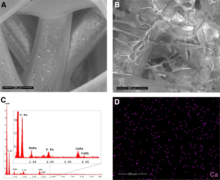
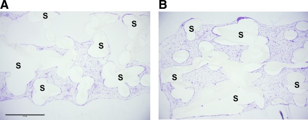
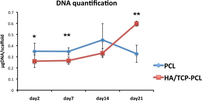
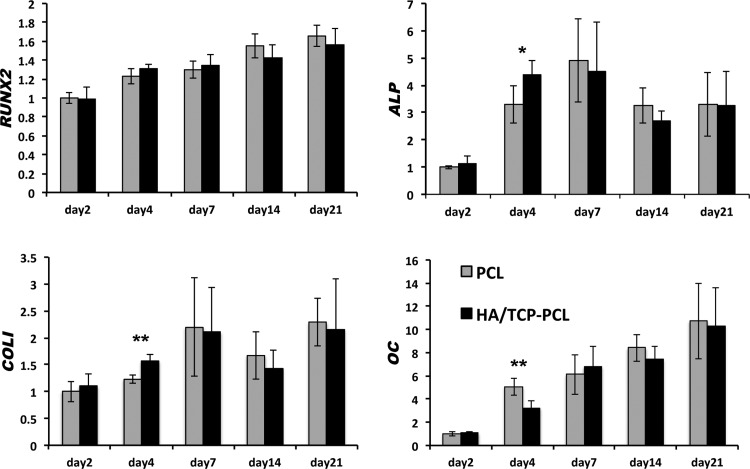
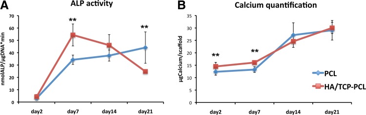
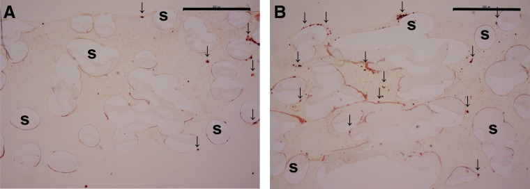
Bone tissue engineering requires a well-designed scaffold that can be biodegradable, biocompatible, and support the stem cells to osteogenic differentiation. Porous polycaprolactone (PCL) scaffold prepared by fused deposition modeling is an attractive biomaterial that has been used in clinic. However, PCL scaffolds lack biological function and osteoinductivity. In this study, we functionalized the PCL scaffolds by embedding them with a matrix of hyaluronic acid/β-tricalcium phosphate (HA/TCP). Human mesenchymal stem cells (MSCs) were cultured on scaffolds with and without coating to investigate proliferation and osteogenic differentiation. The DNA amount was significantly higher in the HA/TCP-coated scaffold on day 21. At the gene expression level, HA/TCP coating significantly increased the expression of ALP and COLI on day 4. These data correlated with the ALP activity peaking on day 7 in the HA/TCP-coated scaffold. Scanning electron microscope and histological analysis revealed that the cell matrix and calcium deposition were distributed more uniformly in the coated scaffolds compared to scaffolds without coating. In conclusion, the HA/TCP coating improved cellular proliferation, osteogenic differentiation, and uniform distribution of the cellular matrix in vitro. The HA/TCP-PCL scaffold holds great promise to accommodate human bone marrow-derived MSCs for bone reconstruction purposes, which warrants future in vivo studies.
Keywords: bone tissue engineering; cell distribution; human mesenchymal stem cell; osteogenic differentiation; scaffold.
Figures

Similar articles
-
Functionalization of polycaprolactone scaffolds with hyaluronic acid and β-TCP facilitates migration and osteogenic differentiation of human dental pulp stem cells in vitro.Tissue Eng Part A. 2015 Feb;21(3-4):729-39. doi: 10.1089/ten.TEA.2014.0177. Epub 2014 Nov 11. Tissue Eng Part A. 2015. PMID: 25252795 Free PMC article.
-
Effects of three-dimensionally printed polycaprolactone/β-tricalcium phosphate scaffold on osteogenic differentiation of adipose tissue- and bone marrow-derived stem cells.Arch Craniofac Surg. 2018 Sep;19(3):181-189. doi: 10.7181/acfs.2018.01879. Epub 2018 Sep 20. Arch Craniofac Surg. 2018. PMID: 30282427 Free PMC article.
-
Osteogenesis of adipose-derived stem cells on polycaprolactone-β-tricalcium phosphate scaffold fabricated via selective laser sintering and surface coating with collagen type I.J Tissue Eng Regen Med. 2016 Oct;10(10):E337-E353. doi: 10.1002/term.1811. Epub 2013 Aug 16. J Tissue Eng Regen Med. 2016. PMID: 23955935
-
Osteogenic differentiation and proliferation potentials of human bone marrow and umbilical cord-derived mesenchymal stem cells on the 3D-printed hydroxyapatite scaffolds.Sci Rep. 2022 Nov 14;12(1):19509. doi: 10.1038/s41598-022-24160-2. Sci Rep. 2022. PMID: 36376498 Free PMC article.
-
Osteogenic and angiogenic potentials of monocultured and co-cultured human-bone-marrow-derived mesenchymal stem cells and human-umbilical-vein endothelial cells on three-dimensional porous beta-tricalcium phosphate scaffold.Acta Biomater. 2013 Jan;9(1):4906-15. doi: 10.1016/j.actbio.2012.08.008. Epub 2012 Aug 16. Acta Biomater. 2013. PMID: 22902820 Free PMC article.
Cited by
-
Characterization and Optimization of the Seeding Process of Adipose Stem Cells on the Polycaprolactone Scaffolds.Stem Cells Int. 2019 Feb 20;2019:1201927. doi: 10.1155/2019/1201927. eCollection 2019. Stem Cells Int. 2019. PMID: 30915123 Free PMC article.
-
Modifications in Gene Expression in the Process of Osteoblastic Differentiation of Multipotent Bone Marrow-Derived Human Mesenchymal Stem Cells Induced by a Novel Osteoinductive Porous Medical-Grade 3D-Printed Poly(ε-caprolactone)/β-tricalcium Phosphate Composite.Int J Mol Sci. 2021 Oct 18;22(20):11216. doi: 10.3390/ijms222011216. Int J Mol Sci. 2021. PMID: 34681873 Free PMC article.
-
Evaluation of the osteogenic potential of rat adipose-derived stem cells with different polycaprolactone/alginate-based nanofibrous scaffolds: an in vitro study.Stem Cell Investig. 2020 Aug 7;7:14. doi: 10.21037/sci-2020-015. eCollection 2020. Stem Cell Investig. 2020. PMID: 32964007 Free PMC article.
-
Osteogenic and Chondrogenic Potential of the Supramolecular Aggregate T-LysYal®.Front Endocrinol (Lausanne). 2020 May 5;11:285. doi: 10.3389/fendo.2020.00285. eCollection 2020. Front Endocrinol (Lausanne). 2020. PMID: 32431670 Free PMC article.
-
Polycaprolactone nanofiber scaffold enhances the osteogenic differentiation potency of various human tissue-derived mesenchymal stem cells.Stem Cell Res Ther. 2017 Jun 24;8(1):148. doi: 10.1186/s13287-017-0588-0. Stem Cell Res Ther. 2017. PMID: 28646917 Free PMC article.
References
-
- Murphy CM, O'Brien FJ, Little DG, et al. . Cell-scaffold interactions in the bone tissue engineering triad. Eur Cell Mater. 2013;26:120–132 - PubMed
-
- Garg T, Goyal AK. Biomaterial-based scaffolds—current status and future directions. Expert Opin Drug Deliv. 2014;11:767–789 - PubMed
-
- Mourino V, Cattalini JP, Roether JA, et al. . Composite polymer-bioceramic scaffolds with drug delivery capability for bone tissue engineering. Expert Opin Drug Deliv. 2013;10:1353–1365 - PubMed
LinkOut - more resources
Full Text Sources
Other Literature Sources