

Case Reports
doi: 10.12998/wjcc.v3.i10.894.
Littoral cell angioma: A case report
Affiliations
- PMID: 26488026
- PMCID: PMC4607808
- DOI: 10.12998/wjcc.v3.i10.894
Item in Clipboard
Case Reports
Littoral cell angioma: A case report
World J Clin Cases.
.
Abstract
Primary splenic lesions are rare entities among which littoral cell angioma (LCA) is a recently described, uncommon vascular lesion that is unique to the spleen. It has heretofore been described primarily in pathologic series and has been found mostly to behave as a benign entity. A few reports of malignant variants have been reported. We present a case report of a solitary LCA discovered after splenectomy for an incidentally discovered splenic lesion, along with a literature review.
Keywords: Littoral cell angioma; Splenic tumor.
Figures
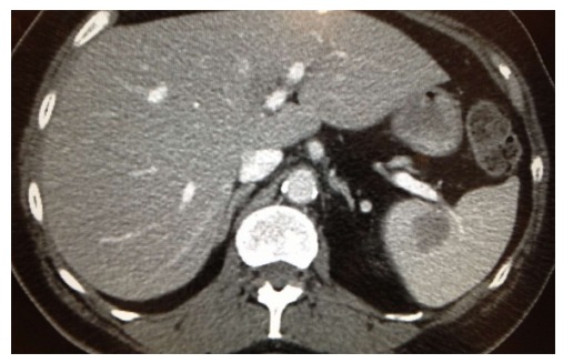
Computed tomography abdomen and pelvis, axial view of hypodense splenic lesion.
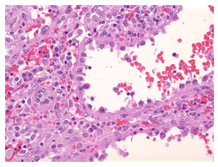
High power view of the tumor demonstrates tall columnar endothelial cells that line the cyst-like spaces. These cells show no cytologic, nuclear atypia or mitotic figures (H and E stain, × 400).
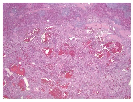
Low power view of the well-demarcated tumor with uninvolved spleen. The tumor has anastomosing vascular channels and cyst-like hemorrhagic spaces.
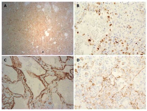
Endothelial cells lining the cyst-like spaces are immunoreactive. A: CD68 (CD68 stain, × 100); B: Histiocytic marker lysozyme (lysozyme stain, × 400); C: Endothelial marker CD34 and the histocytoid cells are negative for CD34 (CD34 stain, × 400); D: Endothelial marker CD31 (CD31 stain, × 400).
References
-
- Abbott RM, Levy AD, Aguilera NS, Gorospe L, Thompson WM. From the archives of the AFIP: primary vascular neoplasms of the spleen: radiologic-pathologic correlation. Radiographics. 2004;24:1137–1163. - PubMed
Publication types
LinkOut - more resources
Full Text Sources
Other Literature Sources
Research Materials
