. 2015 Oct 21;10(10):e0141127.
doi: 10.1371/journal.pone.0141127. eCollection 2015.

A Simple Method for Discovering Druggable, Specific Glycosaminoglycan-Protein Systems. Elucidation of Key Principles from Heparin/Heparan Sulfate-Binding Proteins

Affiliations

A Simple Method for Discovering Druggable, Specific Glycosaminoglycan-Protein Systems. Elucidation of Key Principles from Heparin/Heparan Sulfate-Binding Proteins

Aurijit Sarkar et al. PLoS One. .

Abstract

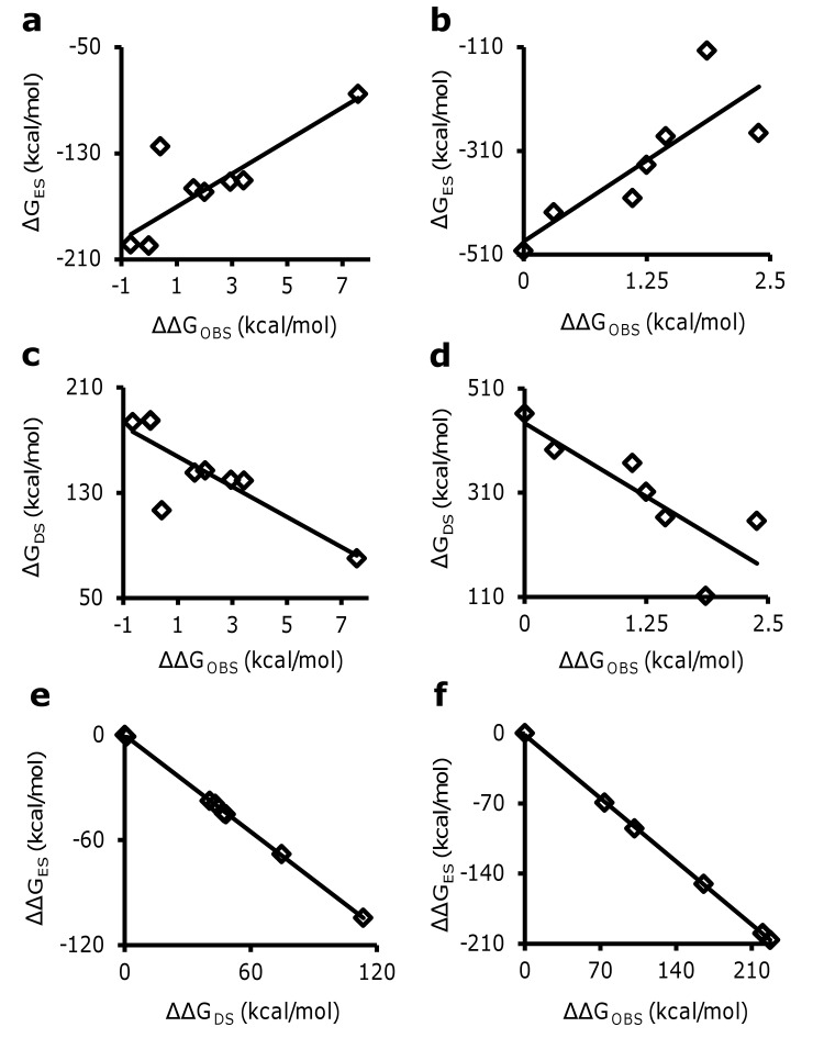
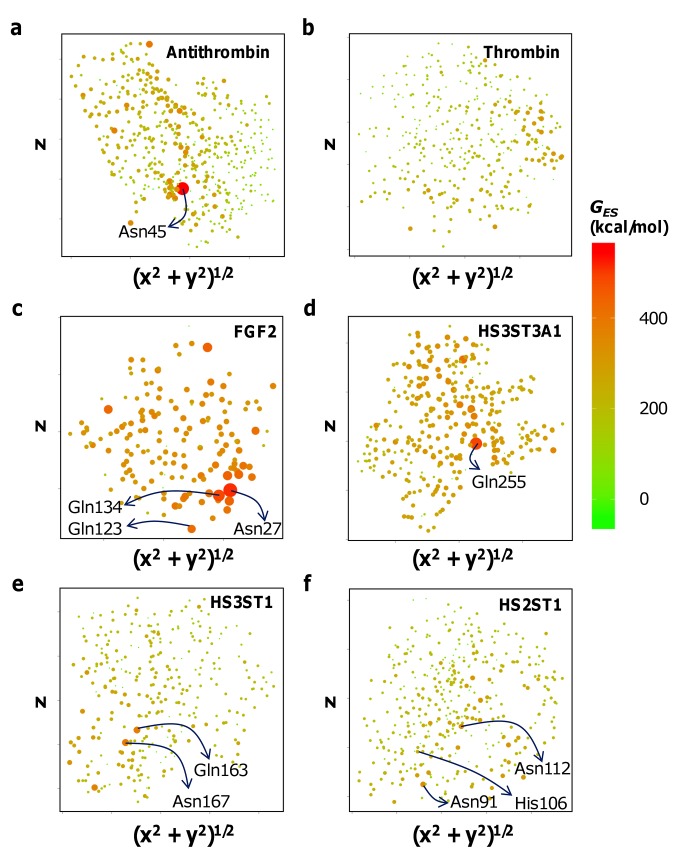
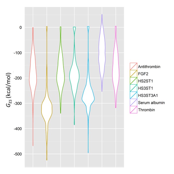
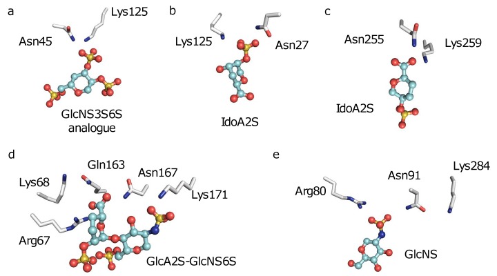
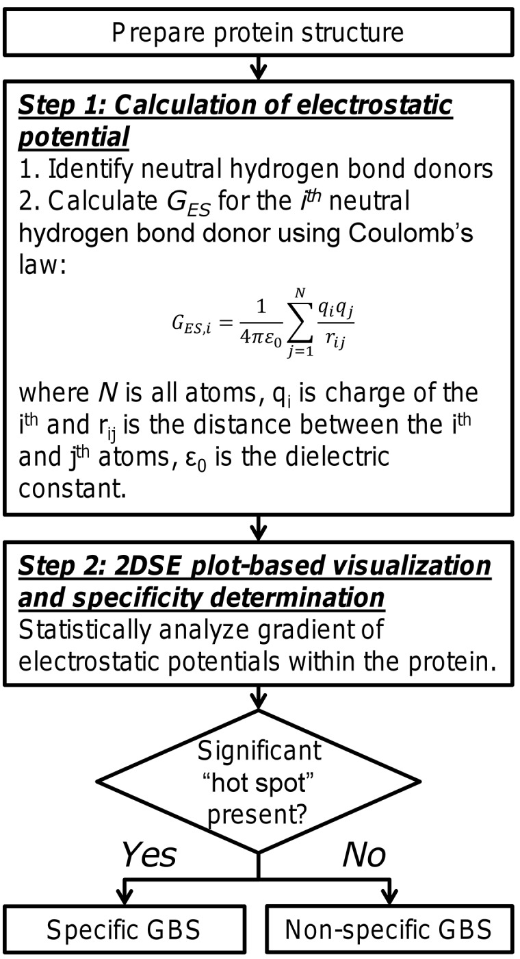
Glycosaminoglycans (GAGs) affect human physiology and pathology by modulating more than 500 proteins. GAG-protein interactions are generally assumed to be ionic and nonspecific, but specific interactions do exist. Here, we present a simple method to identify the GAG-binding site (GBS) on proteins that in turn helps predict high specific GAG-protein systems. Contrary to contemporary thinking, we found that the electrostatic potential at basic arginine and lysine residues neither identifies the GBS consistently, nor its specificity. GBSs are better identified by considering the potential at neutral hydrogen bond donors such as asparagine or glutamine sidechains. Our studies also reveal that an unusual constellation of ionic and non-ionic residues in the binding site leads to specificity. Nature engineers the local environment of Asn45 of antithrombin, Gln255 of 3-O-sulfotransferase 3, Gln163 and Asn167 of 3-O-sulfotransferase 1 and Asn27 of basic fibroblast growth factor in the respective GBSs to induce specificity. Such residues are distinct from other uncharged residues on the same protein structure in possessing a significantly higher electrostatic potential, resultant from the local topology. In contrast, uncharged residues on nonspecific GBSs such as thrombin and serum albumin possess a diffuse spread of electrostatic potential. Our findings also contradict the paradigm that GAG-binding sites are simply a collection of contiguous Arg/Lys residues. Our work demonstrates the basis for discovering specifically interacting and druggable GAG-protein systems based on the structure of protein alone, without requiring access to any structure-function relationship data.


Conflict of interest statement

Competing Interests: The authors have declared that no competing interests exist.

Figures

Fig 1
Fig 1. G ES at arginines and/or lysines does not identify the GBS on a protein.
G ES for Arg/Lys residues are mapped using 2DSE plots for multiple GAG-binding proteins including (a) antithrombin, (b) thrombin, (c) FGF2, (d) HS3ST3A1, (e) HS3ST1 and (f) HS2ST1. The maps reveal that GAG-binding site Arg/Lys residues may not always possess high G ES and not all Arg/Lys with high G ES on a protein are part of the GAG-binding site.
Fig 2
Fig 2. Desolvation energy is critical for quantitative analysis of GAG–protein interaction.
Neither ΔGES (a and b) nor ΔGDS (c and d) alone explain the change in ΔGOBS for antithrombin (a and c) and thrombin (b and d) mutants studied to date. Any enthalpic gain due to electrostatics is opposed by desolvation (R2 = 0.99) in antithrombin (e) as well as in thrombin (f), suggesting that desolvation is critical for quantitative analysis of GAG-protein interactions. In all cases, the correlation was found to be significant at α = 0.05.
Fig 3
Fig 3. 2DSE plots for G ES at neutral hydrogen bond donors.
GAGs bind neutral hydrogen bond donors on the protein that possess significantly high GES. (a) Asn45 of antithrombin GAG-binding site possesses the highest GES within the structure. (b) In contrast, the nonspecific thrombin GAG-binding site demonstrates a diffused GES. Similarly, significantly high G ES are observed at (c) Asn27 of the FGF2 GBS; Asn27Ala mutation affects GAG-binding (ΔΔG~1.1 kcal/mol) almost as much as K125A (ΔΔG~1.7 kcal/mol), which had the largest effect, (d) Asn255 of the HS3ST3A1 GAG-binding site; the N255A mutant is inactive, and (e) Gln163 of HS3ST1; Gln163Ala mutant loses ~65% activity. (f) Diffused GES of HS2ST1 may represent its ability to bind low-sulfated GAGs. However, Asn91 and 112 of the HS2ST1 GAG-binding site possess a potential higher than His106, mutation of which is already known to affect GAG-binding.
Fig 4
Fig 4. Specific proteins demonstrate unique, non-uniform distributions of electrostatic potential across neutral hydrogen-bond donors.
Specific proteins such as antithrombin, FGF2, HS3ST1 and HS3ST3A1 demonstrate at least one location of electrostatic potential that deviates significantly from the mean. Nonspecific GAG-binding sites on proteins such as thrombin and serum albumin demonstrate a uniform, Gaussian distribution of the same, so no location is preferred significantly over another.
Fig 5
Fig 5. The structural basis for existence of hot spots in GBSs.
Nature has designed specific GBSs by placing neutral hydrogen bond donors such as the ND2 and NE2 atoms of Asn and Gln respectively in close proximity to charged Arg or Lys residues, as seen in (a) antithrombin, (b) FGF2, (c) HS3ST3A1, (d) HS3ST1 and (e) HS2ST1. This close proximity maximizes the GES at these residues, thereby generating a specific GBS. Not all atoms are displayed, for the sake of visual clarity.
Fig 6
Fig 6. The two-step algorithm for identification of GBSs on proteins and elucidating their specificity.
The process involves preparation of protein; identification of neutral hydrogen bond donors in the structure; calculation of 2DSE plots for the protein; and evaluation of ‘hot spots’ for deduction of specificity of GAG–protein interaction.

References

    1. Yamada S, Sugahara K, Ozbek S. Evolution of glycosaminoglycans: Comparative biochemical study. Commun Integr Biol. 2011;4: 150–8. 10.4161/cib.4.2.14547 - DOI - PMC - PubMed
    1. Ori A, Wilkinson MC, Fernig DG. A systems biology approach for the investigation of the heparin/heparan sulfate interactome. J Biol Chem. 2011;286: 19892–904. 10.1074/jbc.M111.228114 - DOI - PMC - PubMed
    1. Raman R, Sasisekharan V, Sasisekharan R. Structural insights into biological roles of protein-glycosaminoglycan interactions. Chem Biol. 2005;12: 267–77. 10.1016/j.chembiol.2004.11.020 - DOI - PubMed
    1. Cardin A, Weintraub H. Molecular modeling of protein-glycosaminoglycan interactions. Arter Throm Vas. 1989;9: 21–32. 10.1161/01.ATV.9.1.21 - DOI - PubMed
    1. Sobel M, Soler DF, Kermode JC, Harris RB. Localization and characterization of a heparin binding domain peptide of human von Willebrand factor. J Biol Chem. 1992;267: 8857–62. - PubMed

Publication types