. 2015 Oct 21;10(10):e0139882.
doi: 10.1371/journal.pone.0139882. eCollection 2015.

Heterodimerization of Two Pathological Mutants Enhances the Activity of Human Phosphomannomutase2

Affiliations

Heterodimerization of Two Pathological Mutants Enhances the Activity of Human Phosphomannomutase2

Giuseppina Andreotti et al. PLoS One. .

Abstract

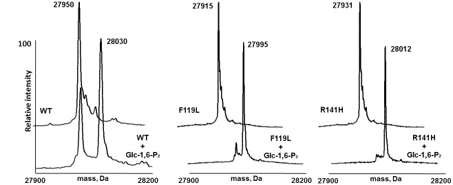
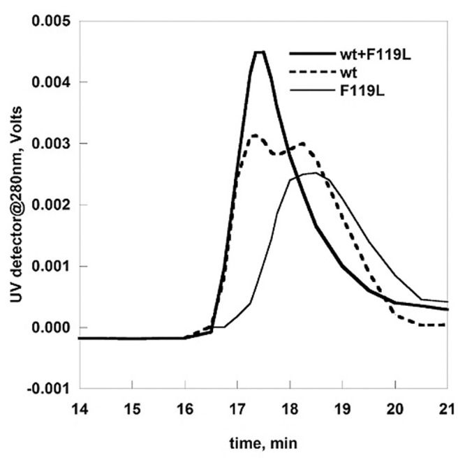
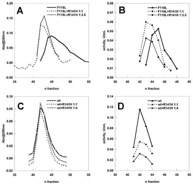
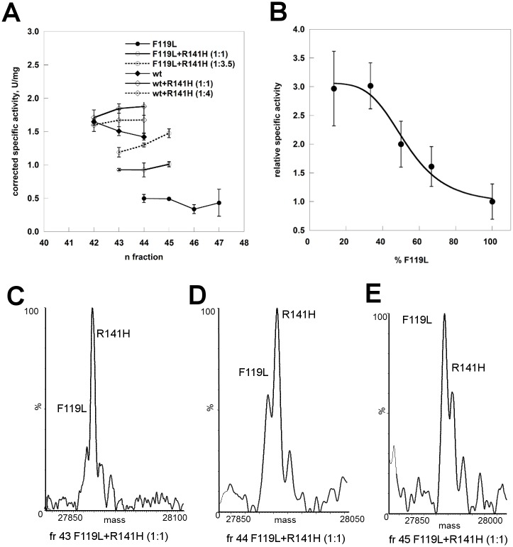
The most frequent disorder of glycosylation is due to mutations in the gene encoding phosphomannomutase2 (PMM2-CDG). For this disease, which is autosomal and recessive, there is no cure at present. Most patients are composite heterozygous and carry one allele encoding an inactive mutant, R141H, and one encoding a hypomorphic mutant. Phosphomannomutase2 is a dimer. We reproduced composite heterozygosity in vitro by mixing R141H either with the wild type protein or the most common hypomorphic mutant F119L and compared the quaternary structure, the activity and the stability of the heterodimeric enzymes. We demonstrated that the activity of R141H/F119L heterodimers in vitro, which reproduces the protein found in patients, has the same activity of wild type/R141H, which reproduces the protein found in healthy carriers. On the other hand the stability of R141H/F119L appears to be reduced both in vitro and in vivo. These findings suggest that a therapy designed to enhance protein stability such as those based on pharmacological chaperones or modulation of proteostasis could be beneficial for PMM2-CDG patients carrying R141H/F119L genotype as well as for other genotypes where protein stability rather than specific activity is affected by mutations.


Conflict of interest statement

Competing Interests: The authors have declared that no competing interests exist.

Figures

Fig 1
Fig 1. Mass spectrometry analysis of wild type phosphomannomutase2, F119L and R141H, with and without Glucose-1,6-bisphosphate.
Panel A shows the deconvoluted mass spectrum of wt-PMM2 (2 μM) upon RP-HPLC-ESI-MS analysis of the incubation mixture in absence (back peaks) and in presence of Glc-1,6-P2 (250 μM, front peaks), showing an increment of 80 Da attributed to a single phosphorylation event. Panels B and C show the same experiment carried out on F119L and R141H, respectively.
Fig 2
Fig 2. Analytical gel filtration analysis of wild type and F119L phosphomannomutase2.
wt-PMM2 (10 μg), F119L (7μg) and a mixture of them (5 μg of wt-PMM2 and 3.5 μg of F119L) were analyzed by gel filtration on BioSep-SEC-S3000 column at 0.5 ml/min in Hepes 20 mM pH 7.5, NaCl 150 mM, MgCl2 5 mM.
Fig 3
Fig 3. Preparative gel filtration of wild type and F119L phosphomannomutase2.
wt-PMM2 and F119L, alone and in the presence of R141H were fractionated on a Superdex75 column equilibrated in Hepes 20 mM pH 7.5, NaCl 150 mM, MgCl2 1 mM. The fractions were analyzed for the protein content and for the phosphoglucomutase activity (in the presence of 40 μM Glc-1P and 5 μM of Glc-1,6-P2). Before loading, each protein sample was pretreated with EDTA on ice. Panel A and B: F119L and F119L+R141H (1:1 and 1:3.5). Panel C and D: wt-PMM2 and wt-PMM2+R141H (1:4).
Fig 4
Fig 4. Specific activity analysis of phosphomannomutase2.
Panel A) The samples (wt-PMM2, F119L, wt-PMM2 plus R141H (1:1 or 1:4), F119L plus R141H (1:1 or 1:3.5) were fractionated on a Superdex75 column. The specific protein ratio of the sample loaded was taken into account in order to calculate the corrected specific activity shown. The protein content of F119L plus R141H (1:1) fr.43,44 and 45 were analyzed by ESI-MS and the deconvoluted mass spectra are shown in panel C, D and E respectively. Panel B) F119L, F119L plus R141H (1:0.5; 1:1; 1:2; 1:6.5) were analyzed in batch and the activity expressed as fold increase relative to pure F119L. The phosphoglucomutase activity was measured in the presence of 40 μM Glc-1P and 5 μM of Glc-1,6-P2.
Fig 5
Fig 5. FRET measurements.
The dye-protein conjugates (AF555-R141H 0.15 mg/ml, AF488-F119L 0.26 mg/ml) were analyzed by UV-spectroscopy (Panel A). Normalized fluorescence emission spectra of a mixture of AF488-F119L (9.75 μg/ml) and AF555-R141H1 (16.9 μg/ml) recorded upon excitation of 470nm is shown (Panel B, emply circles). The emission spectra obtained when the protein mixture was pre-treated with thermolysin is showed for comparison (Panel B, filled circles).
Fig 6
Fig 6. Effect of glucose1,6-bisphosphate concentration on the phosphoglucomutase activity of wild type phosphomannomutase2, F119L and F119L+R141H.
Protein samples from the Superdex75 fractionations were used. The phosphoglucomutase activity was measured in the presence of 40 μM Glc-1P. The specific protein ratio of the sample F119L+R141H was taken into account in order to calculate the corrected specific activity.
Fig 7
Fig 7. Comparison of the stabilities of phosphomannomutase2.
Panel A: wt-PMM2, F119L, and R141H were treated with trypsin (1:50 protease:enzyme ratio) and the time-course of the reaction was monitored by SDS-PAGE. Panel B: wt-PMM2, F119L, and R141H (0.275 mg/ml) were incubated in the presence of increasing concentration of urea. The incubation was conducted at room temperature in the presence of Sypro Orange 4x, the emitted fluorescence was recorded and data were normalized. Panel C: temperature melting profiles of wt-PMM2, F119L and R141H were recorded by thermal shift assay. The proteins (0.2 mg/ml) were heated from 25 to 80° at 0.5°C/min in the presence of Sypro Orange 2.4x. Panel D: temperature melting profiles of wt-PMM2, F119L and R141H recorded by circular dichroism. The proteins (0.2 mg/ml) were heated from 20 to 70° at 0.5°C/min and the signal at 220nm was recorded. All the experiments were conducted in Hepes 20 mM pH 7.5, NaCl 150 mM, MgCl2 1 mM.
Fig 8
Fig 8. Urea-induced melting profile of phosphomannomutases2.
The proteins (wt-PMM2, F119L, wt/R141H 1:6, and F119L/R141H 1:5, 0.2 mg/ml in Hepes 20 mM pH 7.5, MgCl2 1 mM, NaCl 150 mM) were equilibrated with urea (from 0 to 6 M) for 2 hours at 10°C after which the residual phosphoglucomutase activity was measured under standard condition. Data were expressed as residual enzymatic activity.
Fig 9
Fig 9. Comparison of phosphomannomutase2 contents by western-blotting analysis.
Cell extracts (two healthy control fibroblasts and three primary fibroblasts of patients, 10 μg of each) were analyzed and protein were visualized by incubation with polyclonal anti-PMM2 antibody (A).Cell extracts of Cos7 transiently transfected with wt-PMM2-IRES2-EGFP vector or with F119L-IRES2-EGF (10 μg of each), were analyzed and protein were visualized by incubation with polyclonal anti-GFP antibody (B) and polyclonal anti-PMM2 antibody (C) Beta-actin (Panel A and B) or Alpha-tubulin were used as loading controls and purified wt-PMM2 as a standard (Panels B and C).
Fig 10
Fig 10. F119L/R141H structural model.
The two chains are represented as cartoons in light or dark grey. F119 on one chain and L119 on the other chain, are shown as spheres in green or red respectively. R141on one chain and H141 on the other chain, are shown as sticks in green or red respectively. The side chains of E93 and R116 forming a salt bridge at the interface, are shown as sticks in orange or blue respectively. The side chains of K115 and N101 forming a H-bond at the interface, are shown as sticks cyan or magenta respectively.

References

    1. Pirard M, Achouri Y, Collet JF, Schollen E, Matthijs G, Van Schaftingen E. Kinetic properties and tissular distribution of mammalian phosphomannomutase isozymes. Biochem J. 1999;339 (Pt 1):201–7. . - PMC - PubMed
    1. Krissinel E, Henrick K. Inference of macromolecular assemblies from crystalline state. J Mol Biol. 2007;372(3):774–97. . - PubMed
    1. Andreotti G, Pedone E, Giordano A, Cubellis MV. Biochemical phenotype of a common disease-causing mutation and a possible therapeutic approach for the phosphomannomutase 2-associated disorder of glycosylation. Mol Genet Genomic Med. 2013;1(1):32–44. 10.1002/mgg3.3 - DOI - PMC - PubMed
    1. HGMD. HGMD: http://www.hgmd.cf.ac.uk/ac/index.php. 2011. 1954227.
    1. Schollen E, Kjaergaard S, Legius E, Schwartz M, Matthijs G. Lack of Hardy-Weinberg equilibrium for the most prevalent PMM2 mutation in CDG-Ia (congenital disorders of glycosylation type Ia). Eur J Hum Genet. 2000;8(5):367–71. . - PubMed

Publication types

Substances