Review
. 2016;10(2):88-100.
doi: 10.1080/19336950.2015.1106654. Epub 2015 Oct 21.

Structures of multidrug and toxic compound extrusion transporters and their mechanistic implications

Affiliations
Review

Structures of multidrug and toxic compound extrusion transporters and their mechanistic implications

Min Lu. Channels (Austin). 2016.

Abstract

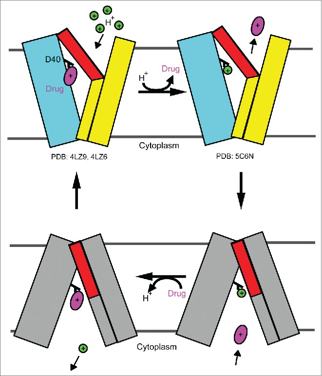
Multidrug resistance poses grand challenges to the effective treatment of infectious diseases and cancers. Integral membrane proteins from the multidrug and toxic compound extrusion (MATE) family contribute to multidrug resistance by exporting a wide variety of therapeutic drugs across cell membranes. MATE proteins are conserved from bacteria to humans and can be categorized into the NorM, DinF and eukaryotic subfamilies. MATE transporters hold great appeal as potential therapeutic targets for curbing multidrug resistance, yet their transport mechanism remains elusive. During the past 5 years, X-ray structures of 4 NorM and DinF transporters have been reported and guided biochemical studies to reveal how MATE transporters extrude different drugs. Such advances, although substantial, have yet to be discussed collectively. Herein I review these structures and the unprecedented mechanistic insights that have been garnered from those structure-inspired studies, as well as lay out the outstanding questions that present exciting opportunities for future work.

Keywords: cation binding; membrane transporter; multidrug efflux inhibitor; multidrug resistance; substrate recognition.


Figures

Figure 1.
Figure 1.
Structure of cation-bound NorM-VC. (A) NorM-VC structure as viewed from the membrane plane. (B) The periplasmic view of the NorM-VC structure. The N and C domains of NorM-VC are colored cyan and yellow, respectively. Rb+ is drawn as a green sphere. Relevant transmembrane helices are numbered.
Figure 2.
Figure 2.
Structure of substrate-bound NorM-NG. The structure is viewed from the membrane plane, and the views in (A) and (B) are related by 180° rotation around the membrane normal. The N and C domains of NorM-NG are colored cyan and yellow respectively. Monobody, a crystallization chaperone, is drawn as a magenta ribbon. Bound substrate is shown as magenta sticks.
Figure 3.
Figure 3.
Structure of the multidrug-binding site in NorM-NG. (A), Structure of NorM-NG (in ribbon rendition) as viewed from the membrane plane and colored as in Figure 2. (B, C and D) Close-up views of the binding site for TPP, ethidium and R6G, respectively. Relevant amino acids are drawn as stick models and labeled.
Figure 4.
Figure 4.
Cation-induced conformational changes in NorM-NG. (A) Structural overlay of substrate-bound NorM-NG (cyan and yellow) and substrate-free NorM-VC (gray). Bound substrate in NorM-NG is drawn as magenta sticks, relevant transmembrane helices are numbered. Red arrows high light the movement of TM7 and TM8 relative to TM10. (B) Close-up view of the cation-binding site in NorM-NG. Cs+ is drawn as a green sphere and overlaid with a difference Fourier map (magenta mesh). A substrate taken from the drug-bound structure of NorM-NG is shown in magenta sticks to indicate the location of the multidrug-binding site. Relevant amino acids are drawn as stick models and labeled. (C) The suggested rearrangement of Na+-coordination in NorM-NG during transport. Na+ is shown as a gray sphere and relevant amino acids are drawn as stick models and labeled.
Figure 5.
Figure 5.
Sequence alignment of representative MATE transporters. Residues that are conserved among the 5 MATE proteins are colored magenta. Regions of secondary structural elements are outlined, with every 10th residue marked. Red and blue dots highlight amino acids that likely bind cations in DinF and NorM transporters, respectively. Residues 1 in PfMATE and NorM-NG, residues 1-20 in eukaryotic hMATE1 are omitted for clarity. Notably, the H+-coupled hMATE1 bears the cation-binding amino acids as found in NorM-NG and NorM-VC (blue dots), while lacking the 2 aspartates (red dots) as seen in DinF-BH and PfMATE.
Figure 6.
Figure 6.
Proposed antiport mechanism for NorM-NG. Briefly, Na+ (green circle) binds to a cation-free, drug-bound, extracellular-facing NorM-NG (pink) and triggers the movement of TM7 and TM8 (red arrow) in the cation-bound, drug-bound state, causing the drug (red) to be dissociated from NorM-NG. The cation-bound, drug-free NorM-NG then switches to an intracellular-facing conformation (gray) to intercept another drug from the cytoplasm. Drug binding subsequently induces the release of Na+ into the cytoplasm, and the protein returns to the drug-bound, extracellular-facing state. The known structures (specified by their PDB codes) correspond to the 3 extracellular-facing states. TM1 and TM2 are simplified as a thick cyan stick, TM7 and TM8 as a thick yellow stick, and TM10 as a thin yellow stick, respectively.
Figure 7.
Figure 7.
Structures of PfMATE obtained at high and low pH. The high (A) and low (B) pH structures are viewed from the periplasm, respectively. N and C domains of PfMATE are colored cyan and yellow, respectively, except in the low pH structure, in which the extracellular half of TM1 is colored red to highlight the kinked helix (B).
Figure 8.
Figure 8.
Structure of DinF-BH. The structure as viewed from the membrane (A) and from the periplasm (B). The N and C domains of DinF-BH are colored cyan and yellow, respectively, except for the extracellular halves of TM7 and TM8, which are colored red to highlight the asymmetric arrangement of the transmembrane helices. Relevant helices are numbered.
Figure 9.
Figure 9.
Structure of the substrate-binding site in DinF-BH. (A) The structure of drug-bound DinF-BH as viewed from the membrane plane, with the N and C domains colored cyan and yellow, respectively. The bound substrate is drawn as magenta sticks and overlaid with experimental electron density map (cyan wire). (B) Close-up view of the substrate-binding site. Relevant amino acids are drawn as stick models and labeled. Bound substrate is shown as magenta sticks.
Figure 10.
Figure 10.
Structural basis for the direct competition in DinF-BH. (A and B) Close-up views of the H-bonding networks in DinF-BHD40N (A) and deprotonated DinF-BH (B), respectively. Relevant amino acids are drawn as stick models and H-bonding interactions are indicated by dotted-lines. Red arrow highlights the interaction between DinF-BHD40N and DinF-BHD184 (A).
Figure 11.
Figure 11.
Proposed antiport mechanism for DinF-BH. In brief, protonation of DinF-BHD40 in the drug-bound, extracellular-facing transporter triggers the release of drug into the periplasm. The protonated, extracellular-facing DinF-BH then changes to the protonated intracellular-facing state. Drug binding to the transporter from the cytoplasm subsequently elicits the deprotonation of DinF-BHD40 and yields a drug-bound, intracellular-facing DinF-BH, which returns to the drug-bound, extracellular-facing state to complete the transport cycle. The N and C domains of the extracellular-facing DinF-BH are simplified as cyan and yellow rectangles, whereas those of the intracellular-facing transporter are colored gray, except for the extracellular portions of TM7 and TM8, which are in red. Drug and proton are drawn as a magenta oval and a green circle, respectively. DinF-BHD40 is colored in black and labeled. The known structures of DinF-BH (as specified by their PDB codes) portray the extracellular-facing states of the transporter.
Figure 12.
Figure 12.
Structure of verapamil-bound DinF-BH. (A) Structure of verapamil-bound DinF-BH (ribbon) as viewed from the membrane plane. The N and C domains are colored cyan and yellow, respectively. Verapamil is drawn as magenta spheres. (B) Close-up view of the verapamil-binding site. Verapamil (magenta) and relevant amino acids are drawn as stick models and labeled.
Figure 13.
Figure 13.
Structure of verapamil-bound NorM-NG. (A) Structure of NorM-NG in complex with verapamil as viewed from the membrane plane. NorM-NG is shown in ribbon rendition and its N and C domains are colored cyan and yellow, respectively. Verapamil is shown as magenta spheres. (B) Close-up view of the verapamil-binding site. Verapamil (magenta) and relevant amino acids are displayed as stick models and labeled.

References

    1. Higgins CF. Multiple molecular mechanisms for multidrug resistance transporters. Nature 2007; 446:749-57; PMID:17429392; http://dx.doi.org/10.1038/nature05630 - DOI - PubMed
    1. Fischbach MA, Walsh CT. Antibiotics for emerging pathogens. Science 2009; 325:1089-93; PMID:19713519; http://dx.doi.org/10.1126/science.1176667 - DOI - PMC - PubMed
    1. Brown MH, Paulsen IT, Skurray RA. The multidrug efflux protein NorM is a prototype of a new family of transporters. Mol Microbiol 1999; 31:394-5; PMID:9987140; http://dx.doi.org/10.1046/j.1365-2958.1999.01162.x - DOI - PubMed
    1. Omote H, Miasa M, Matsumoto T, Otsuka M, Moroyama Y. The MATE proteins as fundamental transporters of metabolic and xenobiotic organic cations. Trends Pharmacol Sci 2006; 27:587-93; PMID:16996621; http://dx.doi.org/10.1016/j.tips.2006.09.001 - DOI - PubMed
    1. Kuroda T, Tsuchiya T. Multidrug efflux transporters in the MATE family. Biochim. Biophys. Acta 2009; 1794:763-8 - PubMed

Publication types

MeSH terms

LinkOut - more resources