

Protonation and Trapping of a Small pH-Sensitive Near-Infrared Fluorescent Molecule in the Acidic Tumor Environment Delineate Diverse Tumors in Vivo
- PMID: 26488921
- PMCID: PMC4673398
- DOI: 10.1021/acs.molpharmaceut.5b00430
Protonation and Trapping of a Small pH-Sensitive Near-Infrared Fluorescent Molecule in the Acidic Tumor Environment Delineate Diverse Tumors in Vivo
Abstract
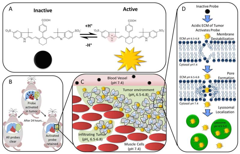
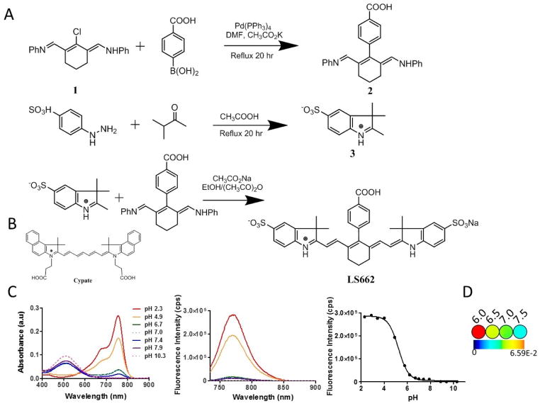
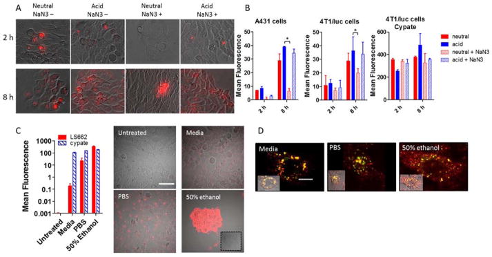
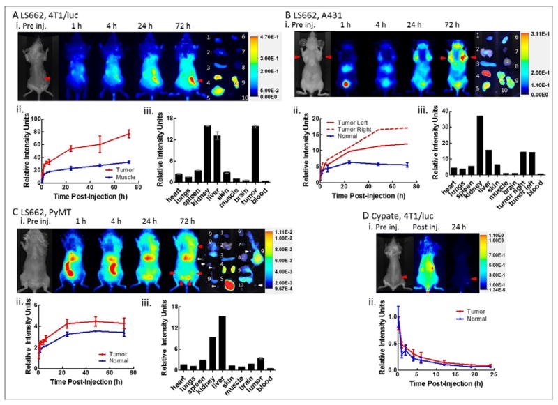
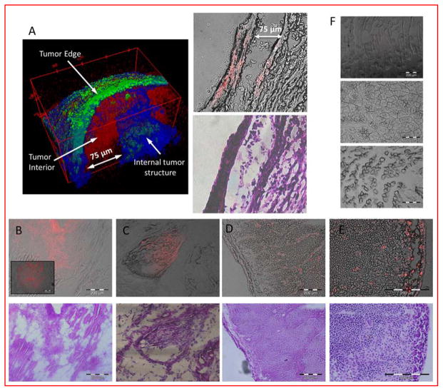
Enhanced glycolysis and poor perfusion in most solid malignant tumors create an acidic extracellular environment, which enhances tumor growth, invasion, and metastasis. Complex molecular systems have been explored for imaging and treating these tumors. Here, we report the development of a small molecule, LS662, that emits near-infrared (NIR) fluorescence upon protonation by the extracellular acidic pH environment of diverse solid tumors. Protonation of LS662 induces selective internalization into tumor cells and retention in the tumor microenvironment. Noninvasive NIR imaging demonstrates selective retention of the pH sensor in diverse tumors, and two-photon microscopy of ex vivo tumors reveals significant retention of LS662 in tumor cells and the acid tumor microenvironment. Passive and active internalization processes combine to enhance NIR fluorescence in tumors over time. The low background fluorescence allows tumors to be detected with high sensitivity, as well as dead or dying cells to be delineated from healthy cells. In addition to demonstrating the feasibility of using small molecule pH sensors to image multiple aggressive solid tumor types via a protonation-induced internalization and retention pathway, the study reveals the potential of using LS662 to monitor treatment response and tumor-targeted drug delivery.
Keywords: cancer imaging; extracellular pH; fluorescence; pH-sensitive probe; small animal; tumor model.
Conflict of interest statement
Notes
The authors declare no competing financial interest
Figures
Similar articles
-
Evaluating tumor metastatic potential by imaging intratumoral acidosis via pH-activatable near-infrared fluorescent probe.Int J Cancer. 2015 Feb 15;136(4):E107-16. doi: 10.1002/ijc.29153. Epub 2014 Sep 4. Int J Cancer. 2015. PMID: 25155456
-
High-sensitivity detection of breast tumors in vivo by use of a pH-sensitive near-infrared fluorescence probe.J Biomed Opt. 2012 Jul;17(7):076028. doi: 10.1117/1.JBO.17.7.076028. J Biomed Opt. 2012. PMID: 22894511
-
Near-Infrared Light and pH-Responsive Polypyrrole@Polyacrylic acid/Fluorescent Mesoporous Silica Nanoparticles for Imaging and Chemo-Photothermal Cancer Therapy.Chemistry. 2015 Nov 2;21(45):16162-71. doi: 10.1002/chem.201502177. Epub 2015 Sep 10. Chemistry. 2015. PMID: 26494031
-
Tumor microenvironment-activated NIR-II reagents for tumor imaging and therapy.J Mater Chem B. 2020 Jun 10;8(22):4738-4747. doi: 10.1039/d0tb00030b. J Mater Chem B. 2020. PMID: 32124909 Review.
-
Recent advances in fluorescent probes for extracellular pH detection and imaging.Anal Biochem. 2021 Jan 1;612:113900. doi: 10.1016/j.ab.2020.113900. Epub 2020 Sep 11. Anal Biochem. 2021. PMID: 32926864 Review.
Cited by
-
Spatiotemporally controlled nano-sized third harmonic generation agents.Biomed Opt Express. 2019 Jun 13;10(7):3301-3316. doi: 10.1364/BOE.10.003301. eCollection 2019 Jul 1. Biomed Opt Express. 2019. PMID: 31360600 Free PMC article.
-
Interactions Between Tumor Biology and Targeted Nanoplatforms for Imaging Applications.Adv Funct Mater. 2020 May 11;30(19):1910402. doi: 10.1002/adfm.201910402. Epub 2020 Mar 3. Adv Funct Mater. 2020. PMID: 34093104 Free PMC article.
-
Dynamic cell-matrix interactions modulate microbial biofilm and tissue 3D microenvironments.Curr Opin Cell Biol. 2016 Oct;42:102-112. doi: 10.1016/j.ceb.2016.05.005. Epub 2016 May 31. Curr Opin Cell Biol. 2016. PMID: 27257751 Free PMC article. Review.
-
Explorations into the Effect of meso-Substituents in Tricarbocyanine Dyes: A Path to Diverse Biomolecular Probes and Materials.Angew Chem Int Ed Engl. 2021 Mar 15;60(12):6230-6241. doi: 10.1002/anie.202008075. Epub 2020 Dec 28. Angew Chem Int Ed Engl. 2021. PMID: 32959963 Free PMC article. Review.
-
Na+-H+ exchanger 1 determines atherosclerotic lesion acidification and promotes atherogenesis.Nat Commun. 2019 Sep 4;10(1):3978. doi: 10.1038/s41467-019-11983-3. Nat Commun. 2019. PMID: 31484936 Free PMC article.
References
Publication types
MeSH terms
Substances
Grants and funding
LinkOut - more resources
Full Text Sources
Other Literature Sources
Miscellaneous
