. 2015 Oct 22:5:15591.
doi: 10.1038/srep15591.

Zebrafish Models for Human Acute Organophosphorus Poisoning

Affiliations

Zebrafish Models for Human Acute Organophosphorus Poisoning

Melissa Faria et al. Sci Rep. .

Erratum in

  • Corrigendum: Zebrafish Models for Human Acute Organophosphorus Poisoning.
    Faria M, Garcia-Reyero N, Padrós F, Babin PJ, Sebastián D, Cachot J, Prats E, Arick M 2nd, Rial E, Knoll-Gellida A, Mathieu G, Le Bihanic F, Lynn Escalon B, Zorzano A, Soares AM, Raldúa D. Faria M, et al. Sci Rep. 2016 Jan 7;6:17244. doi: 10.1038/srep17244. Sci Rep. 2016. PMID: 26738741 Free PMC article. No abstract available.

Abstract

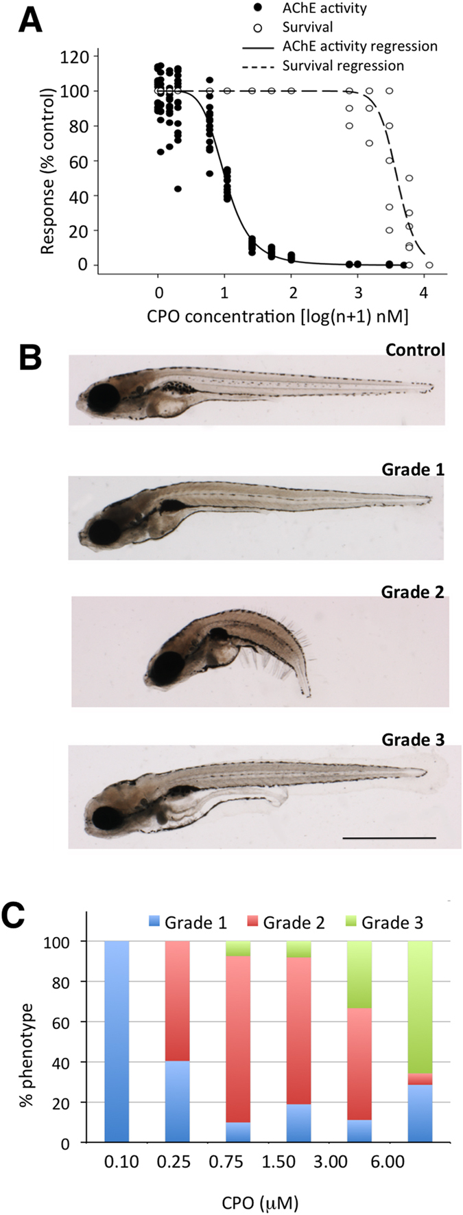
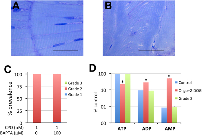
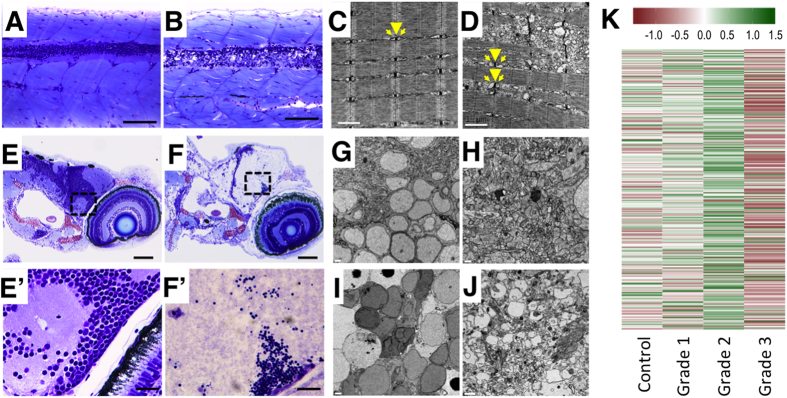
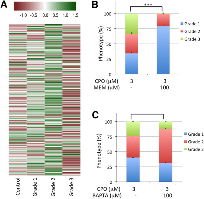
Terrorist use of organophosphorus-based nerve agents and toxic industrial chemicals against civilian populations constitutes a real threat, as demonstrated by the terrorist attacks in Japan in the 1990 s or, even more recently, in the Syrian civil war. Thus, development of more effective countermeasures against acute organophosphorus poisoning is urgently needed. Here, we have generated and validated zebrafish models for mild, moderate and severe acute organophosphorus poisoning by exposing zebrafish larvae to different concentrations of the prototypic organophosphorus compound chlorpyrifos-oxon. Our results show that zebrafish models mimic most of the pathophysiological mechanisms behind this toxidrome in humans, including acetylcholinesterase inhibition, N-methyl-D-aspartate receptor activation, and calcium dysregulation as well as inflammatory and immune responses. The suitability of the zebrafish larvae to in vivo high-throughput screenings of small molecule libraries makes these models a valuable tool for identifying new drugs for multifunctional drug therapy against acute organophosphorus poisoning.


Figures

Figure 1
Figure 1. Chlorpyrifos-oxon (CPO) induces concentration-dependent inhibition of acetylcholinesterase (AChE) and the expression of three different phenotypes in zebrafish larvae.
(A) Concentration-response analysis of CPO regarding the inhibition of AChE activity and survival in zebrafish larvae exposed to different CPO concentrations from 7 to 8 days post fertilization (24 h exposure, 16 larvae per experimental group, p < 0.0001; AChE activity, F(1,143) = 1950.77, r2 = 0.978; survival, F(1,59) = 251.295, r2 = 0.9208). (B) Lateral views of representative 8 days post fertilization larvae control and the mild (grade 1), moderate (grade 2) and severe (grade 3) phenotypes. (C) Prevalence of the three different phenotypes in response to different CPO concentrations (48 larvae per experimental group). Scale bar: 1 mm.
Figure 2
Figure 2. CPO induces concentration-dependent impairment of the retina architecture and the phototransduction pathways in zebrafish larvae.
Retinal histology (transverse plastic semithin sections) of representative control (A), grade 1 (B) grade 2 (C) and grade 3 (D) 8 days post fertilization zebrafish larvae shows a good relationship between the grade of the phenotype and the severity of the effects on the retinal architecture. (E) Heatmap of the phototransduction pathway (dre04744) in zebrafish control and grades 1, 2 and 3 larvae, showing a clear relationship between the grade of phenotype severity and the degree of down-regulation of this pathway. Abbreviations: GCL, ganglion cell layer; INL, inner nuclear layer; IPL, inner plexiform layer; ONL, outer nuclear layer; OPL; outer plexiform layer; RPE, retinal pigment epithelium. Scale bar: 100 μm.
Figure 3
Figure 3. Motor behaviour is strongly impaired by chlorpyrifos-oxon in zebrafish larvae.
(A) VMR profiles, showing that the hyperactivity peak evoked in response to sudden exposure to darkness is reduced until total abolition when 8 days post fertilization zebrafish larvae were previously exposed to increasing concentrations of CPO for 24 h. (B) Locomotion tracking plots for the VMR assay in a 48-well behavioural arena (1 larva per well) recorded a decrease in the swimming activity with increasing chlorpyrifos-oxon concentrations during the first 2 min of the light/dark challenge (30–32 min of the assay). (C,D) Kinematics of TMR traces for control (C) and grade 1 (D) larvae. For each condition, six representative traces are shown from the first 150 ms of the escape response. Each trace is from a different larva. The curvature of the body is represented in degrees, with 0 indicating a straight body. Only grade 1 larvae without any morphological defects were selected for behavioural analyses.
Figure 4
Figure 4. Oxidative stress is only induced in grade 3 larvae.
(A) SOD and catalase activities and GSH and MDA (LPO) levels in control and grade 1–3 zebrafish larvae exposed from 7 to 8 days post fertilization (dpf). The data represent the mean ± SEM. The value on each bar indicates the number of pools for each condition, and bars with different letters are significantly different (p < 0.05, one-way ANOVA with Tukey’s multiple comparison test). (BG) Brightfield (B–D) and fluorescence (E–G) images of the trunks of representative 8 dpf control (B,E), grade 2 (C,F), and grade 3 (D,G) larvae phenotypes. The oxidative fluorescent dye DCFH-DA, which is used for identifying ROS generation in live larvae, showed high ROS generation in grade 3 muscle fibres and spinal cord (G) compared to grade 2 (F) and control (E). Inset at (G) shows brightfield and fluorescence images of the trunk of a grade 3 larva, with the focal plane at the level of the spinal cord. Dorsal and ventral limits of the spinal cord are highlighted by a white dotted line. Abbreviations: sc, spinal cord. (HJ) Grade 3 larvae exhibit a significant reduction in mitochondrial respiration. The basal and maximal respiration (H) and the coupled respiration (I) were strongly reduced in grade 3 larvae with respect to the control larvae (3–11 pools with 20–25 larvae each were analysed for each group; *p < 0.05, ***p < 0.001, Student’s t-test). Moreover, the ratio between coupled respiration and leak was strongly altered (J). (K,L) Prevalence of the grade 3 phenotype can be modified by altering the endogenous levels of GSH. Larvae were pre-incubated with either 50 μM N-acetyl-L-cysteine (NAC) or 0.5 μM diethylmaleate (DEM) for 24 h (from 6 to 7 dpf) followed by co-exposure with 3 μΜ CPO for 24 h (from 7 to 8 dpf). (K) Increasing the GSH levels with N-acetyl-L-cysteine (NAC) decreased the prevalence of the grade 3 phenotype by 67% [3 groups with 8–19 larvae each were analysed for 3 μΜ CPO and 3 groups with 13–23 larvae for 3 μΜ CPO + 50 μΜ NAC; p < 0.05, Student’s t-test]. (L) Decreasing the GSH levels with diethylmaleate (DEM) increased the prevalence of grade 3 larvae by 82% [6 groups with 22–46 larvae each were analysed for 3 μΜ CPO and 6 groups with 21–43 larvae for 3 μΜ CPO + 0.5 μΜ DEM; p < 0.05, Student’s t-test]. (M,N) Prevalence of the grade 3 phenotype in zebrafish larvae exposed to 3 μM CPO is not significantly reduced by either antioxidant. Larvae were pre-treated with the antioxidants vitamin C and MitoQ for 3 h or 24 h, respectively, followed by co-exposure with 3 μM CPO for an additional 24 h (M); 4 groups with 13–22 larvae each per condition; p > 0.05, one-way ANOVA with Dunnett’s multiple comparison test) or MitoQ (N); 4–8 pools with 19–24 larvae each per condition; p > 0.05, Student’s t-test). Abbreviations: n.s., not significant.
Figure 5
Figure 5. Hypercontracture of the axial muscles in grade 2 larvae is not related with calcium overload or ATP depletion.
(A,B) Morphology and distribution of fast-twitch fibres in the trunks of representative control (A) and grade 2 (B) larvae. Notice the regular distribution of the fibres, position of the nuclei, negligible spaces between fibres, and homogeneous distribution of the sarcomeres into the individual fibres in control larvae (A). In contrast, grade 2 larvae (B) present heavily altered myomeric structures, with clear shortening of the myomeres and heterogeneous alignment of the fibres and white spaces. A dense blue band is also present at the myoseptum level. (C) BAPTA-AM, a permeable calcium chelator, is not able to rescue the grade 2 phenotype, indicating that Ca2 + overload is not the mechanism leading to the hypercontracture [3 groups per condition with 16 larvae each group, p = 0.187, Student’s t-test (t(4) = 1.225)]. Six days post fertilization (dpf) larvae were pre-treated for 24 h with 100 μM BAPTA-AM and then co-exposed to 100 μM BAPTA-AM/1 μM CPO for an additional 24 h. (D) Seven days post fertilization larvae that were pre-treated with 40 mM 2-deoxyglucose (2-DOG) for 24 h and then treated with a cocktail of 5 μM oligomycin/40 mM 2-DOG for an additional 2 h exhibited ATP depletion. In contrast, no differences in the content of adenosine phosphates (ATP, ADP, and AMP) were found between the grade 2 and control larvae. Scale bars: 100 μm.
Figure 6
Figure 6. Histopathological assessment of grade 3 larvae shows severe lesions in the central nervous system and muscle fibres.
(A,B) Myotomes with fast-twitch fibres and the spinal cord of representative control (A) and grade 3 (B) larvae in a medial plane section of the trunk. In the grade 3 larva, severe alterations in the fast-twitch fibres and spinal cord are evident. The spinal cord displays severe necrotic changes, with disruption of the normal nuclear distribution in the neuronal bodies (grey matter) and severe vacuolization of axons (white matter). (C,D) Electron micrographs of fast-twitch muscle from control (C) and grade 3 (D) larvae. Whereas control muscles (C) exhibit the normal arrangement of sarcomeres, with well-developed t-tubules (arrowhead) and terminal cisternae (arrows), grade 3 muscles (D) exhibit swelling of the longitudinal sarcoplasmic reticulum and the terminal cisternae. Moreover, irregular spacing of adjacent myofibres and serious disruption of some of the myofibres are evident in grade 3 larvae. (E,F) Retina and brain of representative control (E) and grade 3 (F) larvae. Grade 3 larvae exhibit liquefactive necrosis at the brain level. (E’,F’) Higher magnification of the brain at the optic tectum level corresponding to the area indicated by a frame dashed box at (E) and (F). Whereas the control larva exhibits a normal structure in both neuronal bodies and axons (E’), the grade 3 larva exhibits pyknotic nuclei and striking alteration of the axons (loss of integrity, granular aspect, faint colour) (F’). (GJ) Characteristics of neuronal bodies (G, I) and axons (H,J) in TEM sections of the brain of representative control (GH) and grade 3 (I–J) larvae. Whereas severely altered neuronal bodies (nuclear changes associated with necrotic processes) are evident in (I), altered axons, some of which are extremely enlarged, are present in (J). (K) Heatmap of the neuroactive ligand-receptor interaction pathway shows the strong down-regulation of this pathway found in grade 3 larvae. This result is consistent with the severe disruption of the CNS found at the histological level in this phenotype. Scale bars: (A,B) 100 μm, (C,D) 1 μm, (E,F) 100 μm, (G,H) 2 μm, (E’,F’) 20 μm, (I,J) 2 μm.
Figure 7
Figure 7. Calcium dysregulation is central to grade 3 development.
(A) Heatmap of the calcium signalling pathway (dre04020) in control and the different grades of OPP, showing clear down-regulation of this pathway in grade 3 larvae. (B) Memantine, an antagonist of NMDA receptors, reduced the prevalence of the grade 3 phenotype dramatically [9 groups with 10–18 larvae (total larvae: 132) were analysed for 3 μΜ CPO and 9 groups with 13–25 larvae (total larvae: 200) for 3 μΜ CPO + 100 μΜ memantine; p < 0.001, Mann-Whitney U Statistic (T(9) = 126)]. Seven days post fertilization (dpf) larvae were pre-incubated with 100 μM memantine for 1 h followed by co-exposure with 100 μM memantine/3 μM CPO for 24 h. (C) BAPTA-AM, a permeable calcium chelator, also reduced the prevalence of the grade 3 phenotype significantly [7 groups with 12–42 larvae each (total larvae: 214) were analysed for 3 μΜ CPO and 5 groups with 11–18 larvae (total larvae: 70) for 3 μΜ CPO + 100 μΜ BAPTA-AM; p < 0.005, Student’s t-test (t(12) = 3.287)]. Six days post fertilization zebrafish larvae were pre-incubated with 100 μM BAPTA-AM for 24 h followed by 24 h co-exposure with 3 μM CPO.

References

    1. Bertolote J. M., Fleischmann A., Eddleston M. & Gunnell D. Deaths from pesticide poisoning: a global response. Br J Physchiatry 189, 201–203 (2006). - PMC - PubMed
    1. Peña Llopis S. & Pena L. Antioxidants as Potentially Safe Antidotes for Organophosphorus Poisoning. Current enzyme inhibition 1, 147–156 (2005).
    1. Shih T. Ä. & McDonough J. H. Neurochemical Mechanisms in Soman-induced Seizures. J Appl Toxicol 17, 255–264 (1997). - PubMed
    1. Tryphonas L. & Clement J. G. Histomorphogenesis of soman-induced encephalocardiomyopathy in Sprague-Dawley rats. Toxicol Pathol 23, 393–409 (1995). - PubMed
    1. Weissman B. A. & Raveh L. Therapy against organophosphate poisoning: the importance of anticholinergic drugs with antiglutamatergic properties. Toxicol Appl Pharmacol 232, 351–358 (2008). - PubMed

Publication types

MeSH terms