. 2015 Oct 27;13(4):812-828.
doi: 10.1016/j.celrep.2015.09.026. Epub 2015 Oct 17.

Prolyl Isomerase Pin1 Regulates Axon Guidance by Stabilizing CRMP2A Selectively in Distal Axons

Affiliations

Prolyl Isomerase Pin1 Regulates Axon Guidance by Stabilizing CRMP2A Selectively in Distal Axons

Martin Balastik et al. Cell Rep. .

Abstract

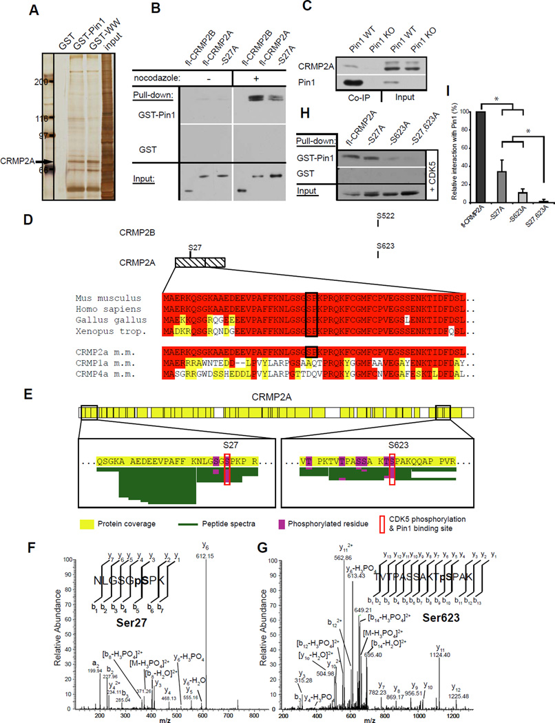
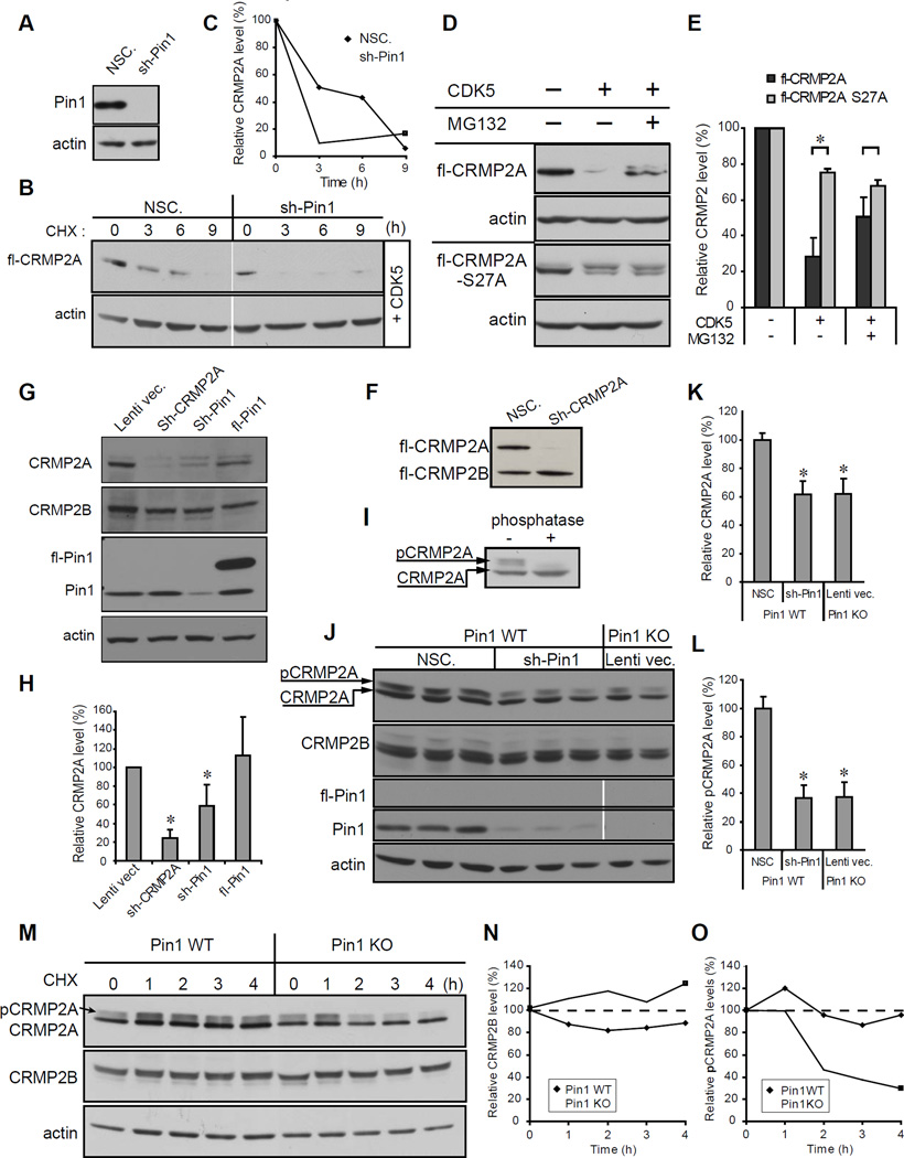
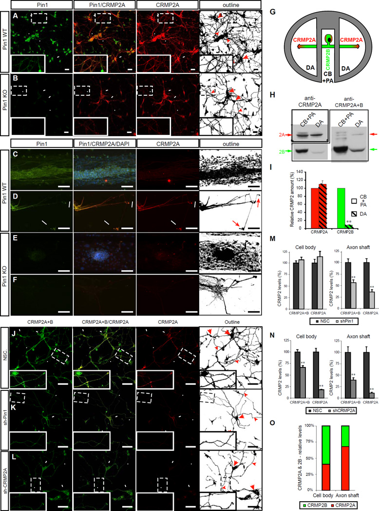
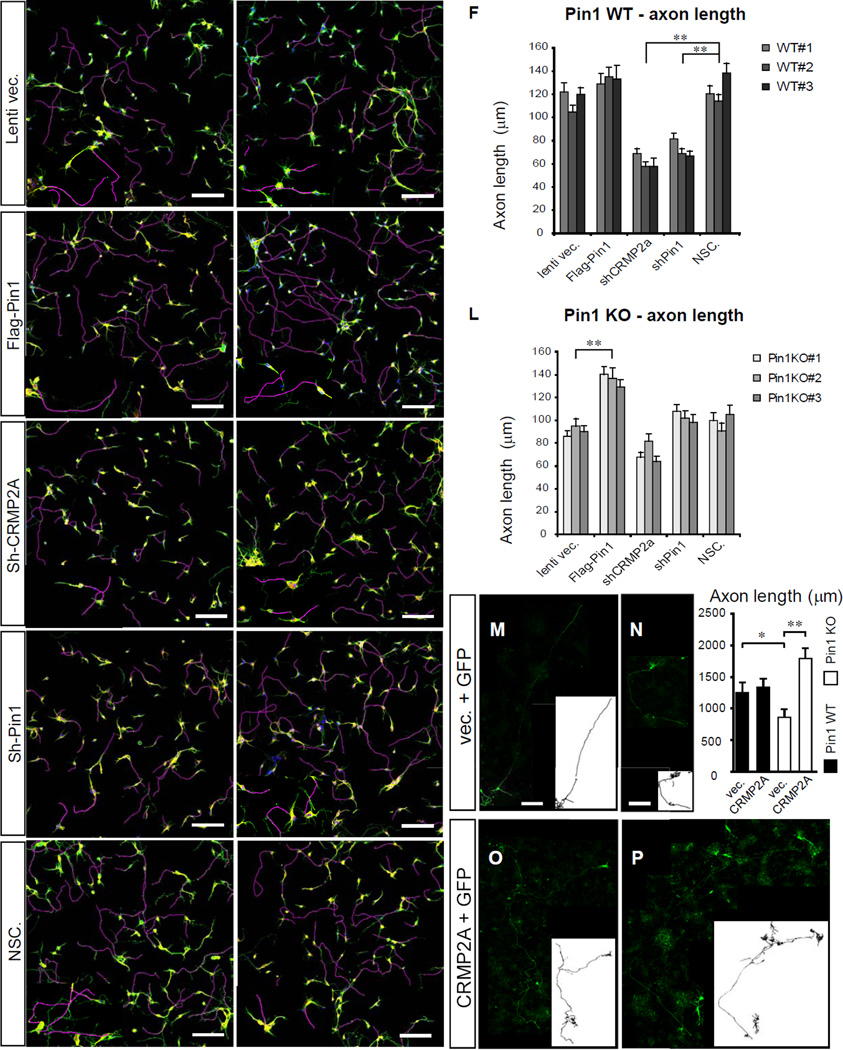
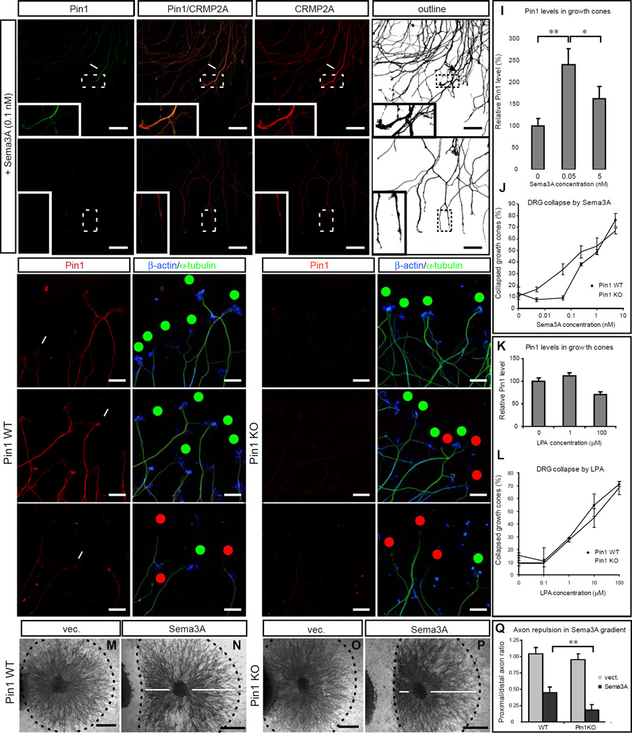
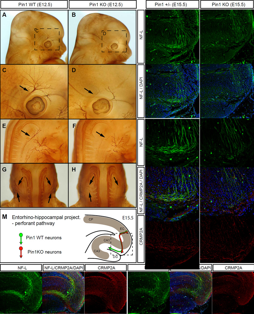
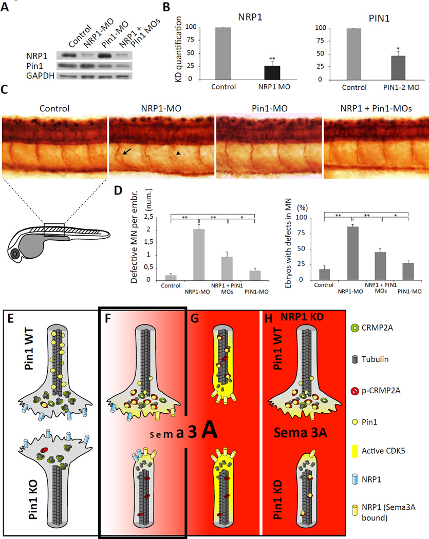
Axon guidance relies on precise translation of extracellular signal gradients into local changes in cytoskeletal dynamics, but the molecular mechanisms regulating dose-dependent responses of growth cones are still poorly understood. Here, we show that during embryonic development in growing axons, a low level of Semaphorin3A stimulation is buffered by the prolyl isomerase Pin1. We demonstrate that Pin1 stabilizes CDK5-phosphorylated CRMP2A, the major isoform of CRMP2 in distal axons. Consequently, Pin1 knockdown or knockout reduces CRMP2A levels specifically in distal axons and inhibits axon growth, which can be fully rescued by Pin1 or CRMP2A expression. Moreover, Pin1 knockdown or knockout increases sensitivity to Sema3A-induced growth cone collapse in vitro and in vivo, leading to developmental abnormalities in axon guidance. These results identify an important isoform-specific function and regulation of CRMP2A in controlling axon growth and uncover Pin1-catalyzed prolyl isomerization as a regulatory mechanism in axon guidance.


Figures

Figure 1
Figure 1. Proteomic identification of CRMP2A as a major Pin1-binding protein in the nervous system
(A) Silver staining of proteins pulled down from postnatal mouse brain lysates by Pin1 (GST-Pin1), its WW-domain (GST-WW) or control GST. CRMP2A band identified by tandem Mass spectrometry indicated. (B) Pin1 binding to mitotically phosphorylated CRMP2A. SH-SY5Y cells expressing FLAG-CRMP2A (fl-CRMP2A) or its S27A mutant (-S27A) were untreated or treated with Nocodazole, followed by pulldown assay with GST-Pin1 (upper panels) or control GST (middle panels). (C) Endogenous Pin1 and CRMP2A form stable complexes in the brain. Pin1 WT and KO brain lysates were subjected to Co-IP with anti-Pin1 antibodies, followed by immunoblotting with anti-CRMP2A antibodies. (D) CRMP2A differs from CRMP2B due to the presence of a 114 long amino acid long N-terminal sequence containing a single Pin1 binding/CDK5 phosphorylation site at Ser27. The site is highly conserved among species but is not present in other CRMP family members that have longer forms. (E–G) Tandem mass spectrometry detected phosphorylation of Ser27 and Ser623 in FLAG-CRMP2A pulled down by Pin1 from HEK-293T cells cotransfected with FLAG-CRMP2A and CDK5/p25 (E). Underlining green lines represent peptide sequence coverage and phosphorylation modifications are highlighted in magenta. The MS/MS spectra are shown for phosphorylation of Ser27 (F) and Ser623 (G). (H) Ala substitution of either Ser27 (-S27A), or Ser623 (-S623A) reduced, but Ala substitution of both sites completely abolishes binding to Pin1. SH-SY5Y cells were cotransfected with FLAG-CRMP2A or its mutants and p25/CDK5, followed by GST-Pin1 pulldown assay. (I) Quantification of Pin1 binding to various CRMP2A mutants. See also Figure S1.
Figure 2
Figure 2. Pin1 stabilizes CRMP2A phosphorylated by CDK5
(A–C) Pin1 KD reduces stability of transfected CRMP2A. SH-SY5Y cells were stably infected with Pin1 silencing (Sh-Pin1) or non-silencing (NSC) lentiviruses (A), and then co-transfected with FLAG-CRMP2A (fl-CRMP2A) and p25/Cdk5, followed by cycloheximide (CHX) chase to measure CRMP2A stability (B) with semi-quantitative results of CRMP2A levels shown in (C). (D) Phosphorylation of CRMP2A on Ser27 by CDK5 increases protein turnover. HEK-293T cells were transfected with fl-CRMP2A or its Ser27Ala mutant with or without p25/Cdk5 in the absence or presence of MG132, followed by immunoblotting with anti-FLAG antibodies (E). (F) Sh-CRMP2A silences CRMP2A but not CRMP2B. SH-SY5Y cells were infected with Sh-CRMP2A lentiviruses, followed by immunoblotting analysis. (G) KD of CRMP2A or Pin1 significantly reduces endogenous CRMP2A but not CRMP2B. SH-SY5Y cells were infected with lentiviruses expressing CRMP2A, Sh-Pin1, or full length Pin1 (Sh-CRMP2A, Sh-Pin1 or fl-Pin1), followed by immunoblotting analysis (H). (I) Incubation with λ phosphatase identifies CRMP2A mobility shift caused by phosphorylation. Primary cortical neuron lysates were incubated without or with λ phosphatase followed by immunoblotting analysis, identifying phosphorylated CRMP2A (pCRMP2A). (J–L) Pin1 KD reduces CRMP2A to levels detected in Pin1-KO cells without affecting CRMP2B in primary neurons. Primary cortical neuron cultures derived from 3 different Pin1 WT embryos and 2 Pin1 KO embryos at E15.5 were infected with Sh-Pin1 lentiviruses, NSC control or lentiviral vector control, followed by immunoblotting analysis (J) with semi-quantification of total (K) or phosphorylated (L) CRMP2A and CRMP2A levels normalized to CRMP2B. (M) Pin1 KO reduces stability of endogenous phosphorylated CRMP2A in primary neurons. Cycloheximide chase was performed on primary cortical neurons at 6DIV with semi-quantitative results of CRMP2B and phospho-CRMP2a levels shown in (N) and (O) respectively (means ± S.D. values shown; * p < 0.05).
Figure 3
Figure 3. Pin1 stabilizes CRMP2A selectively in the distal neurites of primary neurons
(A, B) Pin1 KO reduces CRMP2A selectively in the distal neurites of primary neurons. CRMP2A (red) and Pin1 (Green) immunostaining in Pin1 WT and KO primary cortical neurons at 3 DIV. (A) CRMP2A is expressed strongly in the soma (arrows) as well in the neurites (arrowheads) and co-localize with Pin1 in the neurites (insets). In the Pin1 KO neurons (B), CRMP2A level is lower in the neurites (arrowheads), but its levels in the somas are comparable to WT (arrows). (C–F) Pin1 KO reduces CRMP2A in axons. Using rat Pin1 WT DRG compartmental cultures, Pin1 was detected both in the cell body + proximal axon (CB+PA) compartment (C), as well as in distal axons (DA) (D) by double immunostaining, while higher expression of CRMP2A (co-localizing with Pin1) was detected in DA close to growth cones (D, arrows) when compared to the CB+PA (C). In the Pin1 KO DRG neurons, CRMP2A expression in the DA (F), but not in the CB+PA region (E), is significantly reduced. (G–I) Relative level of CRMP2A increases in distal axon region. Lysates collected from DA and CB+PA compartments of rat primary DRG compartment cultures (G) were analyzed by western blotting using CRMP2A and total CRMP2A+B antibodies (H). Semi-quantitative analysis of the CRMP2A and B levels shows significant reduction of CRMP2B levels in distal axons while no significant reduction of CRMP2A (I). (J–L) Pin1 KD reduces CRMP2A and total CRMP2 selectively in the neurites. Pin1 WT primary cortical neurons were infected with non-silencing (NSC), Sh-Pin1 or Sh-CRMP2A lentiviruses, and immunostained for CRMP2A (red) or total CRMP2 (CRMP2A+B) (Green). In NSC neurons, high levels of CRMP2A and total CRMP2 (CRMP2A+B) were detected both in the neurites (arrowhead) and the soma (arrow) (J). Pin1 KD significantly reduced CRMP2A and total CRMP2 levels in neurites (arrowhead) but not in cell bodies (arrow) (K). CRMP2A KD significantly decreases CRMP2A and total CRMP2 levels in neurites (arrowhead) as well as cell bodies (arrows) (L). (M, N) Quantification of total CRMP2 and CRMP2A levels in Pin1 KD (shPin1) and non-silencing shRNA (NSC) control neurons in neuronal cell body and in the axon shafts (M). (O) Quantification of total CRMP2B and CRMP2A levels in CRMP2A KD (shCRMP2A) and control (NSC) neurons in neuronal cell body and in the axon shafts. (O) Relative distribution of CRMP2A vs. CRMP2B in the cell body and in distal axons calculated from (N). (Scale bars: A, B 20µm ; C-F, J-L 50µm). (means ± SEM; ** p< 0.0001) See also Figure S2.
Figure 4
Figure 4. KD/KO of Pin1 reduces axon growth in primary neurons and is fully rescued by CRMP2A overexpression
Primary cortical neuron cultures were derived from embryos of 3 independent Pin1 WT (A–F) and KO (G–L) mouse littermates and infected with lentiviruses expressing control vector (A, G), Flag-Pin1 (B, H), Sh-CRMP2A (C, I), Sh-Pin1 (D, J), or non-silencing shRNA (NSC) (E, K) lentiviruses. Their axon length was determined at 3 DIV by immunostaining for an axon marker tau (green) and a dendrite marker MAP2 (yellow). Axon tracings are shown in violet. (F) and (L) show the means ± SEM values calculated from quantification of at least 2 optical fields and at least 50 neurons total per each experiment. In Pin1 WT neurons, while overexpression of Pin1 did not have significant effects, KD of CRMP2A or Pin1 in Pin1 WT neurons significantly reduced axon length (** p< 0.0001). In Pin1 KO neurons, overexpression of Pin1 completely rescued axon length, but KD of CRMP2A or Pin1 did not significantly reduce axon length, although Sh-CRMP2A neurons had slightly shorter axon. (M-P) CRMP2A overexpression fully rescues shortened axon length in Pin1 KO neurons. Pin1 WT (M, O) and KO (N, P) primary cortical neurons were co-transfected with GFP and vector control (M, N) or GFP and FLAG-CRMP2A (O, P) and axon growth was analyzed at 7 DIV in GFP-positive neurons. Outlines of the neurons are shown in lower right boxes. Upper right panel indicates quantification of the axon lengths as mean ± SEM. (* p< 0.05, ** p< 0.001), (Scale bars: 100µm) See also Figure S3.
Figure 5
Figure 5. Pin1 KO increases sensitivity to Sema3A-induced growth cone collapse in primary dorsal root ganglia (DRG) neurons
(A, B) Sema3A induces colocalization of high levels of Pin1 and CRMP2A in the vicinity of growth cones (arrows) in Pin1 WT, but not Pin1 KO DRG axons. Pin1 WT (A) and KO (B) primary DRG neurons were treated with 0.1 nM Sema3A for 30 min, followed by double immunostaining for Pin1 (green) and CRMP2A (red). (C–H) Pin1 KO increases sensitivity to Sema3A-induced growth cone collapse. Pin1 WT (C–E) and KO (F–H) primary DRG neurons were treated with different concentrations of Sema3A for 30 min, fixed and triple immunostained with anti Pin1, β-actin and β-tubulin antibodies followed by growth cone collapse analysis, with the percentage of growth cone collapse being shown in (J) Red dots – collapsed growth cones; green dots – intact growth cones. Intensity of Pin1 immunostaining in DRG growth cones (C) significantly increases upon low (non-collapsing) Sema3A stimulation (D) and is reduced upon high Sema3A stimulation (E); the quantification after normalization to β-tubulin levels is shown (I). (K, L) Stimulation with LPA induces similar growth cone collapse in Pin1 WT and KO DRG neurons (L) and does not affect Pin1 levels in the growth cones (K). (M–Q) Pin1 KO significantly increases sensitivity to Sema3A induced growth cone collapse in collagen 3D co-cultures. SH-SY5Y cells were transfected with empty vector (m, o) or Sema3A expression vector (N, P), and co-cultured with Pin1 WT (M, N) or KO (O, P) DRGs. Distance of the collapsed axons from the gradient source was measured (n, p arrows) upon NF-M immunostaining and quantified (Q) (Scale bars: A,B 50µm, C–H 20µm; , M–P 500µm; * p< 0.05; ** p<0.0001). See also Figure S4.
Figure 6
Figure 6. Pin1 KO leads to developmental axon growth defects in the peripheral and central nervous system
(A–H) Axons of the cranial and spinal nerves are less extensive and complex in Pin1 KO embryos. The cranial (A–D) and spinal (E–F) nerves were analyzed in E12.5 Pin1 WT (A, C, E, G) and KO (B, D, F, H) embryos by whole-mount immunohistochemistry for neurofilaments. In Pin1KO embryos stunted neurite processes are found in the ophthalmic branch of the trigeminal nerve (B, D, arrows) and in the lateral branches of the cervical spinal nerves (F, H, arrows). (I–M) Entorhino-hippocampal perforant projections are significantly shorter in Pin1 KO embryos at E15.5. Horizontal sections of E15.5 embryos were stained for neurofilament light subunit (NF-L) (green) to trace growth of entorhinal perforant projections towards the stratum lacunosum-moleculare (s.l-m) of the hippocampus proper in Pin1+/− (I) and KO (J) mice. The dashed lines indicate borders of the developing dentate gyrus. (K, L) Pin1 KO shorter perforant projections are associated with reduced CRMP2A level (red) in growth cones, as pointed by arrows. (M) Schematic drawing of the entorhino-hippocampal perforant pathway at E15.5 in the presence or absence of Pin1 as shown in I-L. (CP - cortical plate, EC – entorhinal cortex, DG – dentate gyrus, CA1, CA3 – hippocampal regions). (N, O) Developmental defects are corrected later during development of Pin1 KO mice. Entorhinal perforant projections are detected in s.l-m of both Pin1 WT (N, arrow) and KO (O, arrow) newborn mice. Lower levels of CRMP2A are present in perforant projections in s. l-m in Pin1 KO mice (O) (Scale bars: I, J, N, O 100µm; K, L , 50 µm). See also Figure S5.
Figure 7
Figure 7. Pin1 regulates Sema3A signaling in vivo
(A, B) Efficiency of single and double morpholino KD of zPin1 and NRP1 in 24-h zebrafish embryos analysed by immunoblotting (A), quantification in (B). (C) Whole mount immunostaining of acetylated tubulin in 1-day-old zebrafish embryos demonstrates increased incidence of developmental motor neurons defects e.g. aberrant branching (arrow) or truncated growth (arrowhead) upon NRP1 KD, which is reduced upon co-injection of Pin1-MO. (D) Quantification of developmental defects in morpholino-injected zebrafish embryos. Control embryos and Pin1-MO injected do not significantly differ in percentage of defective embryos (17% vs. 27% respectively) or average number of defects per embryo (0.22% vs. 0.4% respectively). NRP1 KD induces defective development in 85% of embryos, with the average of 2.05 defects per embryo. Co-injection of Pin1-MO significantly reduces number of defective embryos (44%) as well as the average number of defects per embryo (0.95). (E–H) A model of the Pin1 role in Sema3A-driven axonal growth/retraction.

References

    1. Arimura N, Inagaki N, Chihara K, Menager C, Nakamura N, Amano M, Iwamatsu A, Goshima Y, Kaibuchi K. Phosphorylation of collapsin response mediator protein-2 by Rho-kinase. Evidence for two separate signaling pathways for growth cone collapse. J Biol Chem. 2000;275:23973–23980. - PubMed
    1. Arimura N, Menager C, Kawano Y, Yoshimura T, Kawabata S, Hattori A, Fukata Y, Amano M, Goshima Y, Inagaki M, et al. Phosphorylation by Rho kinase regulates CRMP-2 activity in growth cones. Mol Cell Biol. 2005;25:9973–9984. - PMC - PubMed
    1. Balastik M, Lim J, Pastorino L, Lu KP. Pin1 in Alzheimer's disease: multiple substrates, one regulatory mechanism? Biochim Biophys Acta. 2007;1772:422–429. - PMC - PubMed
    1. Behar O, Golden JA, Mashimo H, Schoen FJ, Fishman MC. Semaphorin III is needed for normal patterning and growth of nerves, bones and heart. Nature. 1996;383:525–528. - PubMed
    1. Cole AR, Knebel A, Morrice NA, Robertson LA, Irving AJ, Connolly CN, Sutherland C. GSK-3 phosphorylation of the Alzheimer epitope within collapsin response mediator proteins regulates axon elongation in primary neurons. J Biol Chem. 2004;279:50176–50180. - PMC - PubMed

Publication types

Substances