. 2015 Oct 21:15:229.
doi: 10.1186/s12862-015-0504-0.

Metamorphic remodeling of morphology and the body cavity in Phoronopsis harmeri (Lophotrochozoa, Phoronida): the evolution of the phoronid body plan and life cycle

Affiliations

Metamorphic remodeling of morphology and the body cavity in Phoronopsis harmeri (Lophotrochozoa, Phoronida): the evolution of the phoronid body plan and life cycle

Elena N Temereva et al. BMC Evol Biol. .

Abstract

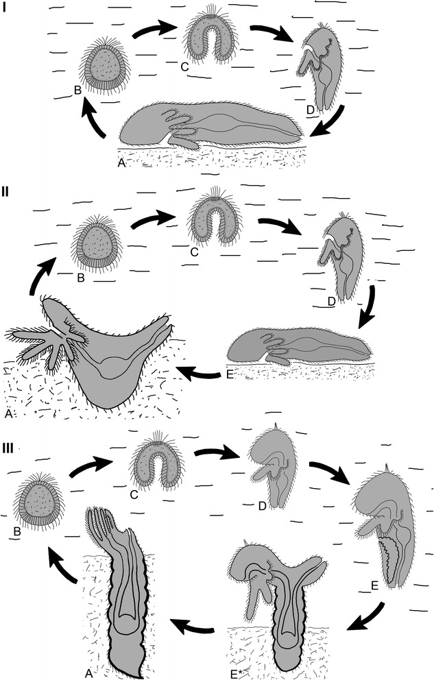
Background: Phoronids undergo a remarkable metamorphosis, in which some parts of the larval body are consumed by the juvenile and the body plan completely changes. According to the only previous hypothesis concerning the evolution of the phoronid body plan, a hypothetical ancestor of phoronids inhabited a U-shaped burrow in soft sediment, where it drew the anterior and posterior parts of the body together and eventually fused them. In the current study, we investigated the metamorphosis of Phoronopsis harmeri with light, electron, and laser confocal microscopy.

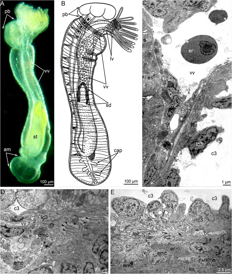
Results: During metamorphosis, the larval hood is engulfed by the juvenile; the epidermis of the postroral ciliated band is squeezed from the tentacular epidermis and then engulfed; the larval telotroch undergoes cell death and disappears; and the juvenile body forms from the metasomal sack of the larva. The dorsal side of the larva becomes very short, whereas the ventral side becomes very long. The terminal portion of the juvenile body is the ampulla, which can repeatedly increase and decrease in diameter. This flexibility of the ampulla enables the juvenile to dig into the sediment. The large blastocoel of the larval collar gives rise to the lophophoral blood vessels of the juvenile. The dorsal blood vessel of the larva becomes the definitive median blood vessel. The juvenile inherits the larval protocoel, mesocoel, and metacoel. Late in metamorphosis, however, the protocoel loses its epithelial structure: the desmosomes between cells and the basal lamina under the cells disappear. This loss may reflect a reduction of the protocoel, which is a characteristic of some recent phoronids.

Conclusions: Based on our investigation of P. harmeri metamorphosis, we hypothesize that the phoronid ancestor was worm-like animal that possessed preoral, tentacular, and trunk coeloms. It lived on the soft sediment and collected food with its tentacles. When threatened, this worm-like ancestor buried itself in the soft sediment by means of the ventral protrusion into which the loop of the intestine and the blood vessels were drawn. We propose that this behavior gave rise to the body plan of all recent phoronids. The evolution of phoronid life cycle seems having more in common with"intercalation" than "terminal addition" theories.


Figures

Fig. 1
Fig. 1
Organization of the competent larvae of Phoronopsis harmeri. In all photographs, the apical side is to the top. a Whole larva viewed from the ventro-lateral side; SEM. b Photograph of a live larva viewed from the left. c Sagittal semithin section of whole larva; the ventral side is to the left. d Longitudinal semithin section of the preoral lobe of larva; the ventral side is to the left. e Longitudinal semithin section of the protonephridium, which bears upper and lower groups of terminal cells. Abbreviations: ao – apical organ; bc – blastocoel; bm – blood mass; bv – blood vessels; c – canal of protonephridium; c1 – protocoel; c2 – mesocoel; c3 – metacoel; d – diaphragm; dv – dorsal blood vessel; es – esophagus; lc2 – mesocoel lining; lg – lower group of terminal cells; m – mouth; mg – midgut; ms – metasomal sac; oms – opening of metasomal sac; pl – preoral lobe; sd – stomach diverticulum; st – stomach; t – tentacle; tt – telotroch; ug – upper group of terminal cells, vm – ventral mesentery
Fig. 2
Fig. 2
A scheme of consecutive stages of metamorphosis of Phoronopsis harmeri. In all panels, the dorsal side is to the top, the ventral side is to the down. a Competent larva. b Start of eversion of the metasomal sac. c The metasomal sac is completely everted. d Stage of eating of the preoral lobe and the postoral ciliated band. e 4-h-old juvenile with completely formed blood system. Abbreviations: am – ampulla; ao – apical organ; bc – blastocoel; bm – blood mass; c1 – protocoel; c2 – mesocoel; c3 – metacoel; ds – dorsal side of the body; dv – dorsal blood vessel; epi – epistome; es – esophagus; lphv – two lophophoral blood vessels; lrpl – two lateral remnants of preoral oobe; lv – ventrolateral blood vessel; mg – midgut; ms – metasomal sac; mv – median blood vessel; nd – nephridial duct; pb – posterior bulge; sd – stomach diverticulum; st – stomach; t – tentacle; tt – telotroch; vm – ventral mesentery; vs – ventral side of the body; vv – ventral blood vessel
Fig. 3
Fig. 3
First steps of metamorphosis of Phoronopsis harmeri; photographs of live animals. In all photographs, the larval preoral lobe with apical organ is to the top. a Competent larva. b First contraction of larval muscles. c Strong contraction of larval muscles: larval length decreases in twice. d Start of eversion of the matasomal sac. e The metasomal sac is everted in half. f The metasomal sac is completely everted. Abbreviations: bm – blood mass; ms – metasomal sac; pl – preoral lobe; st – stomach; tt – telotroch
Fig. 4
Fig. 4
Remodeling of body cavities during metamorphosis of Phoronopsis harmeri. Histological sagittal sections of animals at consecutive stages of metamorphosis. Color code: red – blastocoel, which partly gives rise to the blood system; cyan – protocoel; blue – mesocoel; green – metacoel. a First step of metamorphosis: larva with everted metasomal sac. Stage about 1 min after the onset of metamorphosis. Larval apical organ is at the right; telotroch is at the left; metasomal sac is to the down. b Stage of eating of the preoral lobe; about 10 min after the onset of metamorphosis. Degenerated apical organ is well visible at this stage. c Stage of eating of the postoral ciliated band: stage about 12–15 min after the onset of metamorphosis. d 9-day-old juvenile completely acquired definitive body plan. Abbreviations: ao – apical organ; bc – blastocoel; c1 – protocoel; c2 – mesocoel; c3 – metacoel; es – esophagus; lv – lophophoral blood vessel; m – mouth; mg – midgut; mv – median blood vessel; pl – preoral lobe; pr – proctodaeum; st – stomach; tt – telotroch; vv – ventral vessel
Fig. 5
Fig. 5
First steps of metamorphosis of Phoronopsis harmeri accordingly to SEM. In all photographs, apical is to the top. a Metamorphic animal with completely everted metasomal sac: stage about 1 min after the onset of metamorphosis. Difference in organization of the trunk epithelium allows to mark several zones: anterior trunk part, posterior trunk part, and ampulla. b Metamorphic animal with degenerated preoral lobe, huge posterior bulge, and continuous rope of postoral ciliated band. Stage about 10–12 min after the onset of metamorphosis. c Metamorphic animal is eating the preoral lobe. Macerated epithelium of the preoral lobe is involved into the mouth. d Metamorphic animal without the preoral lobe. e Metamorphic animal at stage of formation and eating of the continuous rope of postoral ciliated band. Abbreviations: am – ampulla; atp – anterior trunk part; m – mouth; pb – posterior bulge; pl – preoral lobe; ptp – posterior trunk part; rpb – rope of postoral ciliated band
Fig. 6
Fig. 6
Details of metamorphic remodeling of external morphology in Phoronopsis harmeri. Photographs according to SEM (A-B, D-G) and semithin section (c). a Tentacles (t) of metamorphic animal with continuous rope of postoral ciliated band (rpb). b A portion of tentacle with degenerated epithelium of postoral ciliated band: line of former location of the postoral ciliated band is indicated by arrowheads. c Pair of latero-frontal ropes of postoral ciliated band is evident on each tentacle. d Tentacles after remodeling: line of former location of the postoral ciliated band is indicated by arrowheads. e Anterior portion of the body in metamorphic animal with partly consumed preoral lobe (pl) and spacious oral disc (od) with peeled epithelium (pe). f A portion of the oral disc is covered by basal lamina (bl). g Cross section of juvenile tentacles (t) with erythrocytes (er). Abbreviations: c2 – mesocoel; m – mouth; tv – tentacle vessel
Fig. 7
Fig. 7
Ultrastructural remodeling of integument during metamorphosis of Phoronopsis harmeri. Stage about 15 min after the onset of metamorphosis. a Epithelium (ep) and basal lamina (bl) of the oral disc. Thick electron dense fibrils are indicated by arrowheads. b Thick extracellular matrix (ECM), numerous protrusions of the basal lamina, and degenerated muscle cells in the preoral lobe near the protocoel. c The basal lamina forms long protrusions, which penetrate profoundly into the ECM. Thick electron dense fibrils are indicated by arrowheads. Abbreviations: dep – degenerated epithelium; lc1 – lining of the protocoel; pao – pilled apical organ; tf – thick electron dense fibrils
Fig. 8
Fig. 8
Newly formed juveniles of Phoronopsis harmeri stained for phalloidin. Stage about 40 min after the onset of metamorphosis. a Whole animal. According to specific organization of the muscles, juvenile body is subdivided into the lophophore (lph) with tentacles (t), anterior trunk part (atp), posterior trunk part (ptp), and ampulla (am). b Anterior part of the body: completely formed epistome (epi) is well visible above the mouth (m)
Fig. 9
Fig. 9
Metamorphic remodeling of the body cavities in anterior part of the body in Phoronopsis harmeri. Histological sagittal sections. The apical is to the top; the oral side is at the right; the anal side is at the left. Color code: red – blastocoel, which partly gives rise to the blood system; cyan – protocoel; blue – mesocoel; green – metacoel. a Metamorphic animal at stage of eating of the preoral lobe and the postoral ciliated band: about 10–15 min after the onset of metamorphosis. The degenerated preoral lobe (dpl) and ropes of the postoral ciliated (rpb) band are visible in the section. b 4-h-old juvenile with completely obliterated blastocoel and thick basal lamina (bl) instead of it. The lining of the posterior portion of the larval metacoel, which separate from the lamina and float in the metacoel, are indicated by double arrowheads. Abbreviations: bc – blastocoel; c1 – protocoel; c2 – mesocoel; c3 – metacoel; es – esophagus; pb – posterior bulge; pr – proctodaeum; tt – telotroch; vv – ventral vessel
Fig 10
Fig 10
Metamorphic remodeling of the circulatory system in Phoronopsis harmeri. a Photographs of live 4-h-old juvenile with formed ventral blood vessel (vv). b Picture of 4-h-old juvenile with lophophoral (lv) and ventral blood vessels. c Ultrastructural organization of the ventral blood vessel. Cells (emc), which form muscular lining of the esophagus (es), form the inner lining of the ventral blood vessel. d Degeneration of muscle cells (dmc), which form the inner lining of the ventral blood vessel; the formation of long protrusions (are shown by arrowheads) of the basal lamina (bl). e The median blood vessel has a very complex wall along the upper portion of the esophagus. The wall is formed by lining of the metacoel (lc3) and two layers of the mesocoel lining (lc2). The muscle cells (emc) of the esophagus (es) form inner lining of the vessel. Desmosomes between cells of mesocoel lining are indicated by double arrowheads. Abbreviations: am – ampulla; cap – blood capillaries; c3 – metacoel; er – erythrocytes; pb – posterior bulge; sd – stomach diverticulum
Fig. 11
Fig. 11
Organization of the protocoel at first stage of metamorphosis of Phoronopsis harmeri. a Sagittal semithin section of the protocoel (c1). Degenerated epithelium of the preoral lobe (dpl) is above the protocoel. Thick protrusions of basal lamina are indicated by arrowheads. b Thin section of the protocoel lateral wall, which includes large bundle of muscle cells. c Thin section of the protocoel lower wall. It contacts the esophagus (es) and muscle cells, which form the musculature (emc) of the esophagus. Desmosomes between cells of the protocoel lining are shown by double arrowheads. d The protocoel upper wall is formed by myoepithelial cells. Abbreviations: bb – basal body; bc – blastocoel; bl – basal lamina; lc1 – lining of protocoel; mc – muscular basal protrusions of cells of protocoel lining; rer – rough endoplasmic reticulum; sr – striated rootlet
Fig. 12
Fig. 12
Organization of the protocoel at later stages of metamorphosis of Phoronopsis harmeri. a Sagittal semithin section of the protocoel (c1) at stage of eating of the peoral lobe. Degenerated epithelium of the preoral lobe (dpl) is above the protocoel. b Sagittal semithin section of the protocoel in 1-h-old juvenile. c Thin section of the protocoel of 1-h-old juvenile. The lumen of protocoel is filled with thick apical protrusions (plc1) of cells of the protocoel lining (lc1). Desmosomes between cells are indicated by double arrowheads. Abbreviations: bc – blastocoel; bl – basal lamina; c3 – trunk coelom; ECM – extracellular matrix; emc – muscle cells, which form esophageal musculature; es – esophagus; m – mouth; mc – muscle cells in blastocoel; n – nucleus; nu – nucleolus; mv – median blood vessel; rer – rough endoplasmic reticulum; sr – striated rootlet
Fig. 13
Fig. 13
Organization of the protocoel in 4-day-old juvenile of Phoronopsis harmeri. Color code: red – median blood vessel; cyan – protocoel; blue – mesocoel. a Sagittal semithin section of the epistome. b Sagittal thin section of the protocoel (c1), mesocoel (c2), and median blood vessel (mv). Desmosomes are indicated by double arrowheads. c Portion of the protocoel lining (lc1). Large cell with phagosome (ph), which contains degenerated myofilaments (mf). Abbreviations: bb – basal body; bl – basal lamina; c – cilium; c3 – trunk coelom; emc – muscle cells, which form musculature of esophagus; er – erythrocyte; es – esophagus; G – Golgi apparatus; lc3 – lining of trunk coelom; m – mouth; n – nucleus; pmc – projections of muscle cells
Fig. 14
Fig. 14
Ultrastructural details of the lining of meso- and metacoel during metamorphosis of Phoronopsis harmeri. Sagittal thin sections. First stage of metamorphosis (A, F); juvenile in 15 min after metamorphosis start (B); 1-h-old juvenile (D); 4-day-old juvenile (C, E, G). a Proliferating cell (cc) of coelomic lining. Well developed larval muscles (mc) are retained. b Presence of subperitoneal neurites (pn), which contain dense-core synaptic vesicles, under the mesocoel lining (lc2). c Completely established mesocoel lining. d Degenerated muscle cells (dnc) within cells of the mesocoel lining. e Degenerated muscle cells (dnc) within cells of the metacoel lining. f Metacoel lining: myoepithelial cells form long basal projections, which are covered by thick basal lamina (bl). g Somatopleura of the juvenile consists of myoepithelial cells, which form circular musculature (cm). Abbreviations: bc – blastocoel; bb – basal body; c2 – mesocoel; c3 – metacoel; ECM – extracellular matrix; emc – muscle cells, which form musculature of esophagus; mf – myofilaments; rer – rough endoplasmic reticulum; te – epidermis of tentacle
Fig. 15
Fig. 15
Hypothetical scenario of evolutionary formation of the phoronid body plan. a The last common phoronid ancestor, which was worm-like animal, had tentacles for food capture, and moved on the ventral side of the body. b Had being in danger, the worm-like ancestor buried itself in soft sediment by means of the ventral protrusion, to which the loop of the intestine was drawn. Abbreviations: dt – digestive tract; pr – hypothetical predator; ss – soft sediment; t – tentacle; vp – ventral protrusion
Fig. 16
Fig. 16
Hypothetical scenario of evolutionary formation of the phoronid life cycle. I Life cycle of the last common phoronid ancestor, which was a benthic animal and had planktotrophic larva (lophotrochophore). II Life cycle of hypothetical ancestor, which was a sessile animal with pelagic larva and benthic creeping juvenile. III A generalized scheme of the life cycle of recent phoronids, which have pelagic larva and pelagic competent larva that is a juvenile. Abbreviations: a – adult animal; b – blastula; c – gastrula; d – planktotrophic larva; e – pelagic juvenile; e* – settled juvenile

References

    1. Hadfield MG, Carpizo-Ituarte EJ, del Carmen K, Nedved BT. Metamorphic competence, a major adaptive convergence in marine invertebrate larvae. Am Zool. 2001;41:1123–31.
    1. Sly BJ, Snoke MS, Raff RA. Who came first—larvae or adults? Origins of metazoan bilaterian larvae. Int J Dev Biol. 2003;47:623–32. - PubMed
    1. Raff RA. Origins of the other metazoan body plans: the evolution of larval forms. Philos Trans R Soc Lond B. 2008;363:1473–9. doi: 10.1098/rstb.2007.2237. - DOI - PMC - PubMed
    1. Page LR. Molluscan larvae: pelagic juveniles or slowly metamorphosing larvae? Biol Bull. 2009;216:216–25. - PubMed
    1. Nielsen C. How did Indirect development with planktotrophic larvae evolve? Biol Bull. 2009;216:203–15. - PubMed

LinkOut - more resources