Review
. 2015 Oct 20;6(5):e01289-15.
doi: 10.1128/mBio.01289-15.

Evolutionary landscape of the Mycobacterium tuberculosis complex from the viewpoint of PhoPR: implications for virulence regulation and application to vaccine development

Affiliations
Review

Evolutionary landscape of the Mycobacterium tuberculosis complex from the viewpoint of PhoPR: implications for virulence regulation and application to vaccine development

Esther Broset et al. mBio. .

Abstract

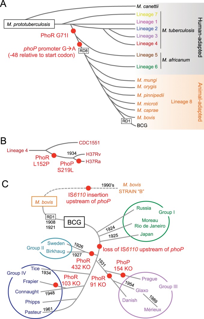
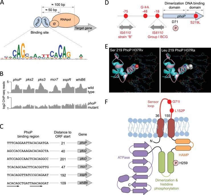
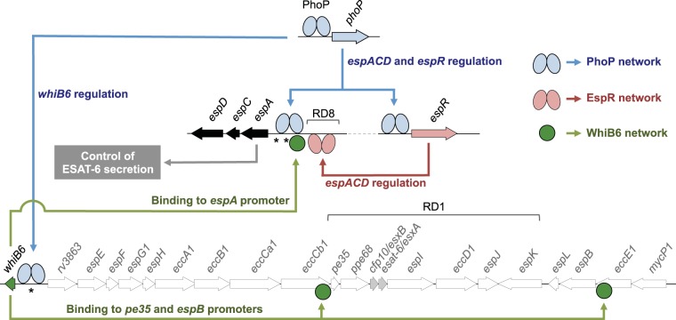
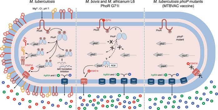
Different members of the Mycobacterium genus have evolved to cause tuberculosis in diverse human populations and in a variety of animal species. Our cumulative knowledge of mycobacterial genomes indicates that mutations in the PhoPR two-component virulence system were acquired not only during the natural evolution of mycobacterial species but also during in vitro subculture, which has given rise to the attenuated reference strain H37Ra or to different daughter strains of Mycobacterium bovis BCG. PhoPR is a well-known regulator of pathogenic phenotypes, including secretion of the virulence factor ESAT-6, biosynthesis of acyltrehalose-based lipids, and modulation of antigen export, in members of the Mycobacterium tuberculosis complex (MTBC). Evolutionarily conserved polymorphisms in PhoPR from Mycobacterium africanum, M. bovis, or M. tuberculosis H37Ra result in loss of functional phenotypes. Interestingly, some members of the MTBC have acquired compensatory mutations to counteract these polymorphisms and, probably, to maintain their pathogenic potential. Some of these compensatory mutations include the insertion of the IS6110 element upstream from phoPR in a particular M. bovis strain that is able to transmit between humans or polymorphisms in M. africanum and M. bovis that affect the regulatory region of the espACD operon, allowing PhoPR-independent ESAT-6 secretion. This review highlights the increasing knowledge of the significance of PhoPR in the evolution of the MTBC and its potential application in the construction of new attenuated vaccines based on phoPR inactivation. In this context, the live attenuated vaccine MTBVAC, based on a phoP fadD26 deletion mutant of M. tuberculosis, is the first vaccine of this kind to successfully enter into clinical development, representing a historic milestone in the field of human vaccinology.


Figures

FIG 1
FIG 1
The phylogenies of the Mycobacterium tuberculosis complex and BCG include several polymorphisms in the PhoPR virulence system. (A) Evolutionary scenario of the MTBC showing the presence of the different members belonging to specific lineages. Adaptation of these species to humans and animals is also indicated. (B) Expanded view of lineage 4, showing representative strains. The Leu152Pro substitution in PhoR, exclusively in H37Rv and its attenuated form H37Ra, and a Ser219Leu substitution in PhoP from H37Ra are indicated. (C) Genealogy of M. bovis BCG daughter vaccine strains grouped by different phylogenetic markers. Deletion of RD1, responsible for BCG attenuation, and dates indicating genetic drifts are shown. Note the evolution of M. bovis strain B by acquisition of an IS6110 insertion upstream from the phoP gene. Other polymorphisms and RD deletions present in these phylogenies have been omitted for clarity; red-filled circles indicate phylogenetic locations of phoPR polymorphisms.
FIG 2
FIG 2
Molecular characteristics of the PhoPR two-component system. (A) Positioning of the PhoP transcription factor relative to the RNA polymerase and its target genes measured as the most probable values from ChIP-seq data. The PhoP consensus motif [TCACAG(N5)TCACAG] is also indicated. (B) ChIP-seq reads from representative promoters of PhoP-regulated genes. Note the significant increase in ChIP-seq peaks in the wild-type strain relative to those in its phoP mutant, indicative of a specific interaction of PhoP with these regions. (C) PhoP binding motifs elucidated from ChIP-seq data from the analyses whose results are shown in panel B. Distances to the start codons of target genes are also indicated. ORF, open reading frame. (D) Genomic locations of relevant phoP polymorphisms discussed throughout the text. Insertions of IS6110 in the promoter region and their positions relative to the phoP start codon are shown. Amino acid positions of the Asp71 residue, which is involved in the phosphotransfer reaction, and the Ser219Leu substitution in H37Ra are also indicated. (E) Ribbon models of the DNA binding domain of PhoP superimposed over the structure of a PhoB-DNA complex. Solid spheres show the wild-type serine 219 in H37Rv (left) or the leucine residue that appears in H37Ra (right) (adapted from reference 40). The mutant leucine residue is expected to interfere with DNA binding and/or recognition. (F) Secondary structure of the PhoR sensor kinase indicating its membrane topology. Each domain has been colored individually, and α-helices (ovals) and β-strands (arrows) are indicated. Note the presence of PhoR polymorphisms in the sensor loop, which is located in the periplasmic space. The position of the histidine involved in the phosphotransfer reaction is also indicated.
FIG 3
FIG 3
PhoP-dependent regulatory networks implicated in the control of ESAT-6 secretion. Different genes from the ESX-1 (whiB6 to mycP1) and the extended ESX-1 (espACD and espR) regions involved in ESAT-6 export are indicated. PhoP (blue ellipses) interacts with the espR, espA, and whiB6 promoters and controls the expression of these genes. EspR (pink ellipses) interacts with and activates the espACD locus. WhiB6 (green circles) also interacts with the promoter regions of espA, pe35, and espB genes. Overall, the PhoP-dependent EspR and WhiB6 regulatory circuits activate the espACD locus, which is required for ESAT-6 secretion. Locations of RD1, absent in BCG, and of RD8, absent in L6 and L8 lineages, as well as polymorphisms in espACD and whiB6 promoters (asterisks), are also indicated.
FIG 4
FIG 4
Comparative illustration of PhoPR-regulated phenotypes in M. tuberculosis, M. bovis, M. africanum L6, and an M. tuberculosis phoP mutant. M. tuberculosis, carrying a functional PhoR, is able to sense its cognate stimulus and subsequently phosphorylate PhoP. Phosphorylated PhoP regulates three well-known phenotypes, including synthesis of SL and DAT/PAT (via pks2 and pks3 regulation), secretion of ESAT-6 (through espA regulation, as depicted in Fig. 3), and posttranscriptional regulation of tatC (mediated by the mcr7 noncoding RNA). M. bovis and M. africanum L6, carrying a defective PhoR G71I allele, are expected to have defects in PhoP phosphorylation, as a consequence of which these strains lack SL, DAT, and PAT. However, ESAT-6 secretion in these strains is restored by compensatory mutations in the espACD promoter region that include RD8 deletion and species-specific polymorphisms (asterisks). M. tuberculosis phoP mutants lack the aforementioned PhoP-regulated phenotypes and consequently do not synthesize SL, DAT, and PAT or secrete ESAT-6. These mutants also have a deregulated TAT system and, consequently, secrete larger amounts of TAT substrates, including the antigens Ag85A and Ag85C. Consequently, adequately attenuated M. tuberculosis phoP-based vaccine strains, such as MTBVAC, are expected to induce improved and longer-lasting immunogenicity compared to that of BCG in clinical trials.

References

    1. Brosch R, Gordon SV, Marmiesse M, Brodin P, Buchrieser C, Eiglmeier K, Garnier T, Gutierrez C, Hewinson G, Kremer K, Parsons LM, Pym AS, Samper S, van Soolingen D, Cole ST. 2002. A new evolutionary scenario for the Mycobacterium tuberculosis complex. Proc Natl Acad Sci U S A 99:3684–3689. doi:10.1073/pnas.052548299. - DOI - PMC - PubMed
    1. Comas I, Coscolla M, Luo T, Borrell S, Holt KE, Kato-Maeda M, Parkhill J, Malla B, Berg S, Thwaites G, Yeboah-Manu D, Bothamley G, Mei J, Wei L, Bentley S, Harris SR, Niemann S, Diel R, Aseffa A, Gao Q, Young D, Gagneux S. 2013. Out-of-Africa migration and Neolithic coexpansion of Mycobacterium tuberculosis with modern humans. Nat Genet 45:1176–1182. doi:10.1038/ng.2744. - DOI - PMC - PubMed
    1. Fabre M, Hauck Y, Soler C, Koeck J, van Ingen J, van Soolingen D, Vergnaud G, Pourcel C. 2010. Molecular characteristics of “Mycobacterium canettii” the smooth Mycobacterium tuberculosis bacilli. Infect Genet Evol 10:1165–1173. doi:10.1016/j.meegid.2010.07.016. - DOI - PubMed
    1. de Jong BC, Antonio M, Gagneux S. 2010. Mycobacterium africanum—review of an important cause of human tuberculosis in West Africa. PLoS Negl Trop Dis 4:e744. doi:10.1371/journal.pntd.0000744. - DOI - PMC - PubMed
    1. Comas I, Chakravartti J, Small PM, Galagan J, Niemann S, Kremer K, Ernst JD, Gagneux S. 2010. Human T cell epitopes of Mycobacterium tuberculosis are evolutionarily hyperconserved. Nat Genet 42:498–503. doi:10.1038/ng.590. - DOI - PMC - PubMed

Publication types

LinkOut - more resources