. 2015 Oct 20;6(5):e01552-15.
doi: 10.1128/mBio.01552-15.

Binding of alphaherpesvirus glycoprotein H to surface α4β1-integrins activates calcium-signaling pathways and induces phosphatidylserine exposure on the plasma membrane

Affiliations

Binding of alphaherpesvirus glycoprotein H to surface α4β1-integrins activates calcium-signaling pathways and induces phosphatidylserine exposure on the plasma membrane

Walid Azab et al. mBio. .

Abstract

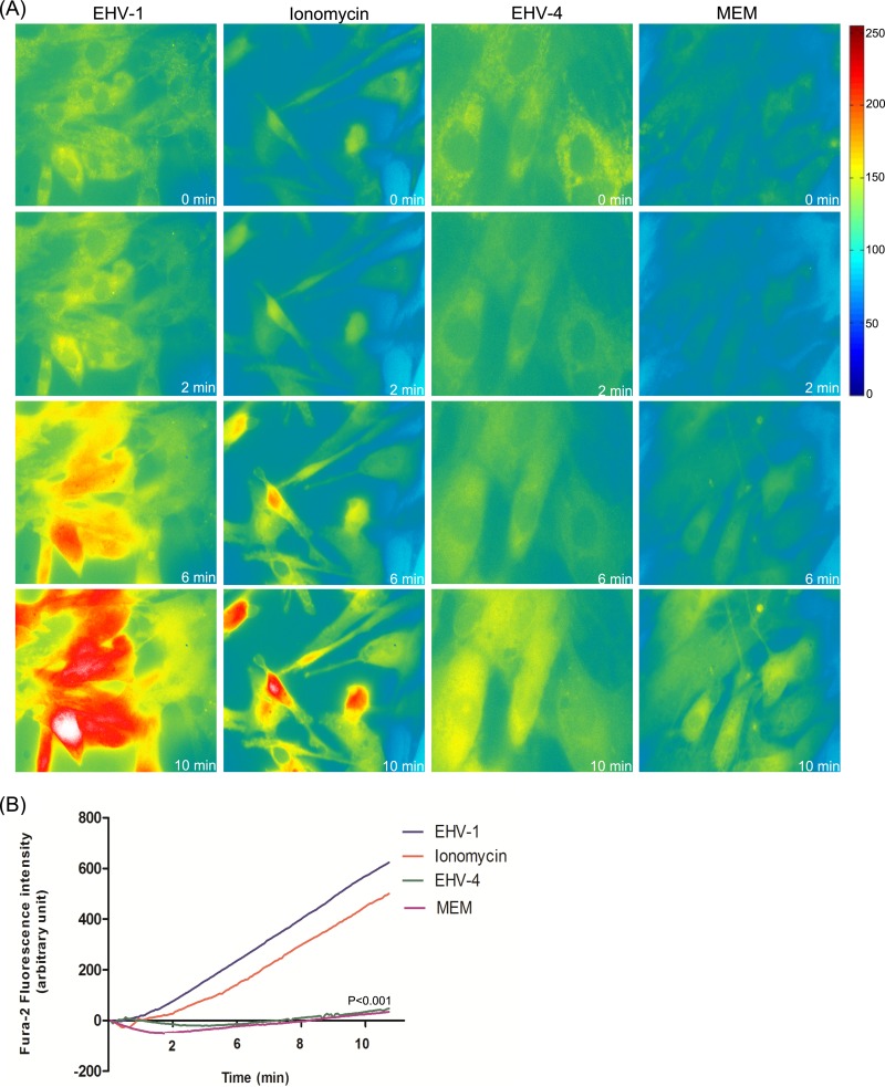
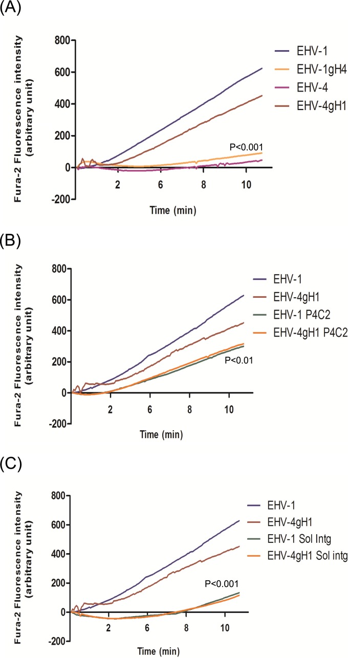
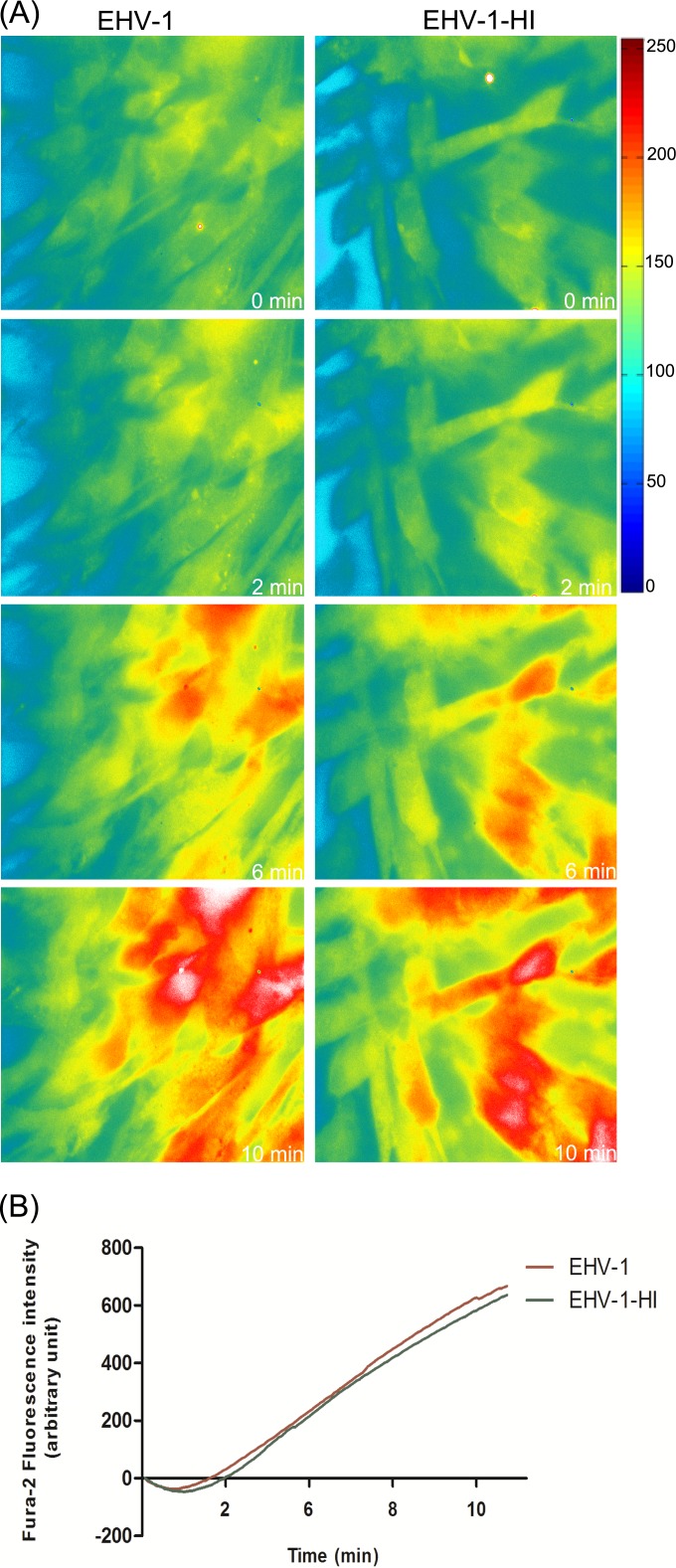
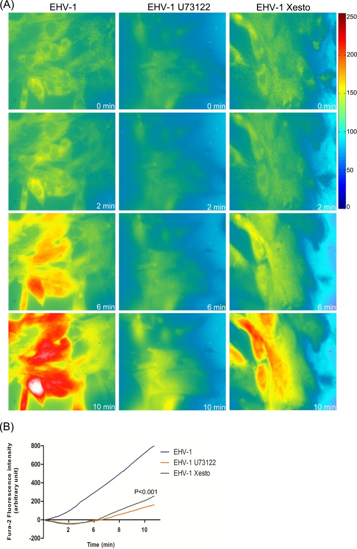
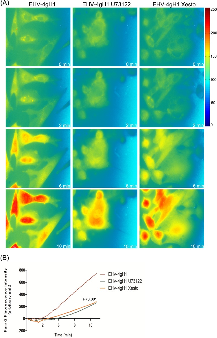
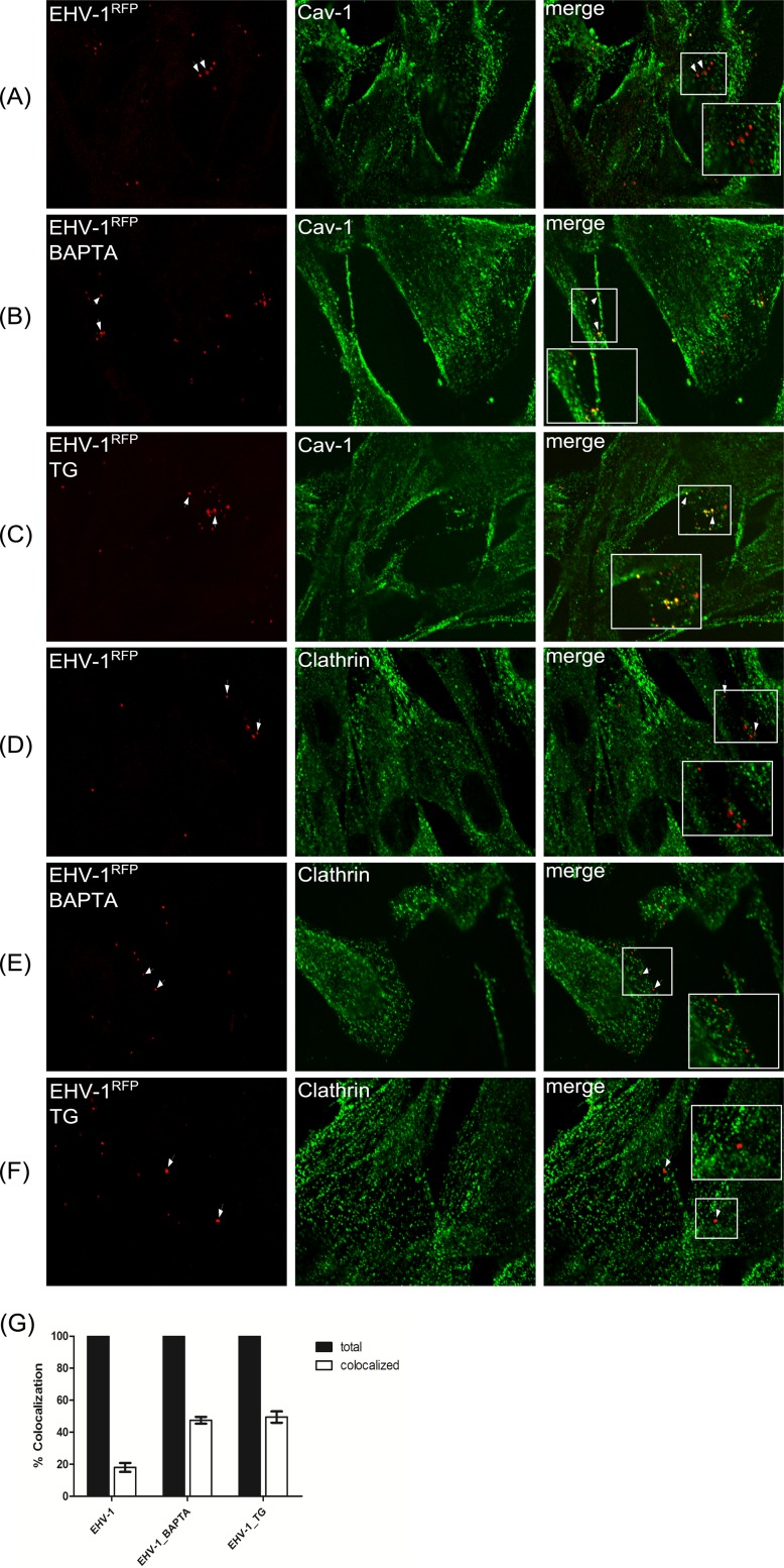
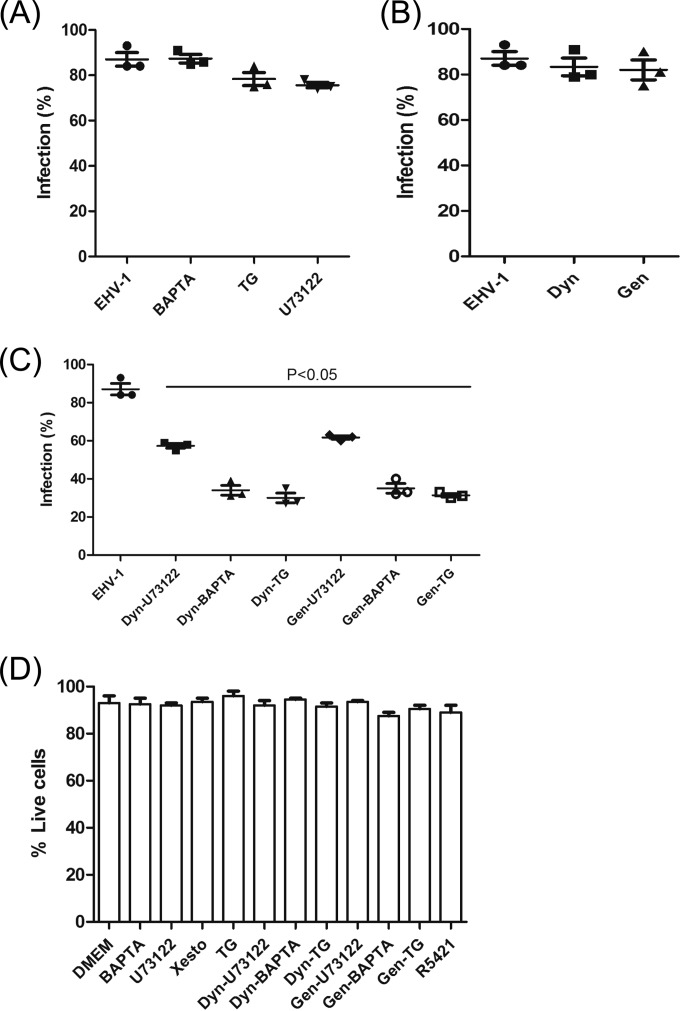
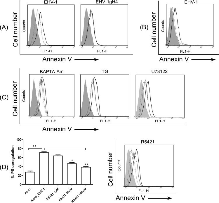
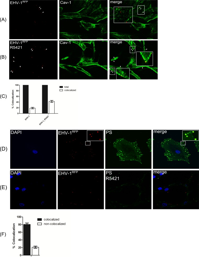
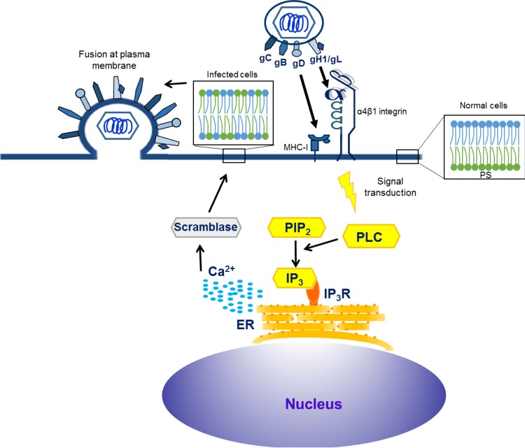
Intracellular signaling connected to integrin activation is known to induce cytoplasmic Ca(2+) release, which in turn mediates a number of downstream signals. The cellular entry pathways of two closely related alphaherpesviruses, equine herpesviruses 1 and 4 (EHV-1 and EHV-4), are differentially regulated with respect to the requirement of interaction of glycoprotein H (gH) with α4β1-integrins. We show here that binding of EHV-1, but not EHV-4, to target cells resulted in a rapid and significant increase in cytosolic Ca(2+) levels. EHV-1 expressing EHV-4 gH (gH4) in lieu of authentic gH1 failed to induce Ca(2+) release, while EHV-4 with gH1 triggered significant Ca(2+) release. Blocking the interaction between gH1 and α4β1-integrins, inhibiting phospholipase C (PLC) activation, or blocking binding of inositol 1,4,5-triphosphate (IP3) to its receptor on the endoplasmic reticulum (ER) abrogated Ca(2+) release. Interestingly, phosphatidylserine (PS) was exposed on the plasma membrane in response to cytosolic calcium increase after EHV-1 binding through a scramblase-dependent mechanism. Inhibition of both Ca(2+) release from the ER and scramblase activation blocked PS scrambling and redirected virus entry to the endocytic pathway, indicating that PS may play a role in facilitating virus entry directly at the plasma membrane.

Importance: Herpesviruses are a large family of enveloped viruses that infect a wide range of hosts, causing a variety of diseases. These viruses have developed a number of strategies for successful entry into different cell types. We and others have shown that alphaherpesviruses, including EHV-1 and herpes simplex virus 1 (HSV-1), can route their entry pathway and do so by manipulation of cell signaling cascades to ensure viral genome delivery to nuclei. We show here that the interaction between EHV-1 gH and cellular α4β1-integrins is necessary to induce emptying of ER calcium stores, which induces phosphatidylserine exposure on the plasma membrane through a scramblase-dependent mechanism. This change in lipid asymmetry facilitates virus entry and might help fusion of the viral envelope at the plasma membrane. These findings will help to advance our understanding of herpesvirus entry mechanism and may facilitate the development of novel drugs that can be implemented for prevention of infection and disease.


Figures

FIG 1
FIG 1
EHV-1 triggers the increase of cytosolic Ca2+. (A) ED cells were loaded with Fura-2AM, and live fluorescent images were taken every 5 s prior to and following the addition of EHV-1, ionomycin, EHV-4, or Ca2+-free medium (MEM) at time point 50 s. Shown is one representative image captured at each of the indicated time points. (B) The curves shown refer to the average of three independent experiments of fluorescence intensities of Fura-2AM versus time of excited ED cells being exposed to EHV-1, EHV-4, ionomycin, or MEM. P < 0.001 indicates a significant difference between EHV-1 and ionomycin compared to EHV-4 and MEM.
FIG 2
FIG 2
EHV-1 gH and cellular α4β1-integrin mediate cytosolic Ca2+ increase during virus entry. (A) ED cells were loaded with the Ca2+ indicator Fura-2AM and exposed to EHV-1, EHV-4, EHV-1gH4, or EHV-4gH1. (B) ED cells were incubated with 20 µg/ml of anti–α4β1-integrin MAb P4C2 for 1 h at 37°C. After washing, cells were loaded with Fura-2AM and exposed to EHV-1 and EHV-4gH1. (C) EHV-1 and EHV-4gH1 were incubated with soluble α4β1-integrin for 1 h at 37°C. The cells were loaded with Fura-2AM and exposed to the viruses. Changes in cytosolic Ca2+ levels were monitored using epifluorescence microscopy. Viruses were added at 50-s time point. The average from three independent experiments of fluorescence intensities of Fura-2AM versus time of exposure of ED cells to the viruses is shown. (A) P < 0.001 indicates a significant difference between EHV-1gH4 and EHV-4gH1 compared to parental EHV-1 and EHV-4, respectively. (B) P < 0.01 indicates a significant difference between EHV-1 and EHV-4gH1 viruses in the presence or absence of the integrin antibodies. (C) P < 0.001 indicates a significant difference between EHV-1 and EHV-4gH1 viruses in the presence or absence of soluble α4β1-integrin (Sol intg).
FIG 3
FIG 3
Inactivated virions trigger cytosolic Ca2+ increase. (A) EHV-1 and heat-inactivated EHV-1 (EHV-1-HI) were added to Fura-2AM-loaded ED cells. Live images were monitored and captured at the indicated time points. Viruses were added at the 50-s time point. (B) Fluorescence intensities of Fura-2AM as a function of time after exposure of ED cells to EHV-1 and EHV-1-HI are shown. Data are represented as the averages from 3 independent experiments.
FIG 4
FIG 4
PLC and IP3R are required for Ca2+ release from ER. (A) ED cells were treated with U73122 or (−)-xestospongin C (Xesto) for 30 to 60 min at 37°C prior to infection. The cells were washed, loaded with Fura-2AM, and exposed to EHV-1. Release of Ca2+ was monitored, and images were taken at the indicated time points. Viruses were added at the 50-s time point. (B) Fluorescence intensity of Fura-2AM as a function of time after exposure of ED cells to EHV-1 in the presence or absence of U73122 or (−)-xestospongin C. The lines show averages from 3 independent experiments. P < 0.001 indicates a significant difference between EHV-1 in the presence and absence of the used inhibitors.
FIG 5
FIG 5
EHV-1 gH activates PLC-IP3R to induce Ca2+ release from ER. (A) ED cells were treated with U73122 or (-)-xestospongin C (Xesto) for 30 to 60 min at 37°C. The cells were washed and loaded with Fura-2AM before being exposed to EHV-4gH1. Release of Ca2+ was monitored, and images were captured at the indicated time points. Viruses were added at the 50-s time point. (B) Fluorescence intensity of Fura-2AM as a function of time after exposure of ED cells to EHV-4gH1 in the presence or absence of U73122 or (−)-xestospongin C. The data show averages from 3 independent experiments. P < 0.001 indicates a significant difference between EHV-4gH1 in the presence and absence of the used inhibitors.
FIG 6
FIG 6
Colocalization of viral particles with caveolin. ED cells were incubated with EHV-1RFP (MOI of 20) at 4°C for 2 h. Cells were incubated with either BAPTA-AM (B and E) or thapsigargin (TG [C and F]) before infection. The medium was replaced with preheated medium at 37°C, and cells were fixed at 5 min after shifting the temperature. Cells were stained with anti-Cav-1 (green [A to C]) or anti-clathrin (green [D to F]). (G) The percentages of virus particles colocalizing with caveolin after infection with EHV-1RFP and in the presence of inhibitors were determined in randomly selected fields of infected ED cells.
FIG 7
FIG 7
Effect of different inhibitors on EHV-1 infection. For treatment with a single inhibitor, ED cells were treated with BAPTA-AM, thapsigargin (TG), or U73122 (A) or dynasore (Dyn), or genistein (Gen) (B). (C) In the case of two inhibitors, the different inhibitor combinations are indicated. The cells were then infected with EHV-1 (MOI of 5) for 8 to 12 h. The percentage of infected cells was determined by flow cytometry. Error bars represent the means ± standard deviations from 3 independent experiments. P < 0.05 indicates a significant difference for means compared to the parental virus without inhibitor treatment. (D) Toxicity assays of pharmacological inhibitors on ED cells. Propidium iodide (PI) uptake is shown in cells following incubation for 8 to 12 h with the indicated inhibitors. The number of live cells (no PI uptake) relative to total cell numbers was determined after flow cytometric analysis and is given as a percentage. Error bars represent the means ± standard deviations from 2 independent experiments.
FIG 8
FIG 8
EHV-1 binding induces PS scrambling. (A) ED cells were incubated with EHV-1 or EHV-1gH4 and stained with FITC-labeled annexin V for detection of surface PS levels. Dotted lines, mock-infected cells stained with annexin V; solid black lines, virus-infected cells stained with annexin V. (B) Cells were incubated with EHV-1 and stained with FITC-labeled annexin V at 4°C. Dotted lines, mock-infected cells; solid black lines, EHV-1-infected cells. (C) Cells were treated with BAPTA-AM, thapsigargin (TG), or U73122 before infection with EHV-1. Cells were stained with FITC-labeled annexin V. Dotted lines, mock-infected cells; solid black lines, EHV-1-infected cells; gray lines, EHV-1-infected cells treated with different inhibitors. (D) Dose-dependent effect of the scramblase inhibitor. Cells were either mock infected or infected with EHV-1 in the presence of increasing concentrations of R5421 (left panel [*, P < 0.05; **, P < 0.001]) or with 100 µM R5421 (right panel). Surface-exposed PS was detected with FITC-labeled annexin V. Dotted line, mock-infected cells; solid black line, EHV-1-infected cells; gray line, EHV-1-infected cells treated with R5421. Data are from one representative experiment out of three.
FIG 9
FIG 9
Colocalization of EHV-1-labeled particles with caveolin after scramblase inhibition. (A and B) ED cells were incubated with EHV-1RFP (MOI of 20) at 4°C for 2 h in the absence (A) or presence (B) of R5421, and cells were stained with anti-Cav-1 antibody. (C) The percentage of EHV-1-labeled particles colocalizing with caveolin in the presence of R5421 was determined in randomly selected fields of infected ED cells. (D and E) Colocalization of EHV-1 with PS. ED cells were incubated with EHV-1RFP in the absence (D) or presence (E) of R5421. Surface PS was stained with FITC-labeled annexin V. (F) The percentage of EHV-1-labeled particles colocalizing with PS was determined in randomly selected fields of infected ED cells.
FIG 10
FIG 10
Proposed model of EHV-1 entry into equine epithelial cells. Virions initially bind to target (ED) cells through gD–MHC-I interaction, followed by activation of the gH/gL complex that interacts with α4β1-integrin. Interaction of gH with α4β1-integrin results in the activation of PLC and generation of IP3 that binds to IP3R and mobilizes Ca2+ from the ER. The release of Ca2+ activates scramblase, which exposes PS on cell surface. Scrambling of PS may facilitate fusion of EHV-1 at the plasma membrane. Blocking the gH-integrin interaction inhibits PS exposure and reroutes the virus to the endocytic pathway.

References

    1. Campadelli-Fiume G, Menotti L, Avitabile E, Gianni T. 2012. Viral and cellular contributions to herpes simplex virus entry into the cell. Curr Opin Virol 2:28–36. doi: 10.1016/j.coviro.2011.12.001. - DOI - PubMed
    1. Gianni T, Cerretani A, Dubois R, Salvioli S, Blystone SS, Rey F, Campadelli-Fiume G. 2010. Herpes simplex virus glycoproteins H/L bind to cells independently of αVβ3 integrin and inhibit virus entry, and their constitutive expression restricts infection. J Virol 84:4013–4025. - PMC - PubMed
    1. Sasaki M, Hasebe R, Makino Y, Suzuki T, Fukushi H, Okamoto M, Matsuda K, Taniyama H, Sawa H, Kimura T. 2011. Equine major histocompatibility complex class I molecules act as entry receptors that bind to equine herpesvirus-1 glycoprotein D. Genes Cells 16:343–357. doi: 10.1111/j.1365-2443.2011.01491.x. - DOI - PMC - PubMed
    1. Spear PG. 2004. Herpes simplex virus: receptors and ligands for cell entry. Cell Microbiol 6:401–410. doi: 10.1111/j.1462-5822.2004.00389.x. - DOI - PubMed
    1. Azab W, Harman R, Miller D, Tallmadge R, Frampton AR Jr, Antczak DF, Osterrieder N. 2014. Equine herpesvirus type 4 (EHV-4) uses a restricted set of equine major histocompatibility complex class I proteins as entry receptors. J Gen Virol 95:1554–1563. doi: 10.1099/vir.0.066407-0. - DOI - PubMed

Publication types

LinkOut - more resources