. 2015 Oct 20;6(5):e01578-15.
doi: 10.1128/mBio.01578-15.

The human skin double-stranded DNA virome: topographical and temporal diversity, genetic enrichment, and dynamic associations with the host microbiome

Affiliations

The human skin double-stranded DNA virome: topographical and temporal diversity, genetic enrichment, and dynamic associations with the host microbiome

Geoffrey D Hannigan et al. mBio. .

Abstract

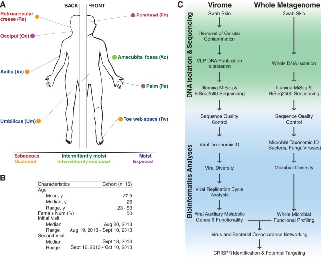
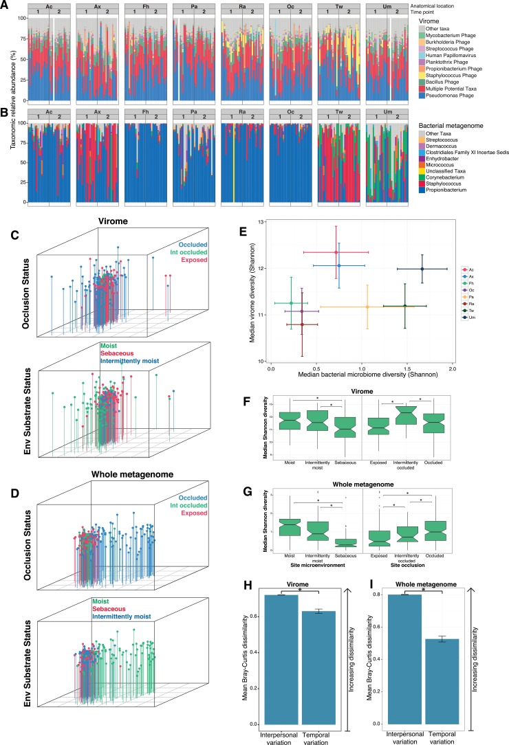
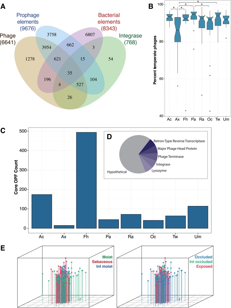
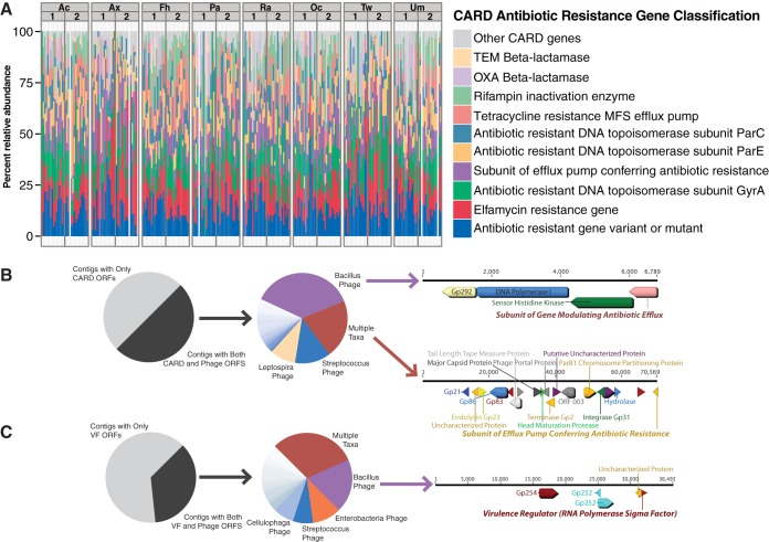
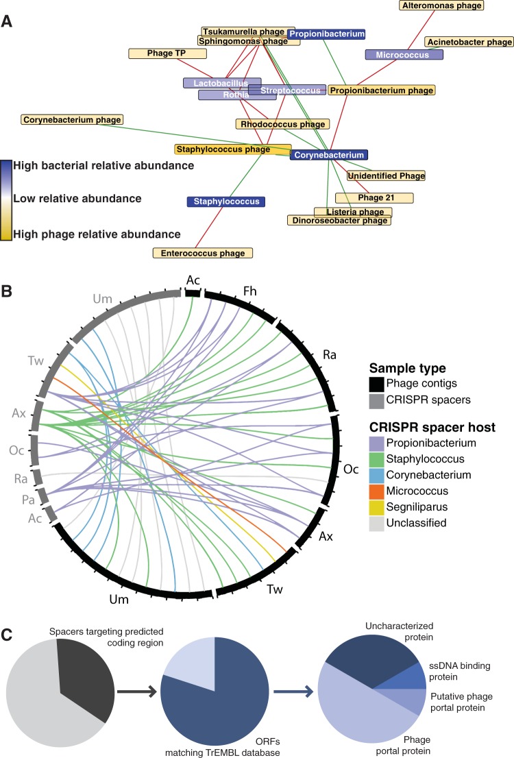
Viruses make up a major component of the human microbiota but are poorly understood in the skin, our primary barrier to the external environment. Viral communities have the potential to modulate states of cutaneous health and disease. Bacteriophages are known to influence the structure and function of microbial communities through predation and genetic exchange. Human viruses are associated with skin cancers and a multitude of cutaneous manifestations. Despite these important roles, little is known regarding the human skin virome and its interactions with the host microbiome. Here we evaluated the human cutaneous double-stranded DNA virome by metagenomic sequencing of DNA from purified virus-like particles (VLPs). In parallel, we employed metagenomic sequencing of the total skin microbiome to assess covariation and infer interactions with the virome. Samples were collected from 16 subjects at eight body sites over 1 month. In addition to the microenviroment, which is known to partition the bacterial and fungal microbiota, natural skin occlusion was strongly associated with skin virome community composition. Viral contigs were enriched for genes indicative of a temperate phage replication style and also maintained genes encoding potential antibiotic resistance and virulence factors. CRISPR spacers identified in the bacterial DNA sequences provided a record of phage predation and suggest a mechanism to explain spatial partitioning of skin phage communities. Finally, we modeled the structure of bacterial and phage communities together to reveal a complex microbial environment with a Corynebacterium hub. These results reveal the previously underappreciated diversity, encoded functions, and viral-microbial dynamic unique to the human skin virome.

Importance: To date, most cutaneous microbiome studies have focused on bacterial and fungal communities. Skin viral communities and their relationships with their hosts remain poorly understood despite their potential to modulate states of cutaneous health and disease. Previous studies employing whole-metagenome sequencing without purification for virus-like particles (VLPs) have provided some insight into the viral component of the skin microbiome but have not completely characterized these communities or analyzed interactions with the host microbiome. Here we present an optimized virus purification technique and corresponding analysis tools for gaining novel insights into the skin virome, including viral "dark matter," and its potential interactions with the host microbiome. The work presented here establishes a baseline of the healthy human skin virome and is a necessary foundation for future studies examining viral perturbations in skin health and disease.


Figures

FIG 1
FIG 1
Study design for the analysis of cutaneous viral and whole metagenomic communities. (A) Eight skin sites of 16 subjects were sampled. Colored text indicates the microenvironment classification, and each colored ball represents the occlusion status of the anatomical site. (B) Characteristics of the cohort sampled. (C) Flowchart illustrating the procedures by which DNA was isolated from cutaneous swabs and sequenced for downstream bioinformatic analyses.
FIG 2
FIG 2
Taxonomy and diversity of cutaneous viral and bacterial metagenomic communities. (A and B) Taxonomic relative abundance of the viral (A) and bacterial (B) communities by site over time. The viral relative abundance plots show the 10 most abundant taxa according to virus TrEMBL annotated contigs. The bacterial communities show the 10 most abundant taxa according to MetaPhlAn analysis. Each bar represents a single sample from a subject, and the bars are separated by time point and anatomical location as indicated at the top. (C and D) Nonmetric multidimensional scaling (NMDS) ordination plots of Bray-Curtis dissimilarities between virome (C) and whole-metagenome (D) samples, showing significant clustering (P < 0.001, Adonis test) by occlusion status and environmental substrate. (E) Alpha diversity (Shannon diversity metric) of the virome and bacterial metagenome for each anatomical site. The x axis represents median bacterial metagenome diversity, and the y axis represents median virome diversity. Each point is the median diversity of the two communities, and error bars indicate the population notch deviation of the median. (F and G) Viral (F) and microbial (G) Shannon diversity is presented by site microenvironment and occlusion, with asterisks indicating statistical significance (P < 0.05) by the Kruskal-Wallis and multiple-comparison post hoc tests. Box plots were calculated with the ggplot2 R package. (H and I) Intrapersonal variance compared to temporal variance of the virome (D) and the whole metagenome (E) as calculated by the mean (± the standard error of the mean) Bray-Curtis dissimilarity metric. A higher value indicates higher dissimilarity. An asterisk indicates statistical significance (P < 1.0−10).
FIG 3
FIG 3
Replication cycle and functional enrichment of bacteriophages on the skin. (A) Euler diagram of the phage contigs (yellow) that also contain an integrase gene (green), at least one prophage element per 10 kb (blue), homology to a known bacterial genome (red), or a combination of these markers. (B) Box plot illustrating the percent relative abundances of predicted temperate phages per body site. Temperate phage contigs were defined as those that contained both a phage gene at least every 10 kb and one of the other three temperate markers. Relative abundance was calculated as the relative number of reads per kilobase of transcript per million mapped unassembled reads that mapped back to the assembled contigs. An asterisk indicates statistical significance at P < 0.05 by the Kruskal-Wallis and multiple-comparison post hoc tests. (C) The distribution of exclusive OPFs associated with each anatomical site. (D) The distribution and UniProt annotation of the 15 core OPFs found across the entire virome. (E) Bray-Curtis dissimilarity of the virome samples by OPF relative abundance. Clustering was statistically significant (P < 0.001) by the Adonis test for both environmental substrate and occlusion.
FIG 4
FIG 4
Antibiotic resistance and bacterial virulence in the skin virome. (A) Relative abundances of predicted ARGs according to the CARD. Each bar represents a subject, and the bars are separated by time point and anatomical location as indicated at the top. (B) Flow diagram of the ARGs associated with bacteriophage contigs. The leftmost part shows the proportions of ARGs that colocalize on contigs with other phage genes or are themselves known phage-associated genes. The middle part shows the distribution of phage taxa that contain predicted ARGs. The rightmost part shows two annotated examples of ARGs colocalized on phage contigs, with the CARD-predicted ARGs in bold italics. (C) Similar to panel B, a flow diagram of the VFs associated with phages. As in panel B, the leftmost part shows the distribution of predicted VFs associated with phages, the middle part shows the taxonomic distribution of those phages, and the rightmost part shows an annotated example.
FIG 5
FIG 5
Modeled bacteriophage-host co-occurrence associations and CRISPR targets within the skin virome. (A) Network analysis of the correlations between bacteriophages of the virome and bacteria of the whole metagenome. Bacteriophages are represented by yellow boxes, while the bacterial genera are represented by blue boxes. The color intensity indicates the overall relative abundance of the taxon. The red lines represent a negative correlation, and the green lines represent a positive correlation. (B) Radial table showing bacterial CRISPR spacers (grey) that target viral phage contigs (black). The line colors represent the CRISPR spacer bacterial hosts. (C) Flow chart depicting the phage genome regions targeted by skin bacterial CRISPRs. The leftmost part shows the abundance of spacers that target a predicted coding region (ORF) within the phage genomes. The middle part is the distribution of ORFs matching a gene in the TrEMBL reference database. The rightmost part is the distribution of annotated coding region CRISPR targets.

References

    1. Hannigan GD, Grice EA. 2013. Microbial ecology of the skin in the era of metagenomics and molecular microbiology. Cold Spring Harb Perspect Med 3:a015362. doi: 10.1101/cshperspect.a015362. - DOI - PMC - PubMed
    1. Oh J, Byrd AL, Deming C, Conlan S, Program NCS, Kong HH, Segre JA. 2014. Biogeography and individuality shape function in the human skin metagenome. Nature 514:59–64. doi: 10.1038/nature13786. - DOI - PMC - PubMed
    1. Bohannan BJM, Lenski RE. 1997. Effect of resource enrichment on a chemostat community of bacteria and bacteriophage. Ecology 78:2303–2315. doi: 10.1890/0012-9658(1997)078[2303:EOREOA]2.0.CO;2. - DOI
    1. Rodriguez-Brito B, Li L, Wegley L, Furlan M, Angly F, Breitbart M, Buchanan J, Desnues C, Dinsdale E, Edwards R, Felts B, Haynes M, Liu H, Lipson D, Mahaffy J, Martin-Cuadrado AB, Mira A, Nulton J, Pašić L, Rayhawk S, Rodriguez-Mueller J, Rodriguez-Valera F, Salamon P, Srinagesh S, Thingstad TF, Tran T, Thurber RV, Willner D, Youle M, Rohwer F. 2010. Viral and microbial community dynamics in four aquatic environments. ISME J 4:739–751. doi: 10.1038/ismej.2010.1. - DOI - PubMed
    1. Tyson GW, Banfield JF. 2008. Rapidly evolving CRISPRs implicated in acquired resistance of microorganisms to viruses. Environ Microbiol 10:200–207. - PubMed

Publication types

LinkOut - more resources